The community site for and by
developmental and stem cell biologists

This is what a scientist looks like

Posted by , on 8 February 2012

What do you look like? The website This Is What A Scientist Looks Like wants to know. The site, run by science writer Allie Wilkinson, is collecting photos of scientists to show people what we look like. It’s an attempt to combat the very stereotypical view of scientists many people have. Just do a Google Image search for the word “scientist”, and you’ll find many messy-haired men in white lab coats. While some scientists may indeed look like the stereotype, most others don’t! This Is What A Scientist Looks Like shows that scientists come in all shapes and sizes, have hobbies and families, and look like everyone else:

(If you’d like to submit your own photo to the project, submission info is on the site.)

Thumbs up (3 votes)
Loading...

Tags: ,
Categories: Images, News

Navigate the archive

Use our Advanced Search tool to search and filter posts by date, category, tags and authors.

In Development this week (Vol. 139, Issue 5)

Posted by , on 8 February 2012

Here are the highlights from the current issue of Development:

ROCKed to the heart

Noonan syndrome – a common cause of congenital heart disease – is often associated with missense mutations in the protein phosphatase SHP-2. Interestingly, some types of leukaemia are associated with another subset of SHP-2 missense mutations. Here (p. 948), Frank Conlon and colleagues introduce SHP-2 that contains Noonan-associated mutations or juvenile myelomonocytic leukaemia (JMML)-associated mutations into Xenopus embryos to investigate how SHP-2 regulates heart development. Embryos that express SHP-2 containing Noonan-associated mutations have morphologically abnormal hearts, they report, whereas embryos that express SHP-2 containing JMML-associated mutations have normal hearts. The cardiac abnormalities caused by the Noonan-associated mutations are coupled with a delay or arrest in the cardiac cell cycle and with defective incorporation of cardiomyocyte precursors into the developing heart. Notably, these defects, which are caused by disruptions in the polarity of cardiac actin fibres and in F-actin deposition, can be rescued by inhibition of the Rho-associated, coiled-coil-containing protein kinase 1 (ROCK), which indicates that SHP-2 acts via ROCK to regulate the cardiac actin cytoskeleton during heart development.

Breaking symmetry during lateral root formation

Lateral root (LR) formation, which is essential for the construction of plant root systems, is initiated by coordinated asymmetric cell divisions (ACDs) of LR founder cells in existing roots. In Arabidopsis, LR formation is regulated by auxin signalling through the auxin response factors ARF7 and ARF19, which transcriptionally activate the plant-specific transcriptional regulator LBD16/ASL18 and other LBD/ASL genes. Hidehiro Fukaki and colleagues now provide new insights into the biological role of LBD/ASL genes in LR formation (see p. 883). They show that LBD16/ASL18 is expressed in Arabidopsis LR founder cells prior to ACD and that the spatiotemporal expression of LBD16/ASL18 during LR initiation is dependent on a specific auxin signalling module. Moreover, inhibition of LBD16/ASL18 and related LBD/ASL proteins in LR founder cells blocks nuclear migration, ACD and LR initiation. These results indicate that the localised activity of LBD16/ASL18 and related LBD/ASL proteins establishes the asymmetry of LR founder cells that is required for LR initiation, a key step in the construction of the plant root system.

Untangling the Hairy segmentation clock

During somitogenesis, an oscillating gene network generates a segmental pattern in the presomitic mesoderm. In zebrafish, this segmentation clock centres on cycles of transcription and self-repression of numerous hairy-enhancer of split related (her) genes, which encode proteins that dimerise, bind DNA and repress transcription. On p. 940, Scott Holley and co-workers systematically examine the physical interactions between Her proteins and test the ability of various Her protein dimers to bind to cis regulatory sequences. Dimerisation of Her proteins is specific, they report, with Hes6 serving as the hub of the network. Not all dimers bind to DNA, they note, but those that do so have distinct preferences for different cis regulatory sequences. Finally, Her7 disproportionately influences the availability of Hes6 for heterodimerisation with other Her proteins. The researchers propose, therefore, that Her7 has two functions within the zebrafish segmentation clock – direct repression of transcription through formation of a DNA-binding heterodimer with Hes6 and modulation of the network topology via sequestration of the network hub.

Diet and stem cells TORtuously linked

Nutritional status must be coupled to stem/progenitor cell proliferation and differentiation to ensure the proper growth and homeostasis of tissues, but how does diet regulate stem cell behaviour? On p. 859, E. Jane Albert Hubbard and colleagues show that rsks-1 [the homologue of mammalian p70 ribosomal S6 kinase (S6K), a target of the serine/threonine kinase TOR, which regulates cell growth and proliferation in response to nutritional cues] links cell fate, cell cycle and nutrient response in C. elegans germline stem/progenitor cells. They show that rsks-1 is required germline-autonomously to establish the proper number of germline progenitors, a role that requires a conserved TOR phosphorylation site in RSKS-1. Their analysis also reveals genetic interactions between rsks-1 and Notch, which suggest a prominent role for rsks-1 in cell fate control. Furthermore, dietary restriction causes germline defects similar to those observed in rsks-1 mutants and loss of rsks-1 renders the germline largely insensitive to dietary restriction. The researchers propose, therefore, that TOR-S6K signalling is a key nutrient-responsive regulator of germline progenitors.

Apical ECM and epithelial junction integrity

Polarised epithelial cells form many of the body’s surfaces, including the outer epidermis and the lining of several internal tubular organs. The apical surfaces of these epithelial sheets secrete a specialised extracellular matrix (ECM) that is generally viewed as a passive protective layer against pathogens, but does apical ECM have any other roles? According to Meera Sundaram and colleagues, the apical ECM in C. elegans might help to maintain epithelial junction integrity and, consequently, epithelial tissue integrity (see p. 979). The researchers report that the extracellular leucine-rich repeat only (eLRRon) proteins LET-4 and EGG-6 are expressed on the apical surfaces of epidermal cells and some tubular epithelia, including those of the worm’s excretory system. Mutants lacking one or more of these proteins, they report, have multiple defects in apical ECM organisation and, although epithelial junctions initially form correctly in these mutants, they subsequently rupture. Together, these results suggest that eLRRon-dependent apical ECM organisation might modulate epithelial junction dynamics and integrity.

Bimodal control of HoxD gene transcription

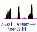
Correct innervation of peripheral muscles by spinal cord motoneurons is required to coordinate body movements in vertebrates. Hox proteins play an important functional role in achieving this innervation by specifying neuronal fates along the anteroposterior axis of the developing spinal cord. However, the mechanisms that generate Hox gene expression patterns are poorly understood. Here (p. 929), Denis Duboule and colleagues use tiling array-based transcriptome analyses and targeted deletions in vivo to investigate the control of HoxD gene transcription in the developing mouse spinal cord. They report that there are two distinct blocks of HoxD transcription that are regulated independently and that define two general expression territories. These territories, they show, are associated with the future nerve plexii at the brachial and lumbar levels. Given these and other results, the researchers propose that the establishment of spatial collinear HoxD domains in the developing mouse spinal cord involves the bimodal control of HoxD gene transcription by two independent ‘enhancer mini-hubs’.

Plus…

Human pre-implantation embryo development

Renee Reijo Pera and colleagues summarize what is currently known about human pre-implantation embryo development and highlight how further studies of human pre-implantation embryos can be used to improve ART and to fully harness the potential of hESCs for therapeutic goals. See the Primer article on p. 829

Retinoic acid signalling during development

Retinoic acid is a vitamin A-derived signaling molecule that acts as ligand for nuclear receptors, converting them from transcriptional repressors to activators. Here, Muriel Rhinn and Pascal Dolle review the main functions of retinoic acid during embryogenesis. See the Primer article on p. 843

Thumbs up (No Ratings Yet)
Loading...

Categories: Research

Freeware for scientists

Posted by , on 6 February 2012

After commenting on a previous post, I’ve decided to make my own post about freeware I use regularly that other scientists might find useful. All of these are available for Windows, Mac and Linux as far as I’m aware and come with various price plans if you want more storage space/functionality. You’ll need to create an account for some of them, but again that’s free.

Libre Office – I use my own laptop in the lab and as a result of that I don’t have some common software like Photoshop, Illustrator and most annoyingly, Microsoft Office. Rather than paying out of my nose for a productivity suite like Office, I chose to have a go at some open source alternatives. LibreOffice is an open source productivity suite that’s spent around 20 years in development, so it’s pretty stable. It comes with the same kind of programs as Office, a word processor, spreadsheets, presentation maker, drawing tools etc. It’s also compatible with MO as well, so you can open documents you created in program with the other. Migrating from Microsoft to LibreOffice was pretty easy and most of the layout and functionality is the same. I’ve been using this for a year or so now and I think whilst it still lacks the polish of Microsoft Office, it works just as well.

AlternativesGoogle Docs, laTEX (this is supposed to be specific for creating manuscripts)

Dropbox – I mentioned this in a comment on a previous post, but I’ll go into a little more detail here. Dropbox is basically a file syncronisation tool that you can download as a client program. Once installed, if you place a file in your Dropbox folder, it syncs it with your account so you can access that file from anywhere with an internet connection. It’s very easy to use and once you set up your Dropbox folder, you can share it with anyone. The syncronisation between folders on different computers is very fast, you can upload a file onto a shared Dropbox folder and within seconds the other person receives the file. This is very useful for collaborations or even just sharing data in a lab.

The servers they use to store files are very secure and you can create multiple sub-folders that you can share with various people. You get 2GB free storage but if you invite other people to share your folders or install it on another computer you get some extra storage.

AlternativesSugarsync

Mendeley – Mendeley is a web based reference manager that also has a desktop app you can download to organise PDFs and documents on your computer. The web app lets you build a library with an easy to use web importer that works as a plug-in to your browser. Mendeley also store pdfs on the (up to 500MB) and you can retrieve them from any computer or share them with other users by forming groups.

The Mendeley desktop app organises and indexed your PDFs that are stored on your computer. You can also annotate, highlight and add sticky notes to your files. As I begin to write my thesis, I’m finding this part increasingly useful. There is also a toolbar you can install in Microsoft Word or LibreOffice to cite papers whilst writing.

Unlike Endote, it’s free (although you can pay for more cloud storage) and unlike Papers, it’s a cross-platform tool (available for Windows, Mac and Linux), making it very useful for collaborations. I find the interface really easy to use and within half-hour of downloading it, I had most of my references stored on the desktop app. Also the ability to annotate and make notes on papers is proving to be invaluable.

AlternativesZotero

Reflect – This is a useful look-up tool when reading papers online. Basically, it’s a plugin for your browser that highlights proteins/molecules/biological concepts in any text. You can click on the highlighted text to show a pop-up window which displays some basic information such as what the molecule is, it’s role, structure and what it interacts with. The information displayed is community driven so for really obscure proteins and molecules there might not be a lot of information displayed, but the makers are keen on the community using it to edit and add more information.

Doodle – Doodle is a web-based time management tool that you can use to co-ordinate meetings. It does this by creating simple polls where everyone can vote on when they are free. You can use various calender programs like iCal, Microsoft Outlook or Google Calender to track dates and organise meetings with other people. I’ve never used it professionally, only with friends to organise some camping trips and it’s a pretty handy tool that saves on a lot of emails.

Alternatives –  Timebridge

OMERO –  OMERO is an microscopy image management tool created especially for scientists. It’s designed by the Open Microscopy Enviroment team which is based in multiple sites across the globe. Once you have an account set up and downloaded the programs, you can upload your images to a central server and process/analyse images and even make them ready for publication with a nice figure making tool. The Journal of Cell Biology has a data viewer based on OMERO that allows authors to upload images as they were acquired and users can look through z-stacks, time lapses and individual channels in these images.

If you have any tips for other useful freeware let us know in the comments section below.

Thumbs up (14 votes)
Loading...

Tags: ,
Categories: Discussion, Resources

Stem cells on the Slovenian slopes

Posted by , on 6 February 2012

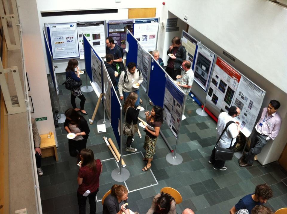
A couple of weeks ago, around 70 stem cell scientists gathered in the beautiful ski resort of Kranjska Gora, Slovenia, for the sixth meeting organised by the European Stem Cell consortium EuroSyStem. Although the snow wasn’t up to much (as the photo proves – just take a look at the opposite side of the valley!), the lack of fresh powder left more time for the science. And there was a lot of great science to be discussed…

Hans Clevers (Hubrecht Institute) kicked things off in the first of two outstanding plenary talks, with the latest developments on intestinal stem cell homeostasis, including beautiful demonstrations of how two innovative technologies – in vitro organoid culture, and the “Brainbow” cell labelling technique – have provided insights into the life of the Lgr5+ crypt stem cell. The name of Charles Leblond came up often in his talk: neither I nor many in the audience had ever heard of him, but his insights into stem cell self renewal, as well as his development of autoradiography, definitely earn him a place in the stem cell Hall of Fame (see here for a summary of his achievements).

The following day took us on a whistle-stop tour of model organisms, with planaria, flies, zebrafish, salamanders and Arabidopsis all taking their turn in the spotlight. After that, mammals took centre stage, with talks covering the whole spectrum of the stem cell field, from lineage determination and ES cell reprogramming, to aging and cancer. Prize for “Unsettling Animal Photo of the Week” (with apologies to the Guardian newspaper for blatant plagiarism of their feature) goes to Tom Rando (Stanford), whose lab has provided striking insights into systemic effects of aging from heterochronic parabiosis experiments – essentially grafting two mice together. Take home message: if you need a blood transfusion, you really want a young person’s blood! Other highlights included a lively debate on the Immortal Strand hypothesis following talks from Shahragim Tajbakhsh (Institut Pasteur) and Peter Lansdorp (Terry Fox Laboratory), and a detour into the molecular mechanisms regulating autophagy from Paul Coffer (University Medical Centre Utrecht).

Finally, the scientific program ended with an impressive demonstration of what money and technical resources can achieve, when coupled with hard work and – most importantly – a sharp nose for sniffing out an interesting story. The second plenary speaker, Huck Hui Ng (Genome Institute of Singapore), presented a tour-de-force analysis of the transcriptional and post-transcriptional networks underlying reprogramming, self-renewal and differentiation.

But the real talking point of the meeting came on the Wednesday evening, when we were fortunate enough to be joined by Arnd Hoeveler from the European Commission, who came to talk about future funding from the EC for stem cell research. While the direction the EC’s framework program is taking – towards funding mainly translational research – may not have gone down universally well with the audience of mostly basic researchers, we were given a fantastic forum to discuss science funding and politics with someone who clearly cares deeply about advancing science in Europe, and who faces a tough challenge to convince the political elite of the importance of the kind of research that this meeting was all about.

I’ve only had the chance to mention a few of the great talks, but all in all this was a fantastic conference, seamlessly organised by the EuroSyStem team. So thanks to them, the speakers and the rest of the participants for putting on an eye-opening and stimulating meeting. Now, if only they could have arranged for better piste conditions, it would have been just perfect!

Thumbs up (2 votes)
Loading...

Tags: ,
Categories: Events, Funding, Research

New Development editor

Posted by , on 1 February 2012

As of today, a new face has joined the team of Development editors, and a familiar one is soon to be leaving us. After 6 very successful years at the journal, Ken Zaret has decided to step down: we will miss him, but we’re sure the extra time he’ll have will be put to great use – both for his research and his other activities. Replacing Ken, and complementing particularly his expertise in epigenetics, we are delighted to have recruited Professor Haruhiko Koseki to the Development editorial team. Haruhiko is Group Director at the RIKEN Center for Allergy and Immunology in Yokohama, Japan. His lab is interested in understanding the molecular basis of epigenetic inheritance, and its consequences on haematopoiesis, stem cell identity and various other aspects of development. We’re excited to have Haruhiko on board, and we wish him luck for his first days and weeks in his new job!

Thumbs up (2 votes)
Loading...

Tags:
Categories: News

A wave from Quintay

Posted by , on 31 January 2012

The International course on Developmental Biology was a great experience, both instructive and mind-opening. All the students were shuttled to the remote and very small fishing village of Quintay, where the CIMARQ, the investigation centre where the course took place, is located. Originally a whaling station, this centre is dedicated to the instruction of professionals in the area of marine resources and has various branches of research mainly based in repopulation strategies of different species ranging from Sea Urchins to the delicious Conger eel or Sole fish. Their main objective is to provide small scale fish-farming to the general community. In fact, on the day of our arrival, after a Lecture on the history of and the main, original questions in Development by Dr. Roberto Mayor, we were given a short practical on Sea Urchin gamete harvesting and fertilization. This was followed by a very instructive tour of CIMARQ and its various projects, from seaweed culture (which is the main source of food for Sea Urchins) to the Conger and Cole fish tanks (see below). This course was unique in that it covered a wide range of developmental models instead of focusing on one or two: Throughout the twelve days of the course we had two days of each: Zebrafish, Xenopus, Planarian, Drosophila and Chick (plus a symposium and a first day tour). While including such a variety of different models may seem too optimistic (especially for just two days of each!), the truth is that the course was a huge success as proved by the fact that most of the experiments were successful. Our day schedule started with lectures and lab work in the morning. Then lunch, after which we spent most of the time in the lab and, after dinner, everyone attended presentations, by students, about their research. This part (the presentations) was a very good innovation this year and, given its success, it will probably continue in future courses. The discussions were very productive, and, from a student’s point of view, it was great having peak scientists listening, criticizing and suggesting experiments for my research. It was also good to share our areas of research between students since it was very different from the casual exchange of area of research in informal gossip. So, on to the course.

Zebrafish module

Zebrafish was coordinated by Dr. Kate Whitlock. The first Lecture was on Zebrafish basics (rearing and genetics) and embryo morphology. We then proceeded to the lab in which work consisted of cataloging the effects of different concentrations of alcohol in zebrafish development by observation under dissecting microscope of live embryo general morphology and craniofacial development. Afterwards, we carried out an immunohistochemistry protocol for the detection of neuron and neural crest markers so as to further characterize the effects of ethanol in early development. To sum up the results, I would say that the message ¨Vertebrate development and alcohol don’t mix¨ was extremely clear: The deleterious effects on general and craniofacial development were patent even without the need for immunohistochemistry. The second lecture by Kate focused on neural crest development and how neural crest cells migrate and interact with the neural tube and placodes to give origin to the olfactory system At the lab, we studied gene expression of three main neuron and neural-crest marker genes (shh, sox10 and six4b) using in-situ hybridization. Finally, we observed fluorescent-tagged transgenic lines and we compared the results with those of immunohistochemistry and hybridization.

Xenopus module

Xenopus was the next chapter in this course and, again, experiments were very successful (albeit with a lot of effort). We began with a lecture from Dr. John Gurdon on the history of Xenopus as a Development model and classic experiments followed by a focus on the regulation of induction by molecule gradients. In the lab, we tried some of those same experiments ourselves: After a brief introduction by Roberto Mayor on egg collection and fertilization, we injected GFP mRNA into two, four and eight cell embryos. The next step was to create Nieuwkoop recombinants by separating vegetable and animal poles from different embryos and then setting them one against the other so that the vegetable pole would induce growth and mesoderm tissue in the animal pole. The following task was to graft neural crest tissue from GFP labeled neurulas into normal ones. Although it took some practice, after a few hours we successfully observed neural crest cells migrating under the ectoderm. On the second day, Roberto took the stand for a lecture on the post-fertilization phenomena of the Xenopus embryo and on the development and function of the neural crest. The final (and most challenging) experiment was to perform a Spemann organizer graft. After about five or ten minutes of dissection, John Gurdon displayed, with a proud smile, a clean and very neat graft. Although John definitely made it look easy, I had like four or five embryos which attest to the contrary. This was the price of success however as, although most of us agreed that it was harder than it looked, we managed to come up with several grafts which, at least, looked quite tidy. Due to a power shortage (and consequent rise in temperature of the incubator) we were unable to photograph many of those embryos, but the truth is that we were all very satisfied with our achievements.

Planarian module

Planarian was an interesting module in that it is a relatively new model and that we didn’t focus on embryogenesis but on regeneration instead (although we did have a very interesting lecture on planarian embryogenesis, which involves very rare and interesting processes). Planarians have unparalleled regeneration capacities and can regenerate a whole organism from a very small portion of the parent planarian. Dr. Alejandro Sánchez Alvarado was the scientist who established planarians as research models and it was great having him! Alejandro’s lecture on the establishment of planarians as regeneration research models and the similarities and differences between regeneration and embryogenesis was astounding. In the lab, we started out by cutting up worms in as many ways as we could think of. Over the following days, we got to see strange or downright weird forms of planarians as they regenerated the parts we had cut off. A second experimental part of this module consisted of dissociating cells, staining with Hoechst and observing  the cellular morphology of neoblasts (stem cells) among other cell types. In the third part we observed the differences in target proteins and tissue-specific markers between worms under normal conditions and worms either treated with RNAi or cut in half. I particularly enjoyed taking photos of these last worms showing the progressive regeneration of these systems and comparing the velocity and sequence of events that lead to the new worms. This was one of my favorite modules since I didn’t practically know anything about planarians past what I studied in an early zoology course (which seemed boring at the time) and, now, I can’t read enough about them!

Drosophila module

This module was taught by Drs Trudi Schüpbach, Eric Wieschaus and John Ewer. The first lecture, by Eric Wieschaus, was an interactive talk about fly genetics and fly crossing. We discussed the screen with which he identified genes that regulated embryogenesis. This was incredible and very instructive, because most of the time, we read about results without taking into account the real work that had to be done to obtain them. In the lab, we carried out several observational experiments: We were given embryos from unknown crosses and had to hypothesize what the parents´ phenotypes were by peeling embryos or bleaching them, followed by immersion in halocarbon oil or fixing in hoyers mountant. Another part of the practical consisted of analyzing mRNA expression (or localization) and observing embryo morphology and movement using transgenic lines. With the help of Trudi Schüpbach, we  also dissected ovaries and looked at oogenesis in transgenic lines with either GFP-tagged histones or a membrane-bound GFP. The second day, lead mainly by John Ewer, we focused on later stages of development. John gave a lecture about larval growth, physiology and metamorphosis concentrating on the reorganizing of the neural system during the pupal stage. In the lab we learned how to locate and remove imaginal discs from 3rd instar larvae and we watched the retraction and regrowth of sensory neuron axonal arbors and dendrites during the pupal stage Worthy of mention was Eric’s incredible enthusiasm with experiments and his loud cheering when the results were revealed (captured in photo). For me, all of the faculty of the course were extremely good professors: Their lectures were very clear and they were all very open to questions or doubts and were very watchful and helpful in the lab. Eric, however, was something else. I can’t actually explain how or why, but, as an example, he took it upon himself to single handedly sharpen most of our pincers to ease embryo peeling and larval dissection!

Chick module

The chick embryo was the last model and one of the most challenging, not only because of the complexity of dissection and grafting, but also because of how tired we were. After learning how to set up New cultures, we performed two experiments: Node grafts and cutting embryos in half. The first experiment, which is analogous to the one done in Xenopus, was intended to demonstrate how Hensen’s Node induces other tissues. In the second experiment we separated posterior and anterior halves of the embryo and observed their development, since the cells of each half reorganized and redefined the embryo axis. As professor Claudia Linker pointed out, in both of these experiments we had an impressive success rate (>90%), something most of us were very proud of! Additionally, we learned two other very useful techniques which were applied on embryos that were not removed from the egg: Embryo injection with either DNA or a fluorescent label and electroporation of the DNA-injected embryos. Although the success rate was lower, we did get to see some embryos with pretty neat dye labels and even a few good electroporations. Claudio Stern gave two more lectures on the molecular regulation and timing of neural specification and induction and a very interesting and comprehensive one integrating molecular and cellular processes that control, occur during and give rise to gastrulation.

Summing up…

As a student, I was extremely grateful to have had the opportunity to participate in this course. All the faculty were extremely helpful, friendly and sympathetic. In my experience, the closest I can get to scientists of the stature as the faculty of this course is by asking questions at lectures (if I’m extremely lucky). Sharing at least two days with them was very productive and actually giving them) a short presentation was incredible! I was given very good advice on how to guide my research and I also had some very interesting questions (the sort of that great minds usually ask)! Apart from the advantages/tricks/advice I learned for the model I currently work with, this course was very mind-opening: I learned about models that I practically had never heard of before and I feel comfortable about working, for example, with Zebrafish , Xenopus or Chick, three models I never though I would do experiments with! I’m currently thinking about how I can relate my research to one of these models and, hopefully, get my hands dirty working a few months in a lab which uses such models. I would strongly recommend this course for anyone with a strong curiosity and willing to take a look ¨outside the box¨. Please contact me at gersabio@gmail.com if you have any particular doubts about the course or this article and this is the course website: http://biodesarrollo.unab.cl/I wanted to shout out a special thanks for the three organizers: Alfredo Molina, Ariel Reyes and Roberto Mayor, without whom this course would not have occurred, for their dedication and very good will.

Germán Sabio

Thumbs up (10 votes)
Loading...

Tags: , , , , , , , , , ,
Categories: Education, Events, News

Society Journals and the Research Works Act

Posted by , on 31 January 2012

This post about the Research Works Act and the effect on society journals was written for, and first published on, Reciprocal Space. Reposted with permission, and edited to add a correction.

Much has been said about the RWA, and the involvement of big name publishers. Less discussed, but very important for many scientists, is the role that scientific societies and their journals have, and the impact of current or future publishing practices. Some societies, such as the American Association of Immunologists (AAI), the American Society of Nephrology (ASN), the American Heart Association (AHA), and the American Society for Clinical Investigation (ASCI), publish their own journals. Of these, very few (in this list only the ASCI), have an open access policy. Other societies, such as the Society for Developmental Biology (SDB), publish in journals operated by big publishers, such as Elsevier (in the case of SDB’s journal, Developmental Biology). So where do these societies and their journals stand on RWA?

Well in some cases it’s crystal clear. In letters that responded to a “Request for Information” (RFI) by the Office of Science and Technology Policy, Executive Office of the President, regarding “Public Access to Peer- Reviewed Scholarly Publications Resulting from Federally Funded Research””, the AAI wrote:

“…working in partnership with professional societies and other scholarly publishers offers the federal government the most cost-effective and efficient way of ensuring that private sector, scholarly journals survive, preserving their crucially important service of providing independent, expert peer review (accomplished at publishers’ expense) of government-funded scientific research.”

This refers to their perceived duplication of efforts in having PubMedCentral duplicating published manuscripts. The statement might make sense, if the journals were to offer free access after 12 months. But they continue with:

“…want to express our clear opposition to government mandates which require private sector publishers to make their legally-owned property (i.e., journal manuscripts, published articles and associated data) available online on sites other than our own, or to comply with a government-determined embargo period. These mandates allow the government to take private property without owner authorization or compensation, and threaten the sustainability of our nation’s premier peer-review publishing system.”

So, not so happy about providing free access. They also claim that PMC is an inferior means of disseminating and archiving published material. This is highly debatable, especially the permanence of published work. Finally, they answer many questions posed by the RFI, and include this delightful gem, which we’ve heard from Congresswoman Maloney:

“…increased “free” access is likely to benefit scientists in other nations, whether allies or enemies. In some instances, this will enhance international cooperation in the sciences, but it is not necessarily beneficial to the U.S. economy as even our friendly competitors will gladly take our research findings for free….. Neither publishers, nor the U.S. scientific enterprise, nor the U.S. taxpayer benefits from the “giving away” of our peer-reviewed publications.”

Oh dear. The point really is, these societies make most of their money from their publications, and of course feel threatened.

But then how does ASCI do it, publishing all papers in JCI for free? I don’t have the answer, but perhaps these societies should talk to each other…

In the case of the SDB, it’s a bit more complicated. They too make most of their revenue from their journal. But in this case, they fall under Elsevier’s control, and only receive a fraction of the journal’s revenues (around 10%). What can they do, stuck between a rock and a hard place? Currently they are debating what to do, so the jury is out. The SDB does provide an OA option stemming from agreements with HHMI and Wellcome Trust, that allows researchers to make their paper available as OA for a fee of $3,000. (The Company of Biologists, who publish Development, have a similar hybrid model and offer OA for a fee.)

Then the final question is for those of us who are members of these societies. Do we boycott our own society journals? Do we engage the leadership to try to convey our views? There is no clear answer, but it should be something. The editors of the journals are scientists, just like us. They understand, and they will listen. The societies have existed for a long time, and are an important part of science, in assembling scientists with shared interests in the form of conferences and journals, promoting scientific education, amongst other laudable goals. But many have their survival inextricably linked with their closed access journals.

As scientists we must ask ourselves how to help our scientific societies, while promoting open access.

Thumbs up (4 votes)
Loading...

Categories: Discussion

This month on the Node – January 2012

Posted by , on 31 January 2012

This round-up also includes a few posts from the end of December that didn’t make it into December’s post due to holiday scheduling.

Developmental Biology Bingo Game
We made a bingo game! You all left many suggestions for words to include in a developmental biology bingo game, and BenchFly turned that into a playable game. Visit their site to download bingo cards for everyone in your lab.

SDB CoRe
The SDB has set up a collaborative resource for teaching materials to use in developmental biology courses, and they are looking for submissions. Do you have great visuals that can be used in undergraduate teaching? Let Marsha Lucas know. She left more information in her post earlier this week.

Research

Just before Christmas, Hillel Kugler, of Microsoft Research, wrote about a project he worked on with Jane Albert Hubbard’s lab at the Skirball Institute.

“In our study, published in Development, we have built a computational model of germline development in C. elegans. In this model, germ cells move, divide, respond to signals, progress through mitosis and meiosis, and differentiate according to a developmental program specified for a “cell”. This developmental program incorporates cellular decision-making that influences germ cell behavior, as defined by a subset of cell components and their dynamic interactions.”

Other research recently covered on the Node included a paper that showed that a small change in bioelectric signals is enough to induce eye development in Xenopus, and an image highlighting the importance of Notch signaling in stem cell self-renewal and intestinal homeostasis.

Write for the Node
If you’re interested in writing for the Node, all you have to do is create an account and wait for approval. But sometimes inspiration is the limiting factor. If you’d love to write, and just want some suggestions and ideas, you can fill out this form, and we’ll occasionally send you some ideas. The first email has gone out this week, but if you sign up now, you’ll get that sent as soon as possible.

Books
Finally, we saw a few more book reviews this month. The last of the Development book reviews went up, in which Wendy Bickmore reviews “The Nucleus” (Edited by Tom Misteli and David L. Spector). We started 2012 with the first Node-exclusive book review: Sasha Terashiva reviewed “The Cell: A Very Short Introduction” (by Terence Allen and Graham Cowling).

Thumbs up (No Ratings Yet)
Loading...

Categories: Highlights

SDB Collaborative Resources (CoRe) Launched

Posted by , on 27 January 2012

The Society for Developmental Biology (SDB) has launched SDB Collaborative Resources (CoRe), an online collection of images, movies, and diagrams for learning and teaching developmental biology.  SDB CoRe is a free and open website developed to help increase understanding of developmental biology at all levels.

SDB CoRe is easily-searchable and can be browsed by topic, organism, or featured objects.  All objects have short descriptions aimed at helping users learn something about development with glossary words highlighted in green.  Object pages contain references as well as  links to related CoRe objects, links to reviews in the soon-to-be-launched WIREs Developmental Biology, and when relevant, to original research papers in SDB’s official journal Developmental Biology.  All users can create a My CoRe account in order to comment on an object or save it in their favorites.

SDB needs your help in building this community resource!  We are looking for visuals that help explain basic concepts in developmental biology across numerous plant and animal species.  Here are the guidelines for submitting to CoRe.  If you are an SDB member you can login to CoRe with your email address to submit.  Non-members that would like to submit to CoRe please contact me at info@sdbcore.org.  If you have any questions or suggestions for the site please email me as well.  Enjoy SDB CoRe!

Thumbs up (5 votes)
Loading...

Categories: Education, Images, Resources, Video

Would you use a digital lab notebook?

Posted by , on 26 January 2012

Nature featured a news article about digital lab notebooks, which launched a discussion in various places (including the comments of the article itself) about whether or not they’re useful.

What do you think? Would you use a digital lab notebook in your lab, or would you rather keep your old paper notebook? Or maybe you already keep all your notes in a digital format only. You’re all web-savvy Node readers, of course, but let’s see how digital you are when it comes to benchwork. Here’s a poll:


(more…)

Thumbs up (2 votes)
Loading...

Categories: Discussion, News