Thanks to everyone who interacted with the Node in 2022, it been another fantastic year for the site. We have a few new initiatives that we’ll be announcing soon, but first let us look back on 2022!
What are you looking at…
>400,000 total page views
18,483 views of our jobs board
4,992 view of our event calendar
What’s being posted…
226 blog posts, including 7 SciArt profiles, 7 Featured resources, 12 behind the paper stories and 12 preprint lists
At number two on our list was the livestream from our Development meeting ‘From Stem Cells to Human Development. Setting this up was a little nerve wracking and it was fantastic to see so many people tuning in live and viewing the recording, which featured talks from Sarah Teichmann and Sergiu Pasca and a panel discussion on ‘Technical, ethical and legal challenges of studying early human development’.
The third most read post started on a whim, with Alex Eve converting his popular #wordcountchop tweetorial into a blog post. If you have written a tweetorial on a topic relevant to developmental and stem cell biologists and want to give it a more permanent home, do considering sharing it as a blog post on the Node
Voting for your favourite Development cover comes in as our 4th most read post. Everyone loves a competition, and we had a worthy winner with the Issue 21, the mouse lung lobe from Prashant Chandrasekaran, Nicholas Negretti, Aravind Sivakumar, Jennifer Sucre, David Frank and colleagues. Keep your eyes out for our next competition, which is coming soon!
Joachim Goedhart’s Protocols for data visualization came in at number five. In this post, Joachim shared an update on his book, which brings aims to lower the barrier for using R and the ggplot2 package for data visualization. Joachim’s post on Data Visualization with Flying Colours, published in 2019, was again our most read post with a massive 49,405 views!
We would love to hear feedback and suggestions on how we can make the Node better in 2023. You can contact us using our contact form and at thenode@biologists.com. We are always happy to discuss ideas, comment on drafts, or help with website gremlins. Finally, remember the Node is your site and, once registered, you can post freely.
“This description of the creation of the first humans – Adam and Eve – from the biblical book of Genesis is a cool story. But in my opinion, the scientific truth about the origins of humans is way cooler – and an awful lot messier”
Dr Kat Arney
In the latest episode of the Genetics Unzipped podcast, we’re going back to the very genesis of our species in search of the genetic Adam and Eve. Who were they? When and where did they live? Were there really just two of them? And how should we really be referring to these ancient ancestors anyway?
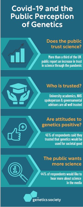
A survey of over 2000 British adults finds that trust in genetics is high, and went up significantly during the pandemic. It also finds that there is a hunger for more coverage of genetics.
The pandemic has gone hand-in-hand with a much-increased public profile of science – genetics in particular. Be it the prominence of PCR testing or the development of vaccines, genetics has been in the spotlight in an unprecedented way. Given this, researchers from the Universities of Bath, Cambridge, Oxford, UCL, and Aberdeen wanted to know what the public felt about genetics and whether this new exposure of the science has made a difference.
In a study funded by the Genetics Society, they commissioned a survey of over 2000 randomly selected British adults through public polling company Kantar Public. The researchers found that as a baseline most people were trusting of genetic technologies before the pandemic. Nearly half (45%) reported they trusted it to work for the societal good. 37% were neutral on this question, while 18% said they did not, and only very few (1-2%) were strongly distrusting. A descriptive report with all the answers from the questionnaire is now available on the Genetics Society website, along the technical report with panel sample and questionnaire: https://genetics.org.uk/public-perception-of-genetics/
When asked if their trust in genetics had gone up through the pandemic, four times more people said their trust had increased than those who reported that it had gone down. (as a control, the same increase in trust was not seen for sciences that were not involved in the pandemic but might be confused with genetics e.g. geologists not geneticists). Trust in science more generally had strongly gone up with a third saying it had increased. Not only has trust in science gone up, but people also want to hear more about it. Less than 10% thought that there is too much coverage of the science in the media, while 44% reported that they want to hear more about it.
Co-lead Professor Laurence Hurst of the Milner Centre for Evolution at the University of Bath commented “this is potentially important to know – scientists have a tendency to stick in their labs, but it looks like, for the most part, public not only trust us but that this trust has gone up somewhat and many want to hear more from us about our work.” As Professor Jonathan Pettitt, co-lead from the University of Aberdeen noted, “It is hard to see any upsides to the pandemic but perhaps this is one? We never knew that so many people wanted to hear more from scientists.” Prof Anne Ferguson-Smith, President of the Genetics Society and Professor in the Department of Genetics at Cambridge University reinforced this: “These results really challenge us to double our efforts. We need to rise to the new opportunity and the challenge created by the outcomes of this survey”.
However, co-lead Prof Alison Woollard of the Department of Biochemistry at the University of Oxford, cautioned: “We think we have established the limits of science communication. Despite all the talk of PCR over the last many months, we found that 30% hadn’t heard the term or knew it was a tool for testing for the virus. It is hard to see how any science can have more exposure than PCR has had. We need to be realistic and understand that, no matter what, we will never reach everyone. For informing people about things like vaccines this is important to know. Dr Adam Rutherford from the UCL department of Genetics, Evolution and Environment, (and prominent public science communicator) notes that ‘We often hear that trust in science is at a low point, but what we found is that most people do trust the science of genetics as the basis of how we address global issues such as pandemics. However, scientists should not be complacent: we also found that the exposure of genetics during the pandemic made those suspicious of science more distrusting, despite the evidence. In a world where these voices can easily be amplified, we must be vigilant that our processes, methodologies and results are clearly and transparently communicated.
Dr Cristina Fonseca, project coordinator for the Genetics Society (the funders of the project), noted that “having a representative random survey is really vital and allows us insight into the true diversity of opinions.”
Why do people hold highly variable attitudes towards well-evidenced science? For many years researchers focused on what people know about science, thinking that “to know science is to love it”. But do people who think they know science actually know science? A new study publishing January 24th in the open access journal PLOS Biology by Cristina Fonseca of the Genetics Society, UK; Laurence Hurst of the Milner Centre for Evolution, University of Bath, UK; and colleagues, finds that people with strong attitudes tend to believe they understand science, while neutrals are less confident. Overall, the study revealed that that people with strong negative attitudes to science tend to be overconfident about their level of understanding.
Whether it be vaccines, climate change or GM foods, societally important science can evoke strong and opposing attitudes. Understanding how to communicate science requires an understanding of why people may hold such extremely different attitudes to the same underlying science. The new study performed a survey of over 2,000 UK adults, asking them both about their attitudes to science and their belief in their own understanding. A few prior analyses found that individuals that are negative towards science tend to have relatively low textbook knowledge but strong self-belief in their understanding. With this insight as foundational, the team sought to ask whether strong self-belief underpinned all strong attitudes. The team focused on genetic science and asked attitudinal questions, such as: “Many claims about the benefits of modern genetic science are greatly exaggerated.” People could say how much they agreed or disagreed with such a statement. They also asked questions about how much they believe they understand about such science, including: “When you hear the term DNA, how would you rate your understanding of what the term means.” All individuals were scored from zero (they know they have no understanding) to one (they are confident they understand). The team discovered that those at the attitudinal extremes – both strongly supportive and strongly anti-science – have very high self-belief in their own understanding, while those answering neutrally do not.
Psychologically, the team suggest, this makes sense: to hold a strong opinion you need to strongly believe in the correctness of your understanding of the basic facts. The current team could replicate the prior results finding that those most negative tend also not to have high textbook knowledge. By contrast, those more accepting of science both believe they understand it and scored well on the textbook fact (true/false) questions.
When it was thought that what mattered most for scientific literacy was scientific knowledge, science communication focused on passing information from scientists to the public. However, this approach may not be successful, and in some cases can backfire. The present work suggests that working to address the discrepancies between what people know and what they believe they know may be a better strategy.
Professor Anne Ferguson-Smith, President of the Genetics Society and co-author of the study comments, “Confronting negative attitudes towards science held by some people will likely involve deconstructing what they think they know about science and replacing it with more accurate understanding. This is quite challenging.” Hurst concludes, “Why do some people hold strong attitudes to science whilst others are more neutral? We find that strong attitudes, both for and against, are underpinned by strong self confidence in knowledge about science.”
The Genetics Society, established 1919, is one of the world’s oldest societies devoted to the study of genetics and to the public understanding of genetics. It is an independent and unaffiliated charity.
The application process for Principal Investigator positions can be daunting, especially if you don’t know what to expect and don’t have the necessary support. For this reason, Development has created a new scheme, our Pathway to Independence (PI) programme, which will provide support, mentorship and networking opportunities for the selected researchers (PI fellows).
While we are only able to select a small number of applicants for the programme, we have been looking for other ways to support those applying for group leader positions. We have come across some fantastic resources that already exist to help candidates through this process (often from new group leaders), and we thought that the Node would be a great place to collate this information. Below, we have included some of the advice that we have come across, and we would like your help in continuing to build this collection. If you have written, used or have come across any useful advice, please get in touch via our contact page, email us at thenode@biologists.com or use the comments section below. Once we have collated this information, we’ll create a new page in our Resources section, which we can continue to update. Thanks to Arjun, Jessica, Kara and Daniel for letting us share their advice!
I sought out a lot of advice last year before starting faculty interviews. After being lucky enough to be invited for several myself I found what worked for me (and what didn’t) so I thought I’d share as the next cohort gets started. In order of my perceived importance:
It's faculty interview season! My ramblings about my experiences on the job market (biology/biomedical tenure-track jobs, 2019-2020) and various things I learned about surviving in-person interviews are here:https://t.co/EkrwyAkQpOhttps://t.co/kMG9KUJIjJ
I wrote a blog post on the academic job market. This is mostly to share my own materials like research/teaching statements, chalk talk slides, startup budgets, etc. But along the way I also give my best notes. Hope it helps some of y'all out there!https://t.co/4TGA1rXYVn
Sfrp2 is a multifunctional regulator of rodent color patterns Matthew R. Johnson, Sha Li, Christian F. Guerrero-Juarez, Pearson Miller, Benjamin J. Brack, Sarah A. Mereby, Charles Feigin, Jenna Gaska, Qing Nie, Jaime A. Rivera-Perez, Alexander Ploss, Stanislav Y. Shvartsman, Ricardo Mallarino
ZBTB20 is Essential for Cochlear Maturation and Hearing in Mice Zhifang Xie, Xian-Hua Ma, Qiu-Fang Bai, Jie Tang, Jian-He Sun, Fei Jiang, Wei Guo, Chen-Ma Wang, Rui Yang, Yin-Chuan Wen, Fang-Yuan Wang, Yu-Xia Chen, Hai Zhang, David Z. He, Matthew W. Kelley, Shiming Yang, Weiping J. Zhang
Pleiotropy of autism-associated chromatin regulators Micaela Lasser, Nawei Sun, Yuxiao Xu, Karen Law, Silvano Gonzalez, Belinda Wang, Vanessa Drury, Sam Drake, Yefim Zaltsman, Jeanselle Dea, Ethel Bader, Kate E. McCluskey, Matthew W. State, A. Jeremy Willsey, Helen Rankin Willsey
Probing the evolutionary dynamics of whole-body regeneration within planarian flatworms Miquel Vila-Farré, Andrei Rozanski, Mario Ivanković, James Cleland, Jeremias N. Brand, Felix Thalen, Markus Grohme, Stephanie von Kannen, Alexandra Grosbusch, Han T-K Vu, Carlos E. Prieto, Fernando Carbayo, Bernhard Egger, Christoph Bleidorn, John E. J. Rasko, Jochen C. Rink
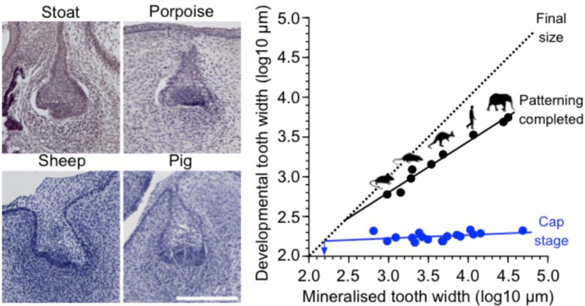
The developmental basis for scaling of mammalian tooth size Mona M. Christensen, Outi Hallikas, Rishi Das Roy, Vilma Väänänen, Otto E. Stenberg, Teemu J. Häkkinen, Jean-Christophe François, Robert J. Asher, Ophir D. Klein, Martin Holzenberger, Jukka Jernvall
Molecular and spatial design of early skin development Tina Jacob, Karl Annusver, Paulo Czarnewski, Tim Dalessandri, Maria Eleni Kastriti, Chiara Levra Levron, Marja L Mikkola, Michael Rendl, Beate M Lichtenberger, Giacomo Donati, Åsa Björklund, Maria Kasper
A cell atlas of human adrenal cortex development and disease Ignacio del Valle, Matthew D Young, Gerda Kildisiute, Olumide K Ogunbiyi, Federica Buonocore, Ian C Simcock, Eleonora Khabirova, Berta Crespo, Nadjeda Moreno, Tony Brooks, Paola Niola, Katherine Swarbrick, Jenifer P Suntharalingham, Sinead M McGlacken-Byrne, Owen J Arthurs, Sam Behjati, John C Achermann
Aging Fly Cell Atlas Identifies Exhaustive Aging Features at Cellular Resolution Tzu-Chiao Lu, Maria Brbić, Ye-Jin Park, Tyler Jackson, Jiaye Chen, Sai Saroja Kolluru, Yanyan Qi, Nadja Sandra Katheder, Xiaoyu Tracy Cai, Seungjae Lee, Yen-Chung Chen, Niccole Auld, Doug Welsch, Samuel D’Souza, Angela Oliveira Pisco, Robert C. Jones, Jure Leskovec, Eric C. Lai, Hugo J. Bellen, Liqun Luo, Heinrich Jasper, Stephen R. Quake, Hongjie Li
Next-generation plasmids for transgenesis in zebrafish and beyond Cassie L. Kemmler, Hannah R. Moran, Brooke F. Murray, Aaron Scoresby, John R. Klem, Rachel L. Eckert, Elizabeth Lepovsky, Sylvain Bertho, Susan Nieuwenhuize, Sibylle Burger, Gianluca D’Agati, Charles Betz, Ann-Christin Puller, Anastasia Felker, Karolína Ditrychová, Seraina Bötschi, Markus Affolter, Nicolas Rohner, C. Ben Lovely, Kristen M. Kwan, Alexa Burger, Christian Mosimann
After a short break, our first ‘Developing news’ post of 2023 focusses on disruption in science.
How good is ChatGPT?
One of the main talking point on Twitter in December was ChatGPT, a chatbot from OpenAI that interacts with the user in a conversational tone. The success of ChatGPT has prompted worries of how the technology could affect student assessment and even write scientific papers!
U.S. universities are starting to overhaul classrooms in response to the new AI chatbot ChatGPT, prompting a potentially huge shift in teaching. Some professors are redesigning their courses, even choosing handwritten assignments over typed ones. https://t.co/5B6eEPyBS1
First article with ChatGPT as coauthor 🤨. Next article: Word, M., Excel, M., and GPT, C. (2023). 'Does Adding Software as Authors Result in More Citations? An Ongoing Experiment' pic.twitter.com/M8IfabC1Tu
A new paper analysing the frequency of major new directions (or disruption) of science over time prompted discussions on i) whether this is true, ii) the causes and iii) whether we should be worried. We have picked out some examples of the chat below and recommend that you click the links for the full discussion.
New Nature paper provides evidence that science has become less innovative since the 1950s. The authors suggest reversing the trend by: 1. reading widely, 2. focusing less on quantity of papers, & more on research quality, 3. taking year-long sabbaticals.https://t.co/oYV5JCXRl8pic.twitter.com/viN2IJ0cSn
This is a thought provoking article, If it is not the quality of published work than what is causing it! 🧐😶🙄🤔thread below 👇🏻 Papers and patents are becoming less disruptive over time | Nature https://t.co/RIwG1VuiMu
Piping hot morning coffee take: if there really is a decline in "disruptive" science, could it be linked to our easy, finger-tip access to *all* the literature? Maybe out-of-the-box thinking is correlated with naivety and ingnorance?
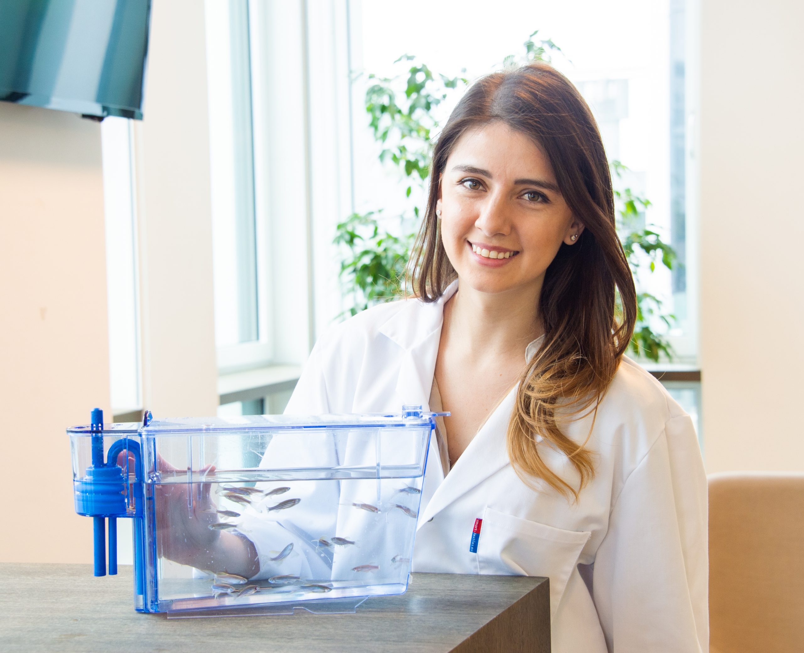
Victoria Deneke, a postdoc in Andi Pauli’s lab at the Vienna BioCenter, was the winner of the 2022 Society of Developmental Biology (SDB) Trainee Science Communication Award. Victoria is passionate about developing communities within the science world and creating opportunities for scientists from low-income countries. We caught up with Victoria to find out about her outreach and communication work, as well as her research career.
Where are you originally from and when did you first get interested in science?
I was born and raised in San Salvador, the capital city of El Salvador in Central America. Whilst growing, I was lucky enough to have access to school, and received a very good education. When I was 18, I applied for university places in the United States and that was when I really picked up science academically, but I’ve always been interested in the natural world. For example, I remember being fascinated by snake scales as a child and one of my favourite experiments in middle school was to dissect a frog to study the vasculature and the amazing organisation of the organism. Despite this early interest in biology, I decided to study chemical engineering as an undergraduate, as I thought that degree would be more versatile, either allowing me to return to my home country, or to develop elsewhere. Biology is not a very developed topic in El Salvador, but after my undergraduate degree I found myself being drawn back to biology. I decided to apply for umbrella PhD programmes, which give you access to a broad range of biology departments. You do rotations in different laboratories and then choose one for your PhD. This was an important aspect of the programme for me, particularly as I didn’t have a strong background in biology as an undergraduate. That’s how I ended up joining Stefano Di Talia’s lab, which was my fourth laboratory rotation. I very quickly fell in love with the research that was ongoing at the lab, which had just started at Duke. I was one of the first two graduate students that joined and the first graduate student that joined a fruit fly project. During my rotation, I was imaging early Drosophila embryos, and in particular the nuclear divisions that occur in the first few hours of development. These events are remarkably synchronous, and the waves of division spread through the embryo. My first PhD project was trying to figure out how these divisions are synchronised in the syncytial embryo. We found that there are waves of chemical activity via cyclin dependent kinase 1 (Cdk1), and through a combination of diffusion and positive feedback, these waves can synchronise the whole cytoplasm within minutes. It was the perfect project for me because it was very visual – I got to see and make these beautiful movies every day of my PhD! At the same time, the project involved not only biological concepts, like kinases and cell cycle regulation, but also concepts from physics and maths. For example, how the signal spreads, and the dynamics and the kinetics of chemical waves. This meant I got to collaborate with physicists, and indeed Stefano is a physicist by training, so I felt this project really suited my engineering background.
You said this was your first PhD project, can you tell us what you worked on for the rest of your PhD?
For the second half of my PhD, I decided to look at the role of cytoplasmic flows in spreading nuclei in the Drosophila embryo. So, originally these nuclei are all clustered in one part of the embryo and then there are beautiful contractions and subsequent flows that emerge and appear to move the nuclei. The regulation of the contractions is fascinating, and we assumed they must be tightly regulated for the nuclei to become so uniformly spaced through self-organisation. I started out by making movies of both the nuclei and the flows, trying to correlate these two movements. I went on to use optogenetics to perturb the contractions and monitor how that affects the nuclear spreading. Another beautiful, visual project where we really benefited from the application of quantitative biology methods. We tracked the cytoplasm and the nuclei and established quantitative relationships between these two movements to build a model. From the model, we made predictions that we then tested experimentally. I loved both of these projects, they allowed me to combine both imaging techniques and mathematical modelling, to address questions about fundamental developmental processes. Overall, I couldn’t have picked a better place for my PhD given my background and the kind of biology that excites me.
For your postdoc, you moved to Andi Pauli’s lab in Vienna, Austria, can you tell us about this move and your research in the Pauli lab?
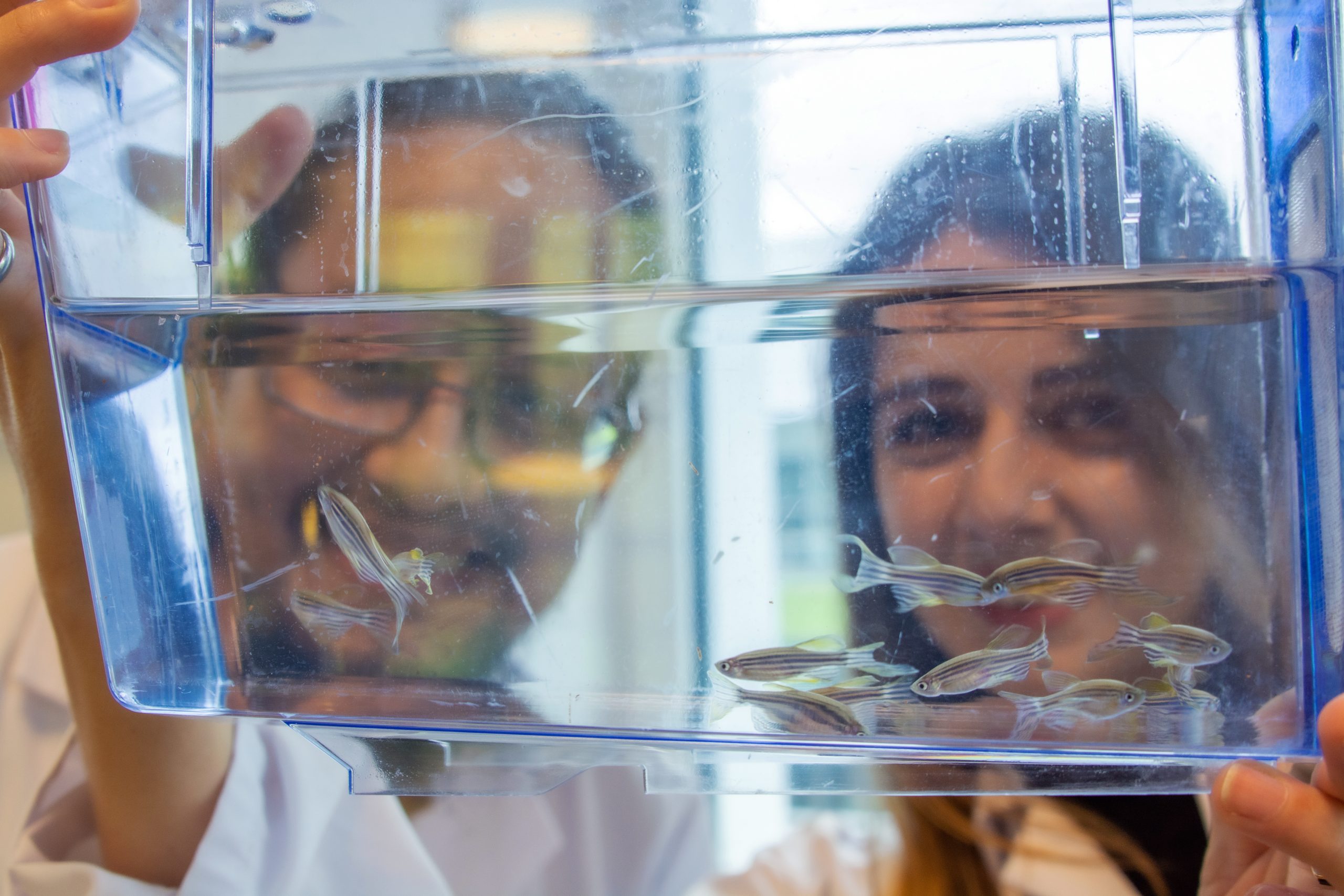
In the final year of my PhD, I started reading more broadly to find other scientific topics that interest me, and I always seemed to be coming back to oocyte and sperm biology. I’ve always thought that those cells are really fascinating and really special, and I was particularly drawn to the process of fertilisation, whereby these two cells have to bind specifically to each other and then fuse. I came across the first paper from Andi Pauli’s lab describing the discovery of a protein called Bouncer, which is on the surface of zebrafish eggs, and is an essential fertilisation factor. The other really cool thing that the Pauli lab found was that if you replace the zebrafish Bouncer with the medaka homologue, you can now fertilise the zebrafish eggs with medaka sperm, changing the compatibility of sperm and egg by just switching this one protein on the surface of the eggs. I thought this was amazing, so I reached out to her, keen to come for a visit. I was excited by the science and felt a good connection with Andi, so I decided to take a plunge and change completely scientific field, change model organism, change continents! It was quite a 180-degree change. I have been here for three years now, and I’ve really had such a good time. I think that picking an area of science that you’re just completely fascinated by for your postdoc is definitely the way to go. I don’t think I’ve ever had a day where I’m bored by my projects, I’m always wondering, what is really going on? How could we figure this out? So, I have been studying the process of fertilisation, mostly using zebrafish, and specifically I’m working on trying to decipher, which proteins, at the molecular level, are mediating this process, both on the sperm side and on the egg side. I love thinking about how these two cells come together and how they can achieve specific cell fusion between them. That’s a concept that really fascinates me and is my research interest in a nutshell!
Did you find switching model organisms a big change, or was that quite easy to do?
It was easier than I expected. I think it helped that the lab here (in Vienna) was already established and there were a lot of people in the lab that helped me pick up the zebrafish model organism. One of the biggest advantages of working with zebrafish is that the embryos are transparent. This means that you can look through a bright field microscope and watch development unfold. You can watch the embryo gastrulating and you can see even the somites forming, which was so exciting to me.
Congratulations on being awarded the 2022 SDB Trainee Science Communication Award, can you tell us what winning this award means to you?
This award means so much to me. I think oftentimes, this kind of work is considered extracurricular, and is overlooked. It’s wonderful that the SDB is recognising that outreach and communication are important aspects of academic science, and I think it shows the direction that we’re moving in. I think that being a well-rounded scientist is not just about making exciting discoveries in biology, but also about mentoring the next generation of scientists, and building an inclusive space for everyone. My short story of how I got involved into outreach work was when, after being in the US for a few years, I realised that I was in a unique position to contribute to my home country of El Salvador. I started doing simple outreach activities whenever I was back in El Salvador in the summertime. I would reach out to a local university and got involved with a workshop encouraging middle school girls to consider STEM fields. I would volunteer to give one of the Saturday morning workshops they do as part of a 12-week programme. One year I organised a workshop on pulleys. I borrowed some material from my high school and we ran this workshop together. So, that’s how it started and then eventually, as I got more involved with the Salvadoran community, I started reaching out to universities to also give motivational talks, to share my story. Then the next step was the fellowship that I created with my postdoc advisor Andi. But as you can hopefully see, the projects started very small, but through the years I built bigger and bigger initiatives.
Can you tell us a little more about the Austria-El Salvador Research Fellowship that you founded?
The idea originated from a yearly mentoring meeting I was having with Andi. We were talking about the Vienna BioCenter Summer School, which is open for students from all over the world to apply to come for a summer research internship at the Vienna BioCenter. It’s an amazing programme, but the realities are that students from lower income countries do not have access to the same opportunities as applicants from other parts of the world. This means that they usually don’t make the cut for the normal programme that we run here. I suggested to Andi that we could try to fill this gap by inviting one person for the summer, see how it goes and take it from there. Since I am very connected to the community in El Salvador, it was easy for me to broadcast the application within El Salvador and then find a student who would be motivated to come join us for a summer. We extended an offer to a student called Eduardo. He was here for three months and I was his direct supervisor. One thing that really stood out was that he was always so eager to come to the lab, and so grateful and amazed by the facilities. He loved the library and would go in the evenings to read books, access that we take for granted. It was really fascinating and inspiring for me as a mentor and made me really start appreciating the resources that we have here. Even though he had never had any research experience, he quickly picked up a lot of concepts. Eduardo is a very talented scientist, and it was a treat to mentor him. I also became aware of a lot of barriers within developing countries that kind of inhibit the progression of science. I’ve heard a lot of stories of having to use makeshift reagents and delayed services to developing countries, which means that science moves a lot slower because you just don’t have access to the same resources. For Eduardo, the fellowship was his first research experience, his first opportunity to try out the techniques he had read about, and it allowed him to be immersed in science. He has also taken his knowledge back to El Salvador. He has started a molecular biology club within his university, where they read papers together and they are creating a scientific community of students back home. It’s a small thing, starting with just one person, but I think in the long run it could really have an impact in how biology develops in in low-income countries, and in El Salvador, in particular.
The bulk of the funding for the fellowship was from the Vienna BioCenter, but I also made a GoFundMe page to have additional funds to use to cover Eduardo’s travel, as well as a small stipend to cover living costs during the three months. I think it is important to remember that if we’re going to bring someone from a low-income country, you cannot expect that that person will be able to cover a roundtrip flight from across the world. We really wanted to be as accommodating as we could and consider the reality of the applicant, including funding travel, visa, but also being aware of access to opportunities when considering the strength of the application. As we move forward in creating an inclusive scientific environment, we have to consider, are we missing out on talent because of barriers to access these kinds of opportunities?
I saw that you also ran a do-it-yourself workshop for teachers in El Salvador, how did this course work and did you intentionally target teachers rather than students?
The do-it-yourself microscopes is a workshop that was developed by Bob Goldstein at UNC Chapel Hill. The premise of his programme was to offer this workshop to teachers, in his case in North Carolina. I came across Bob’s work in a conference where he had a stand with these microscopes. I talked to him about the idea of bringing this workshop to a low-income or an underprivileged country. I thought it would be a perfect fit to introduce both teachers and students to science because it was very affordable, the microscopes are easy to make, but nonetheless, give you access to the microscopic world. I think that all of those factors make it a perfect outreach tool for low-income countries. I saw the value in Bob’s programme to target teachers so that they could expand their knowledge and amplify that effect to their students, so we decided to implement this programme in El Salvador in a very similar fashion. I translated all the material, but Bob has this ‘Ikea-like’ drawing of how to build this microscope, so you don’t need a lot of text. I was also lucky to partner with a local university and we recruited around 40 to 50 teachers from across the country. It was such a good workshop; everyone came out so happy and so proud that they built this microscope. It was fun watching people’s first reaction, when they put say a leaf under this microscope, and all of a sudden, they could see the cells. A lot of these teachers had never seen that, not even in a textbook. The teachers wanted to take the microscopes into to the classroom, but they also wanted to show their families because they were so excited by it!
How can we, as a community, better support and promote scientists from low- and middle-income countries?
We’ve touched on some aspects already, and I think that we are in a very unique position to be able to provide opportunities to students from disadvantaged backgrounds. As a community, we should support open calls, specifically to recruit people from low-income countries, or we could reserve spots within our existing programmes to include talent from disadvantaged backgrounds. And we need to think about the barriers could prevent people from participating in such programmes. The exercise of putting yourself in other people’s shoes could really transform the way that we run a lot of our research programmes and we are currently discussing how we could implement this here at the Vienna BioCenter. We are discussing how, within our current summer programme, we can reserve spots for students from low-income countries, and what would we need to provide to really support the students participating in the programme.
It’s exciting to think that something can start with a small project, inviting one person into the lab, but then if that could be amplified to say 20% of institutes offering a few weeks in the lab, there would be a huge number of scientists benefiting from this kind of experience.
Yes, that’s a really good way of thinking about it. It could be applied to any research institute across the world and could really have an impact. But I think it is good to start with a small ‘experiment’ and then really pitch for implementing something bigger. That’s my hope. I hope that for next summer we can already reserve ~20% of the spots on our research programme for students from underprivileged backgrounds with the idea that we recognise that talent is everywhere, and that science also benefits from a diverse talent pool. And so, in that effort, we have restructured the programme to allow for this enrichment.
You’ve spoken about Andi Pauli and Bob Goldstein as mentors. Do you have any mentors or role models either in research or science communication and outreach?
One person that comes to mind was someone that I intersected with during my time at Duke. I was really lucky to be part of this programme called the Biocore Scholars programme, and this programme is led by an amazing scientist, advocate, and communicator, Sherilynn Black. Sherilynn created this graduate student programme that’s designed to build a supportive community for PhD students of diverse backgrounds. I applied to be part of this programme and remained in the programme throughout my PhD. It became my scientific home during my PhD. It was a cohort of really diverse students that were in the programme to come together and support each other and collectively move through our PhD experiences. I really owe my PhD to this community. It has made me passionate about community building within scientific spaces that allow people to thrive. If I could be a fraction of Sherilynn Black, I will have made it in life!
What’s next for you, both short term and longer term?
In the short term, I’m really enjoying my postdoc here at the Pauli lab. I love mentoring students and I think that the scientific world is the perfect ground for building communities, for mentoring people and for coming together and communicating our science broadly. In the long term, it’s hard to say, but I would be looking for something that allows me to continue this role of creating community, communicating science, and mentoring students. I think that my future role could take shape in many ways for me.
Where do you think developmental biology will be in ten years?
During my PhD, I was introduced to the field of quantitative biology and the additional insights that quantitative methods can provide into the dynamics and the regulation of biological processes within developing organisms. I’m curious to see how this field is going to keep evolving. And not only that, but how interdisciplinary projects that use concepts from physics or concepts from computer science, can bring new insights to biological questions that have been studied for a long, long time. At the SDB meeting, for example, we started seeing how people are using AI to be able to predict differentiation cascades – I think that that is truly fascinating. And combining that with detailed data sets of transcriptional states of cells can really propel the developmental biology field forward. So, those are the things that I’m really excited about, but whether that actually ends up being the crux of developmental biology in 10 years, who knows, but that’s something that I look forward to reading about!
When you’re not in the lab, what do you do for fun?
I really love going on walks. I have a sister that lives with me here in Vienna and every weekend we pick a different district or a different neighbourhood in Vienna to walk around. I think it is a great way to know a city. I also love travelling, and one of the biggest advantages of being in Central Europe is that I have access to a lot of beautiful cities, just amazing historical places. When I travel, I enjoy getting to know the culture and the food of different countries. Those are two of my big ones, but I also really enjoyed dancing, so that’s something I like to do on the weekend.
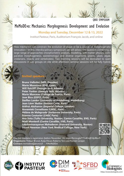
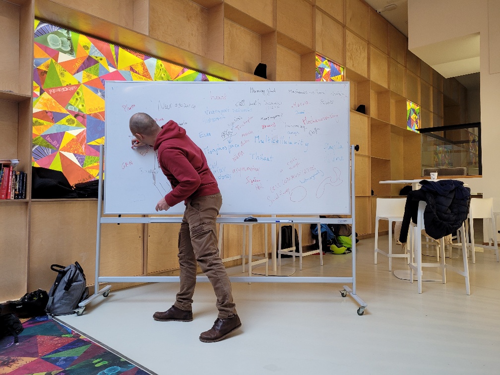
The diversity and complexity of shape in uni- and multicellular organisms has long been a source of fascination and interrogation. Since the seminal work of D’Arcy Wentworth Thompson, the study of the emergence of shape, so called morphogenesis, has been strongly influenced by the concept of emergence whereby complex pattern/shape can be explained by relatively simple mechanical and mathematical laws. In parallel, the rapid progress of the biochemical characterisation of the regulators of cell signalling has opened the possibility to compare developmental programs and dissect the molecular basis of evolution. Yet, how mechanics and mathematical laws may constrain the evolution of new shapes and morphogenesis has only come back to light recently. For two days (12 and 13th of December 2022), the symposium MeMoDEvo taking place in Institut Pasteur in Paris tried to discuss this issue by gathering interdisciplinary speakers and participants on site and online and organising two mornings of open discussions. The discussions and talks covered a large range of approaches (mathematics, fluid mechanics, soft matter physics, genetics, evolution, developmental biology, epistemology…) showcasing a diverse set of organisms, from mammals, birds, reptiles, insects, choanoflagellates, plants, algae, yeasts, and bacteria.
This fully hybrid meeting gathered close to 100 participants on site (mostly from Europe) and 100 participants online and its unusual format led to very interesting discussions and interactions. The conference was possible thanks to the support of several sponsors, including The Company of Biologists, the French Society of Developmental Biology, the Qbio initiative in Pasteur and the Pasteur Institute, as well as the DIM Elicit initiative
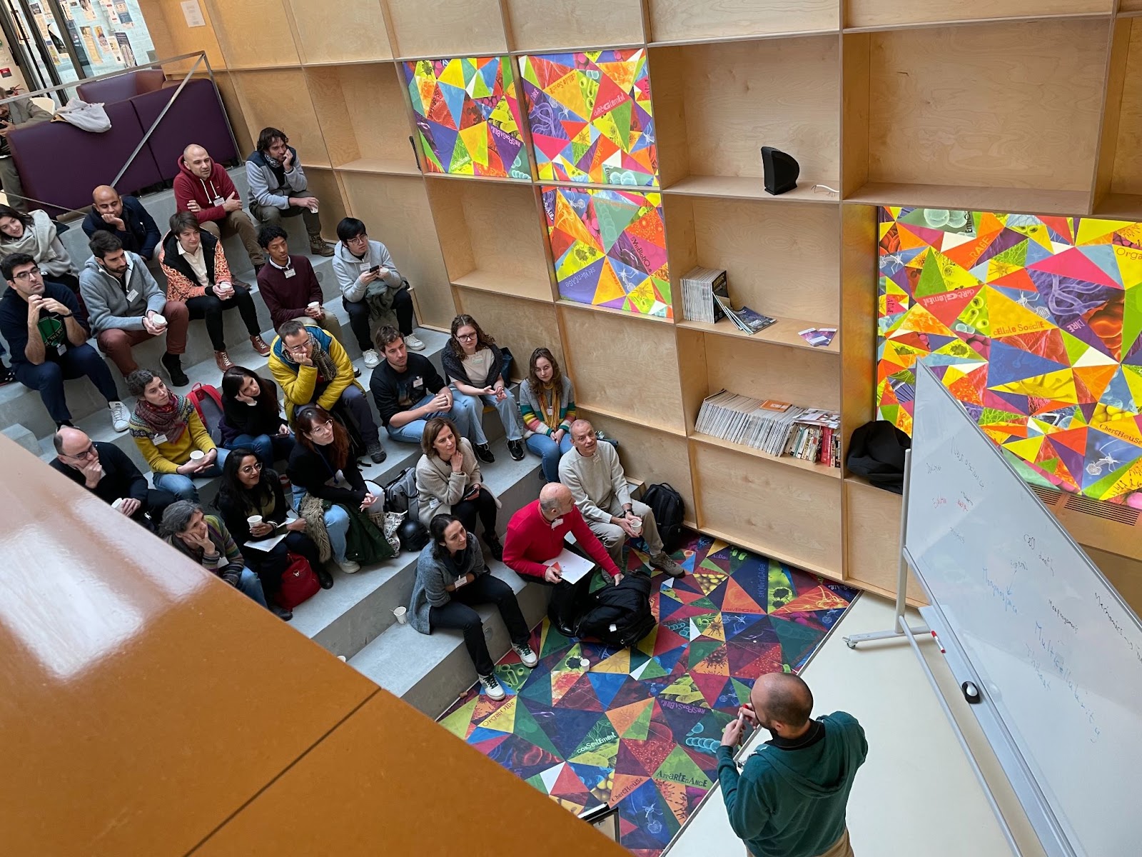
“Unconference” morning sessions and open discussions
One of the best parts of conferences that we had been missing over the past two years is the free time and open discussions in between sessions. We therefore dedicated a significant amount of time to open discussions and round tables during mornings with a subset of participants and the speakers (roughly 40 people). In line with the spirit of the meeting, we relied on a mixture of programmed schedule and self-organization.This engaging time involved first a quick introduction by each participant on their background and interest, followed by open discussions on the first day. This was followed the next morning by three round table discussions.
The first round table addressed constraints on the evolution of shape and how to reveal them experimentally. How can an organ evolve from one shape into another? Are there any limitations regarding shape innovation? The discussion included the distinction between pure physical/environment constraints and developmental hard-wired constraints related to the evolution history of the organism. Exploring the distribution of organ shape in the morphospace (using landmarks and dimension reduction) using intra and inter-specific variability can reveal such constraints by looking at non-occupied zones in the morphospace. The cause of these unoccupied areas can be either selective pressure or funnelling of evolutionary change by developmental constraints. A combination of description of natural variation and exploration of shape variability in the laboratory (including direct perturbation of developmental processes by mutagenesis and experimental evolution) may help to disentangle them. The discussion ended (as expected) with more questions than answers concluding on the mysterious cases of abrupt shape evolution/innovation which have to bypass strong developmental constraints while maintaining proper adaptation.
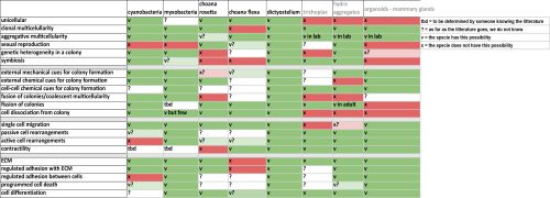
The second discussion was centred on the emergence of multicellularity comparing a large range of organisms which combine single cell and aggregate life mode. By comparing the mode of multicellularity (clonal/aggregative), the signals/conditions triggering aggregation or dissociation and the components that can structure and organise the aggregates (adhesion, contractility, matrix), a very complex pictures emerge with all sort of combination of strategies found in nature, outlining again the diversity of evolutionary path leading to multicellularity (see the table below summarising the comparison).
Finally, the last discussion was centered on sharing experience with various methodologies for mechanical simulation of tissues, ranging from continuous model, object based modeling and finite element modeling.
Afternoon hybrid sessions
The two afternoon sessions were following a more classic conference format alternating talk on site and online with an hybrid crowd. You will find hyperlinks connected to the published works that were discussed during these sessions (preprint and peer-reviewed articles). The meeting was launched by Thibaut Brunet (Institut Pasteur) who made a quick historical overview of the evolution of approaches to understand development and the various phases that brought to the front stage the role of mechanical constraints in shape evolution and its recent rejuvenation. The first talk was then given by Bruno Vellutini from Max Planck CBG who illustrated how the appearance of the cephallic furrow during evolution in Drosophila embryo may have helped to buffer mechanical constraints generated by ectoderm movements and cell division during gastrulation. Marie Monniaux, from the ENS RDP lab in Lyon, described the morphogenesis of petunia petals and sepals and how the comparison of mutants can help to disentangle the regulation of growth and shape by different epidermal layers. Jean-Léon Maître from Institut Curie then provided a quantitative comparison of the mechanisms of embryo compaction in different mammals, illustrating how qualitatively similar mechanisms can yet rely on quite different absolute mechanical properties. Arghyadip Mukherjee from the ENS in Paris described how topological transition through different modes of epithelial fusion can help to describe neuroepithelial organoid shape evolution. This was followed by Jose Bico, from the ESPCI, who illustrated how living matter can be used as an inspiration for generating complex inflatable shape based on local differences of inflation/growth (with a live illustration on stage with cup, saddle or helix shape generating devices). We then came back to insects with Steffen Lemke (from Heidelberg University) who compared gastrulation between different fly species (including Drosophila) and identified essential genes sufficient to explain several morphogenetic innovation in flies (including the mode of mesoderm invagination, cell elongation and the requirement for cephalic furrow). We then finished the first afternoon session with Annemiek Cornelissen from LMSC laboratory in Université Paris Cité, who used cracking theory and differential mechanical properties of tissues to explain the morphogenesis of jellyfish canal network.
The second afternoon session started with Arkhat Abzanof from Imperial College London who illustrated the power of morphospace analysis for the understanding of craniofacial shape evolution in vertebrates (from bats, Darwin finches and crocodiles). He was then followed by Hélène de Maleprade from Sorbonne Université who described various collective and single cell swimming strategies in Chlamydomonas, Volvox and Gonium. We then had an online talk from Lakshminarayanan Mahadevan from Harvard university describing fluid mechanics model of multicellular movements occurring during chick gastrulation and how the variation of initial conditions can recapitulate different modes of gastrulation across Vertebrates. This was followed by conceptual and theoretical considerations from Ana Soto and Maël Montevil from the Centre Cavaillès on the concept of autonomy in living systems and the definition of core principles (default state, variation and organisation) allowing proper understanding of a living system. We then came back to plants with Etienne Couturier from the LMSC lab who applied the Lockhart model (describing cell growth as a function of turgor pressure and viscoelastic deformation of the wall) to describe the dynamics of maïze root growth against a physical obstacle. This was then followed by an online talk from Peter Yunker from Georgia Tech describing his work in collaboration with Will Ratcliff on the ‘long-term experimental evolution’ of multicellular development in yeast. Remarkably, experimentally evolved yeast colonies were recently reported to have acquired macroscopic sizes after years of selection in the lab. The talk dissected the cellular and physical basis of this transition, which turned out to rely on mutations promoting elongated cell shape and reducing the mechanical stress that can cause colony fission. The meeting ended with a broad theoretical view of the evolution of multicellularity and morphogenetic innovation by Stuart Newman from New York Medical College, introducing the concept of dynamical pattern modules (integrating gene regulatory network and associated physical/spatial constraints) and their contribution to developmental and morphological innovation during evolution.
Conclusion: a lively and environmentally friendly format promoting discussions and connections
The meeting managed to gather a diverse crowd leading to a very refreshing and eclectic program. The relative “self-organisation” of the morning sessions actually led to very vivid interactions and deepening of the questions related to the meeting topic. All the participants came to conclusion that we should be ready for a MeMoDEvo#2 ! Of note, the symposium was the living proof of the possibility to organise a diverse, inclusive and very dynamics meeting while limiting environmental impact. Every speakers used the train to commute to the meeting and we could yet gathered a diverse crowd from Europe as well as many online attendees and speakers from other continents. Despite the usual technical hiccup associated with the hybrid format, we can only recommend the application of the same recipe !
Romain Levayer, Thibaut Brunet, Katja Heuer and Roberto Toro, organisers of the MeMoDEvo symposium.
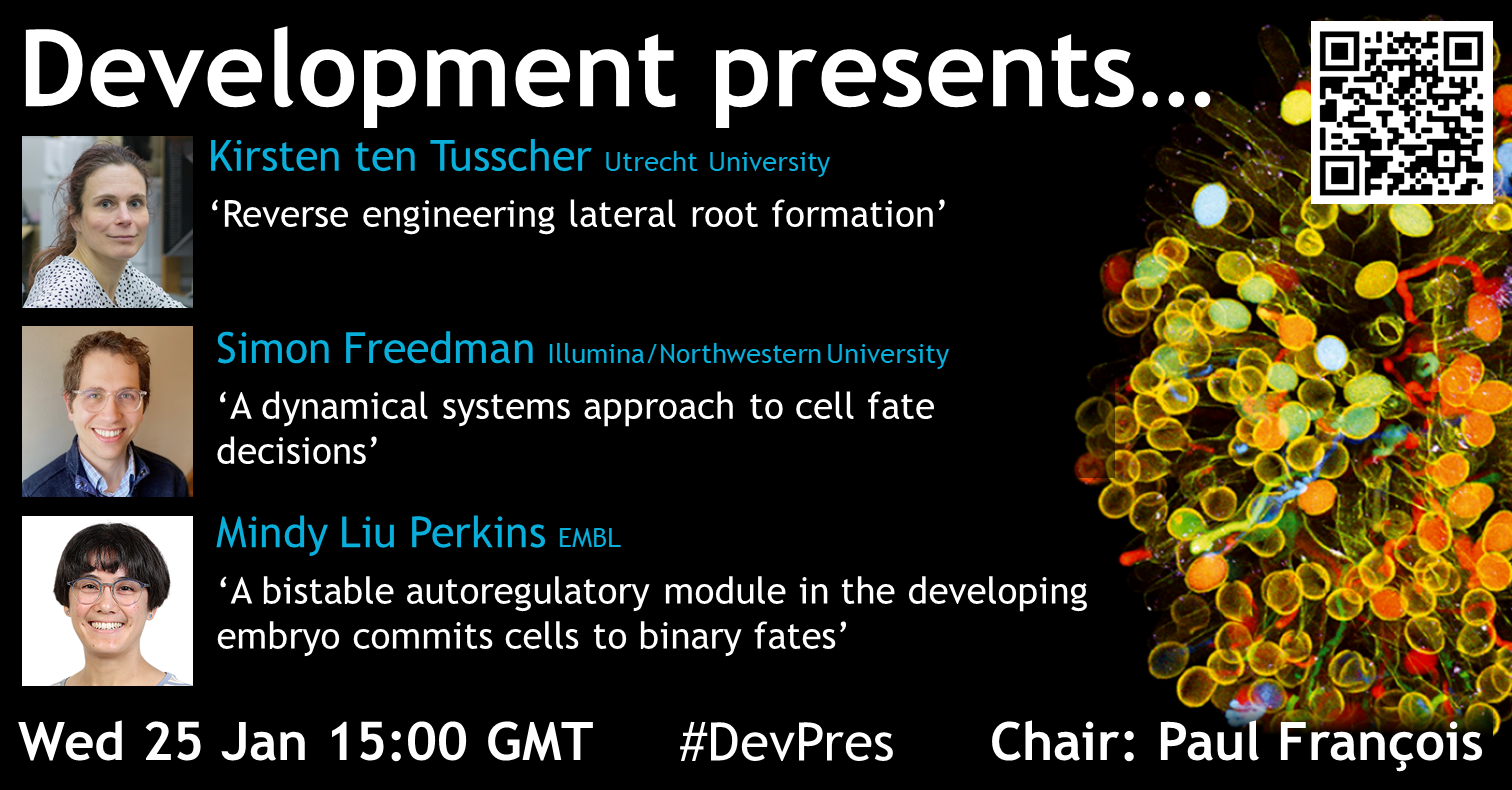
Our next Development presents… webinar will be chaired by Associate Editor Paul François (who recently moved to the University of Montreal from McGill University). Paul has invited three talks on the topic of theoretical and computational modelling of developmental biology.
Kirsten ten Tusscher (Professor of Computational Developmental Biology at Utrecht University) ‘Reverse engineering lateral root formation’
Simon Freedman (Senior Bioinformatics Scientist at Illumina presenting Postdoctoral work from Madhav Mani‘s group at Northwestern University) ‘A dynamical systems approach to cell fate decisions’
Mindy Liu Perkins (Postdoctoral Fellow in Justin Crocker‘s lab at EMBL) ‘A bistable autoregulatory module in the developing embryo commits cells to binary fates’
June 25 – 30 Mount Holyoke College, Massachusetts, USA
It’s back! After 4 years and a global pandemic, the Developmental Biology GRC is on.
This is the premier, international scientific conference for the presentation of cutting-edge and unpublished developmental biology research. The format prioritises discussion and informal interactions among scientists of all career stages after talks, at poster sessions, and during the meeting. We have a great range of speakers, concentrating on the latest developments in the field.
Full speaker list and venue details are also available on the website.
Developmental Biology covers molecular, cellular, tissue and organismal levels, as well as theoretical concepts from physics and mathematics. The 2023 Gordon Conference topics include metabolic fluxes in development, transgenerational inheritance, gene regulation, dynamics of signaling at tissue scale, lineage tracing in the single-cell era, regeneration and tissue mechanics. We have also included a session highlighting the relevance of Developmental Biology to the development of diseases later in life. Because progress in Developmental Biology depends on cross-fertilization of ideas from complementary organisms, presentations will include studies in standard invertebrates such as Drosophila and C. elegans, classic vertebrate models including zebrafish, Xenopus and the mouse, as well as plants, non-classical models and humans. Afternoons and late evenings will be reserved for presentation of posters and informal interactions. The relaxed atmosphere and the rural setting of the meeting will encourage stimulating discussions between established and junior investigators in all aspects of the field.
 (1 votes)
 (No Ratings Yet)