Interested in the cell or molecular mechanisms underlying disease? Do you have an outstanding record and an innovative research plan?
The Dunn School of Pathology at the University of Oxford is looking for outstanding early career researchers seeking a stimulating and supportive environment in which to establish their research group as externally-funded fellows. We are specifically looking for researchers seeking mentoring and sponsorship to apply for career development fellowships (e.g. Wellcome Trust Career Development Award, MRC Career Development Award, CRUK Career Development Fellowship, UKRI Future Leaders, etc). Researchers who succeed in securing a fellowship will then be invited to establish their independent group in the department, benefiting from a generous support package, comprehensive mentorship, career development training and opportunities to recruit Oxford undergraduate and postgraduate students.
Successful candidates will have an outstanding track record in any area of biomedical research, with a particular focus on the fundamental cell and molecular biology underlying disease. The Department celebrates diversity and we welcome applicants from diverse backgrounds that are currently underrepresented at the University of Oxford.
More information and contact for informal queries can be found on our website: https://www.path.ox.ac.uk/content/cdf The deadline for applications is the 12th of October 2022.
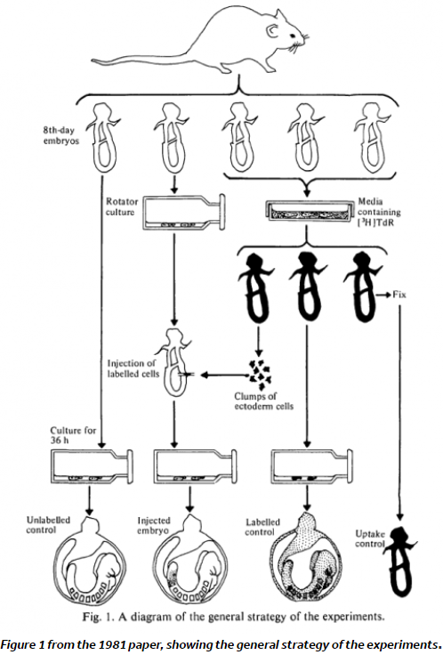
Are you just starting out in your career as a developmental biologist (or feeling nostalgic for those times)? The vast amount of literature can feel a bit daunting when you are tackling a new topic. To help you navigate the field, and to focus on some of the lesser known classics, we asked prominent researchers to recommend their hidden gems from history. Their selection includes Rosa Beddington’s chimeras, Morgan’s planarian experiments and principles of morphogenesis from Gustafson and Wolpert. Let us know if you come across any forgotten classics that we should include in our series.
“Imagine a society where female reproductive rights are a matter of state concern. Where working class females are actively suppressed from having their own children, and only the nobility are allowed to reproduce. I am, of course, talking about honey bees.”
Dr Sally Le Page
In the latest episode of the Genetics Unzipped podcast, Dr Sally Le Page looks at the genetics of societies, exploring how genes underpin the rigid social structures and roles in bees, and how they can rise up the ranks to become queen bee.
If you enjoy the show, please do rate and review on Apple podcasts and help to spread the word on social media. And you can always send feedback and suggestions for future episodes and guests to podcast@geneticsunzipped.com Follow us on Twitter – @geneticsunzip
Read on for our news roundup of the past few weeks, with an emphasis on what has caught our eyes on twitter. This post is focussed on career advice, conferences and related conversations.
— Dr Lisa Nivison-Smith (@LNivisonSmith) June 29, 2022
Can posting a preprint help advance your career in science, especially if you are a student? Learn more in the video below, where Sónia Gomes Pereira discusses her experience with posting a #preprint. https://t.co/9uBhnwWVzA
Numerous faculty job ads are starting to appear, application materials are being prepared – wanted to share advice for applicants on past & future research statements based on my experience as a faculty search chair last year. First, a few thoughts…🧵
— American Society for Cell Biology (@ASCBiology) August 11, 2022
Earlier this year, we heard from Dhruv Raina about his move to industry following his PhD in Dr Christian Schröter’s lab at the Max Planck Institute of Molecular Physiology (MPI).
For the latest job listings at all levels, including an opportunity for a new Features and Reviews Editor at Journal of Cell Science, check out our activejobs board.
Conferences
The return to in-person conferences has been wonderful, but what have we learnt due to the restrictions imposed during lockdowns, and how can we improve future conferencing?
I love going to conferences but i must find a way to bring my family with me otherwise no matter how fun or important it is the guilt is killing me. Perhaps half of a conference day should be free time? (to allow you to be with family)
Today I spoke up at a #FENS2022 discussion event about pandemic effects on mentoring. I spoke about my frustration regarding transmission mitigation at the meeting. I sorta highjacked the discussion and I'm sorry about that, but I just couldn't not say anything and 1/n
— Dr. Maria Veldhuizen 😷💉 (@margaveldhuizen) July 12, 2022
We are delighted to be live–streaming our session on recent recent progress and ethical challenges in studying early human development from our upcoming meeting, From Stem cells to Human Development on Monday 12 September 16:00 BST. More details will follow on the Node shortly.
In vitro models of development have been a talking point on Twitter in the last few weeks. The thread from Dr Martinez Arias sums up some of the key points.
The recent reports on the generation of embryo-like structures from mouse pluripotent stem cells by the @jacob_hanna and @MZG_Lab groups are an important landmark in developmental and reproductive biology. Thread
— Alfonso Martinez Arias (@AMartinezArias) August 7, 2022
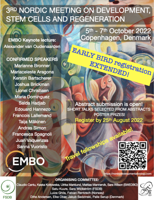
We are arranging a 2-day conference together with the Swedish Society for Developmental Biology (SWEDBO), Finnish Society for Developmental Biologists, and Danish & Norwegian Developmental Biologists the 3rd Nordic Meeting on Development, Stem Cells and Regeneration in Copenhagen in October 5-7th, 2022
The line up of invited speakers is outstanding and brings together experts in developmental and stem cell biology and regeneration! Attending the meeting is a great opportunity to meet with developmental biologists, from the Nordic countries and beyond. All speakers will be there ‘in person’ allowing for lots of networking.
There are many opportunities for short talks, poster prizes, and student and postdoc activities are planned in addition. The registration is now open and it includes membership in one of the Nordic Developmental Biology associations! Follow the link to register: https://nordicdevelopmentalbiology.com
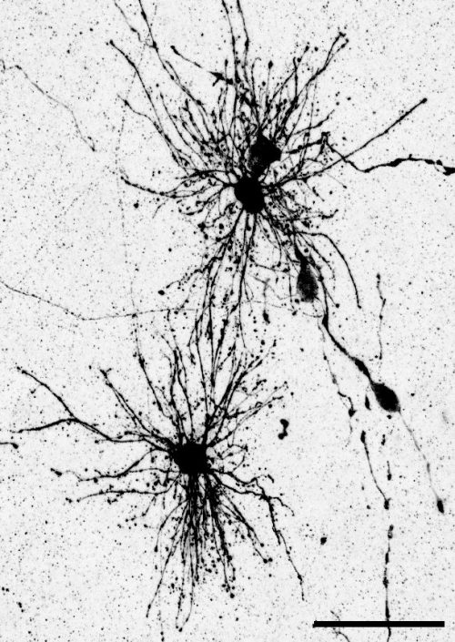
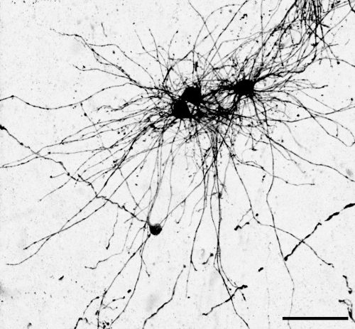
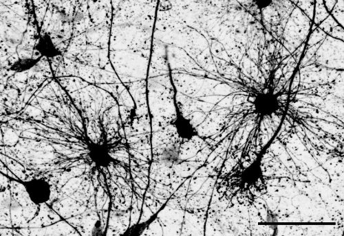
Dr. Denise Allen and Dr. Tomasz Nowakowski at the University of California, San Francisco recently published an article in Science where they revealed a dual origin for astrocytes in the human cortex. Using a combination of fate mapping and single cell analysis, they revealed that the two stem cell niches in the developing cortex give rise to spatially, morphologically, and molecularly distinct populations of astrocytes. The Node asked them to give us a behind the scenes look at how the story came together:
How did you get started on this project?
TJN: For a very long time, we have been interested in the question of why the brains of primates and humans are so much larger and complex than the brains of mice, which we study frequently in the laboratory. Differences in brain size can be found very early on during development, and therefore it was plausible to hypothesize that differences in the way radial glia, which act as neural stem cells, develop could contribute to these differences. In our prior work, we found that animals with large brains, such as primates or humans, contain a greater diversity of radial glia subtypes compared to mice. In particular, we found that based on gene expression profiles, radial glia could be divided into truncated radial glia and outer radial glia, which are located in two anatomically distinct niches of the developing cortex.
DA: During my undergraduate neuroscience classes I was always struck by the deep knowledge we have about the development of neurons in the cerebral cortex, but astrocytes and other glia seemed to be so often overlooked. During my rotation in the Nowakowski lab, I became fascinated with Tom’s preliminary data that suggested distinct subtypes of radial glia could give rise to distinct astrocyte populations. I was really excited by the fact that large brain mammals, including primates and humans, seem to have a different repertoire of radial glia compared to rodents, as well as much more complex astrocytes. So the possibility to study the unique features of human development with a focus on astrocytes was a dream come true.
What was already known about the developmental trajectories of radial glia in the developing brain prior to your work?
A lot of work has been done probing the differentiation of outer radial glia (also known as basal radial glia). Numerous papers have shown that they give rise to neurons, oligodendrocytes and supposedly the majority of astrocytes, but the role of truncated radial glia has not been studied in great detail. Previous studies have suggested that because few mitotic cells can be found in the ventricular zone stem cell niche during midgestation in primates and humans, that the truncated radial glia that reside in this zone are unlikely to serve as a major source of new cells. We decided to challenge this assumption by labeling progenitors in the ventricular zone and determining the fates of the resulting cells. To our surprise, we found that neurons, oligodendrocytes, and astrocytes continue to be produced by ventricular zone progenitors.
Can you summarize your findings? What was the key experiment?
The key experiment involved labeling progenitor cells that occupy anatomically distinct niches that truncated and outer radial glia cells, and tracing the fates of cells that they produce. We found that while both populations broadly produce similar cell types (neurons, astrocytes and oligodendrocytes), they produce very distinct subtypes of astrocytes. In a series of very striking results, we found that truncated radial glia give rise to astrocytes that migrate to the cortical plate, while outer radial glia give rise to astrocytes that do not migrate, and instead differentiate locally in the outer subventricular zone.
We often speak about the diversity of neurons, but classical studies have also shown that astrocytes can be remarkably diverse, even in early development. I wanted to further explore the diversity of the astrocyte subtypes we identified, but it is challenging to connect these classical descriptions of astrocyte subtypes to modern-day descriptions such as those derived from single cell sequencing. To solve this problem, I took advantage of a method called Patch-seq which gave us the ability to aspirate the contents of a cell that was previously defined based on its morphology and position, and then performing sequencing of that cellular contents to determine a molecular identity. This analysis was key for bridging our cellular definitions based on morphology and developmental cell lineage, and linking them to molecular markers. This allowed me to bring the story in full circle.
When doing the research, did you have any particular result or eureka moment that has stuck with you?
The very first experiment I performed that involved labeling these two different stem cell niches resulted in a distribution of cell types that could not have been more different. Many comparisons in developmental biology rest upon small differences between conditions. To see such a stark difference in the distribution of cells–especially of glia–was an exciting moment that defined the course of the project very early on in my PhD.
Another surprising finding was when we started closely comparing classical drawings by Ramon y Cajal and Retzius and to images of our astrocyte subtypes. Remarkably, we found that our “dense bulbous” astrocytes were clearly depicted in those early records, but these cells have rarely been mentioned in modern literature. This realization gave us a lot of confidence that the cells we were observing were a real phenomenon and not an experimental artifact. These cells had just been lying in wait, waiting for someone to put the spotlight on them.
And what about the flipside? Any frustrations or despair?
The Universe really conspired against us when we were trying to finish experiments for the revision of the paper. I set up the last three revision experiments in late December, when we suddenly found out that it was time to move our lab to a new building at UCSF. We came up with an elaborate system to keep the cultures going while we moved and they seemed to have survived, until someone suddenly noticed that the incubator had failed and the alarm hadn’t gone off. What followed was two months of issue after issue trying to repeat these last two experiments, but we finally got there in the end!
Where will this story take the lab?
This work has inspired several new projects in the lab. We are excited to examine if similar findings can be replicated in other models of brain development such as cerebral organoids, what these unique subpopulations look like in the adult brain, and what role they might play in disease states. I’m also hoping this work will also attract more trainees interested in glial development to the lab!
What is next for you/the lab after this paper? Let us know if you are continuing this research, or starting/looking for a new position.
Denise has graduated and is currently interviewing for computational biologist roles in biotech. She is looking forward to delving into the “big data” side of biology, and working towards making a significant impact on patients’ lives.
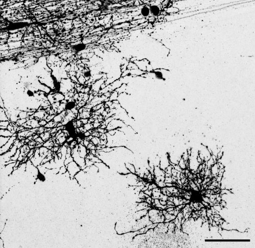
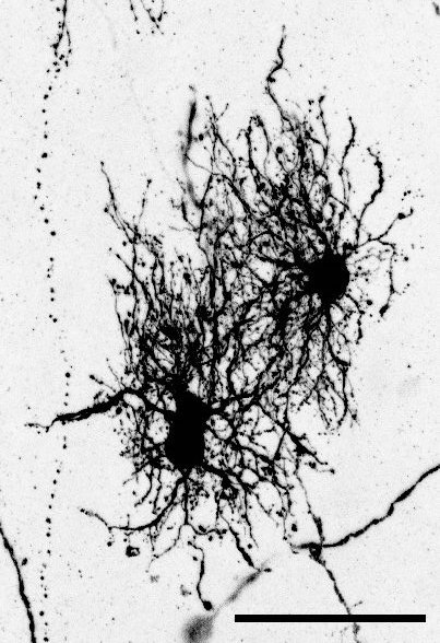
“Dense bulbous” astrocytes derived from the VZ“Dense bulbous” astrocytes derived from the VZ“Dense bulbous” astrocytes derived from the VZ “Dense smooth” astrocytes derived from the OSVZ“Dense smooth” astrocytes derived from the OSVZ (1 votes) Loading...
Development’s biennial meeting, From Stem Cells to Human Development, will be taking place in mid-September (11-14) at beautiful Wotton House in Surrey, UK. After a very successful virtual meeting in 2020, we’re excited to be meeting in person again, but we wanted to explore ways of making part of the meeting accessible to the broader community. We’re therefore delighted to announce that we’ll be livestreaming one session of this meeting, and the recording is available below.
Session details (all times GMT+1): 16:00 Sarah Teichmann (Wellcome Sanger Institute, UK): Human development: one cell at a time 16:30 Sergiu Pasca (Stanford University, USA): From stem cells to assembloids: constructing and deconstructing human nervous system development and disease 17:00 Panel discussion: Technical, ethical and legal challenges of studying early human development Chair: Patrick Tam (University of Sydney, Australia) Panellists: Amander Clark (University of California Los Angeles), Robin Lovell-Badge (The Francis Crick Institute, UK), Sergiu Pasca, Sarah Teichmann, Magdalena Zernicka-Goetz (University of Cambridge, UK and CalTech, USA) 18:00 Close
The method was straightforward: take a bunch of writers, novelists, a playwright, science communicators and scientists from all over the world, from fields as diverse as astrophysics and climate science to materials engineering, neurobiology, and evolution, confine them to a 16th century manor, and get them talking. The result: The Company of Biologists’ whacky and wonderful Creative Science Writing Workshop.
Wiston House, South Downs National Park: the experiment site.
Good scientific writing has the power to help communities make more informed decisions, and a creative route could make it more accessible and exciting for all. But here, without the certainties of a regular science meeting where everyone shares a common interest in a method, organism or question, and data and information that forms the focus of most scientific discussions, what would happen was anyone’s guess.
Any disquiet quickly evaporated in the opening session where everyone introduced themselves through stories, childhood memories, and mementos: a bottled book, a handmade urn, the steady pulse of a ticking metronome, the clinking beads of a treasured necklace, a traditional Indian kolam drawn in flour before our eyes. It was immediately clear that despite our diverse backgrounds and experiences, everyone shared a passion for storytelling and science in all its forms. The Workshop began with a bang!
The programme included a variety of topics and activities. We read S.J. Gould, Primo Levi and the brilliant Karen Joy Fowler, looking for the ingredients of great writing and pondering the authors’ process and intent. We critiqued the work of other delegates, providing structured feedback – a rather nerve-racking exercise when one considers the differences between academic and creative writing. In conventional science writing, the authors present new facts and relationships that help us better understand our world, without ever revealing themselves. In creative writing the opposite holds true; every piece exposes the writer, their style, their quirks of character, beliefs, and passions. This Workshop managed the juxtaposition remarkably well.
There were also sessions on structuring writing, getting published, and the role of agents. Most importantly, there was time to write, review, and revise. Inside oak-panelled rooms, within the verdant grounds, and in the sunny conservatory, relationships grew, word counts climbed, and inspiration abounded.
Over a period of three and a half days, the Workshop exposed delegates to the world of writing and publishing. Aspiring writers learned from one another as well as from the established writers – who were incredible mentors. It was a journey that traversed science and writing, lab and the field, life and the page, lyrical prose and cold hard facts. In doing so, a new community was formed.
Applications will remain open until the position is filled.
The Gambetta lab is recruiting a Postdoctoral researcher to study how genome architecture impacts gene regulation in development.
Project
The advertised project addresses the fundamental question of how regulatory elements are guided to their target genes. You will study a paradigm we uncovered in which genes are controlled by regulatory elements located at flexible and surprising large genomic distances. This model is powerful and unique.
You will use genetics (Drosophila genome-engineering, genetic screens, comparative evolution), genomics (transcriptomics, chromatin accessibility, chromosome conformation capture, genome-wide screens, single-cell genomics), imaging (fixed and live), and biochemistry (proteomics).
This work is expected to continue to reveal new evolutionary perspectives into the relevance of 3D genome folding for correctly wiring genes to their regulatory elements. For more information on our research check our lab website: http://www.gambettalab.org
Job information
Expected start date in position : as soon as possible or to be agreed
Contract length : 1 year, renewable 2 x 2 years, maximum 5 years
Your responsibilities
You will use multidisciplinary approaches such as genomics, genetics, imaging, and/or biochemistry in the fruit fly Drosophila melanogaster. You will present your results during seminars with gene regulation research labs in Lausanne. You will collaborate with other labs in Lausanne and abroad. Full funding for the position is available, but application to fellowships is also expected.
Your qualifications
You are a dynamic and rigorous scientist with a PhD degree in Biology or a related discipline. You have experience in genomics (next-generation sequencing library preparation), molecular biology, genetics, biochemistry or imaging. You are a critical thinker, a team player eager to participate in scientific discussions but able to work independently. You have a strong interest in developing your skills in multidisciplinary experimental strategies to understand basic mechanisms in gene regulation.
Your benefits
The Gambetta lab is hosted at the Center for Integrative Genomics (CIG) at the University of Lausanne (UNIL), a vibrant, well-funded institute with a focus on functional genomics and equipped with modern core facilities (see www.unil.ch/cig).
It is embedded in the broader Lausanne research environment that includes two universities (UNIL, EPFL), the Swiss Institute of Bioinformatics, Ludwig Center for Cancer Research, university hospital, and a cluster of biotech companies flourishing in the larger lake Geneva area.
The Gambetta lab tightly networks with other gene regulation research laboratories at UNIL and EPFL, and collaborates with the on-site Bioinformatics Competence Center. There are regular possibilities to present and participate in local or international conferences and workshops. Hard and soft skill, and career development courses are offered on campus.
We offer a nice working place in a multicultural, diversified and dynamic academic environment.
Your application
Please email lab head Prof. Maria Cristina Gambetta (mariacristina.gambetta@unil.ch). Provide your CV, a brief description of your research experience, and why you think your research interests complement ours.
Posting my lab report from last summer again since I can’t log into my last account. Update: I’ll be starting an Mres looking at the mutual suppression of the cardiac versus skeletal muscle programme!
Biomedical research tries to understand among other things, how during development, gene expression determines cell fates. One of the aspects that we look at is how cells are recruited to the heart and how to apply this knowledge to cardiac therapy. However, cell fate decisions in the head mesoderm, the tissue responsible for delivering the heart, craniofacial muscle, parts of the skull and vasculature, are poorly understood. Last summer, I had the opportunity to work with Dr. Susanne Dietrich, who studies the formation of muscles in early development, including, but not only, genes responsible for committing cells into a mesodermal fate. I was part of ongoing research addressing this question, at her lab in Portsmouth. The Dietrich lab has shown that initially, the entire head mesoderm has cardiac competence. However, at early neurula stages of development, the cardiac inducer Bmp2 fails to induce the cardiac programme and instead, it induces Msc, a craniofacial precursor marker. It is not yet clear how this switch in developmental competence is achieved. We hypothesize that Wnt may be responsible for the switch, inducing an early expression of craniofacial precursor markers and downregulating cardiac markers. To test this idea, I grafted heparin-coated acrylic beads soaked in recombinant Wnt3a, the Wnt inhibitor Sfrp2 and Bmp2 or bovine serum albumin as control, into HH7/8 embryos. Embryos were cultured for 6 hours, the time sufficient for Bmp to induce Msc. I then used In situ hybridization to analyse the expression of Msc, the cardiac marker Nkx2.5 and the Wnt responsive genes Pax3 and Axin2. We found that Wnt did not upregulate and Sfrp2 did not downregulate Pax3 and Axin2 (data not shown), probably because it takes more than 6 hours to change the expression of these genes. However, Wnt3a did downregulate Nkx2.5 as expected (Fig.1). Nonetheless, Wnt did not upregulate Msc (Fig.1), suggesting that the concentration used and the 6-hour time period might not have been enough for Wnt to participate in the activation of Msc. This was against our hypothesis, and we wondered why that might be. So, we decided to test the effect of Wnt on the paraxial head mesoderm marker Cyp26C1, an inhibitor of retinoic acid signalling. We found that Cyp26C1 was suppressed (Fig.1). Thus, Wnt may in fact suppress paraxial head mesoderm features.
I am now faced with new questions: What really is the role of Wnt? Does it suppress heart and paraxial mesodermal features because it posteriorizes the tissue? To answer this question, I will have to analyse if Wnt causes an ectopic expression of posterior information markers (e.g: Raldh2 and Hoxb1). And our original question is not answered: what facilitated the switch from cardiac to skeletal muscle competence? I am intrigued by these results, and I do want to find out the right mechanism that causes this switch. I will be working on this project throughout the next academic year, looking at different embryonic stages, different concentrations of Wnt, different Wnt inhibitors (e.g.: Dkk) and possibly, longer culture periods. I am hoping to find results that tell us if Wnt signalling is or not responsible. If Wnt is not involved, what else could it be? Many other signalling cascades converge on the head mesoderm, and they could be tested using similar approaches. Alternatively, I could use small molecule inhibitors of signalling cascades on embryos cultured as Cornish pasties. We also have to consider that the epigenetic landscape might change over time, and cardiac genes might be put out of use. This would require a different approach, chromatin immunoprecipitation. I would love to learn about chromatin immunoprecipitation and work on this approach during my master’s or PhD. I want to continue working on these questions with Dr. Susanne throughout the next years of my academic life. I hope that with my 3rd year module “genes and development” I will gain more insight into new experimental methods used in developmental biology and maybe use them as an approach in my project. I would love to work with different model organisms and upgrade my knowledge with new techniques that may facilitate the research. Working with Dr. Susanne and her team made me grow as a scientist. I remember the week before starting on my project I was so nervous I even had nightmares about it. But the people in the lab were very kind and helpful and they made me feel at home. During the summer, I was faced with some of the ups and downs of science. In situs that did not work, embryos that were accidentally lost, beads not sticking, and all of that (Particularly the last one), allowed me to develop my problem-solving skills and patience (especially while grafting beads). Being part of a research group made me realize that I do not see myself doing anything else. I love planning my experiments and I love the practical part. I am also very interested in presenting and explaining my results to other people. I look forward to continue working on developmental biology throughout my studies. I plan on continue my education with Dr. Susanne, working on finding the mechanism behind this cardiac to skeletal muscle switch, and other projects.
 (No Ratings Yet)
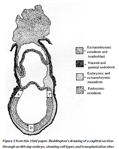
 (3 votes)