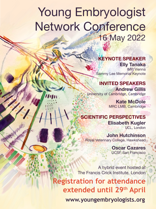
Registration for attendance now extended until the 29th of April!
The Young Embryologist Network conference 2022 (YEN 22) is the 14th iteration of the network’s hugely successful yearly developmental biology meeting. This will be a hybrid conference hosted at the Francis Crick Institute and streamed over Zoom on Monday the 16th of May 2022.
Our meetings provide an opportunity for early-career-stage developmental biologists to share their own research, network and engage with peers and pioneers in the field, with delegates attending from across the globe
We have a varied and exciting programme for YEN 22: Dr. Andrew Gillis (University of Cambridge) and Dr. Kate McDole (MRC LMB) will be delivering talks as two of our invited speakers. Furthermore, we have the pleasure of welcoming Prof. Elly Tanaka (Institute of Molecular Pathology, Vienna) to give the Sammy Lee memorial keynote address. Plus, PhD students and postdocs may submit abstracts for the chance to give a short talk or present a poster.
Moreover, we are immensely proud to be hosting our first “Scientific Perspectives: Working in Science with a Disability” talks at YEN 22. Disability is a tremendous barrier not only to entry, but also to the progression of a scientific career, and researchers with disabilities remain immensely underrepresented at every career stage. Our community could be much better equipped in helping ensure inclusion and equal opportunities for disabled scientists, and so we have invited Dr. Elisabeth Kugler (UCL), Prof. John Hutchinson (RVC) and Dr. Oscar Cazares (UCSF) to share their perspectives and experiences of working in science with a disability. We also have the pleasure of welcoming Dr. Cynthia Andoniadou (King’s College London), who will be delivering the summary address for these sessions.
Abstract submission is now closed but, due to ongoing demand, we have extended the attendance registration deadline to 11:59pm on the 29th of April.
You can register for this free event, and learn a little more about our organisation, via the link below:
In our latest SciArt profile, we hear from Cirenia Arias Baldrich. Cirenia is a freelance illustrator, who has a background in plant physiology and stem cell bioinformatics.
Where are you originally from and what do you work on now?
I am from La Línea de La Concepción (Cádiz), but I lived in Seville for 16 years, where I studied biology and completed my PhD on Molecular Biology and Genetics. After that I moved on in my scientific career to Portugal and then to the UK, where I was rocking bioinformatics as a postdoctoral researcher in the Stem Cell Biology and Evolution Group (Dr Jordi Solana – Oxford Brookes University). Nowadays, I am based in Cádiz, Spain, working as a full time freelance illustrator, helping organisations and other scientists to communicate their work using the power of images.
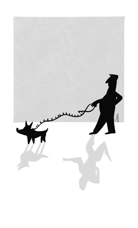
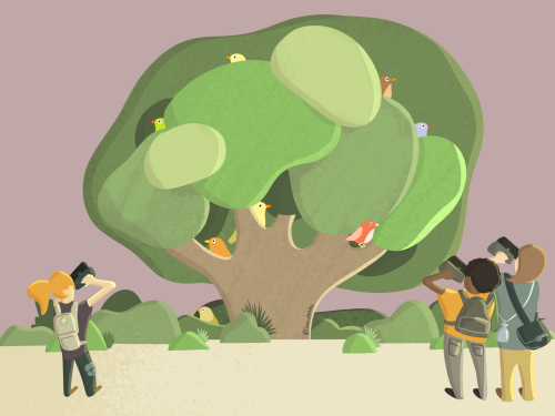
There’s a friend in me, how we domesticate our pets. This is one of the 15 illustrations introducing the 15 chapters of ‘GENES: Escribiendo el guion de la vida’ book (Guadalmazán, Almuzara Libros). With these illustrations, my goal was to introduce the chapters to the reader, to make them curious about what they are about to be immersed in, and at the same time trigger them to rethink what they had already read.Birdwatching. Illustration featured in the paper ‘No Bird Database is Perfect: Citizen Science and Professional Datasets Contain Different and Complementary Biodiversity Information’, Sofía Galván, Rafael Barrientos, Sara Varela (Ardeola, 2022).
Were you always going to be a scientist?
Not really, at least it was not clear in my mind. Though since I learned how to talk, I started asking questions, and replying with ‘but how/why?’ or ‘are you sure of that?’ to every answer. I also loved to decipher how things worked; opening electronic stuff to see its components, making tests, playing with computers – I was pretty curious! I also had other interests like arts, design, informatics, marketing, but I always loved to read the biology notes of my older sister, who is also a biologist. Like her, I decided to study biology. It was amazing to learn about genetics, cell biology, zoology, plant physiology, biochemistry, ecology, to better understand both the world around us and ourselves.
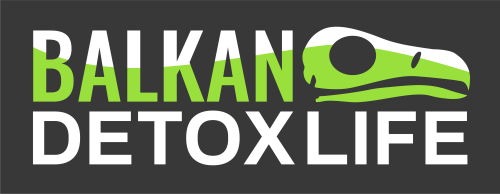
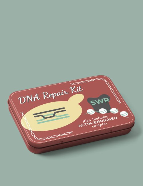
Visual Identity of the BalkanDetoxLIFE project: Strengthening national capacities to fight wildlife poisoning and raise awareness about the problem across seven Balkan countries. The #BalkanDetoxLIFE’s project logotype illustrates the severity of illegal wildlife poisoning, while also signifying hope through the project’s goal to detox the Balkans from this threat.Cover published in GENETICS, Genetics GSA.‘DNA Repair Kit’ highlights the essential role of a chromatin remodelling complex in the DNA repair process. This cover is linked to Morillo-Huesca et al., 2019.
And what about art – have you always enjoyed it?
Definitely. I can’t even remember when I started drawing, what I know is that I never stopped. Since I was a child, I never had enough colours, watercolours, crayons or markers (I still cannot go into an art supply store without going mad)! I loved trying new materials, trying to draw everything I saw, such as everyday objects, or the room where I was. I was always doodling, even in meetings, (I still do) and, funny fact, that is actually how my adventure as a professional illustrator started, taking graphic notes at scientific talks. I discovered that I could join my three passions: science, art and communication, using illustration as a science communication service.
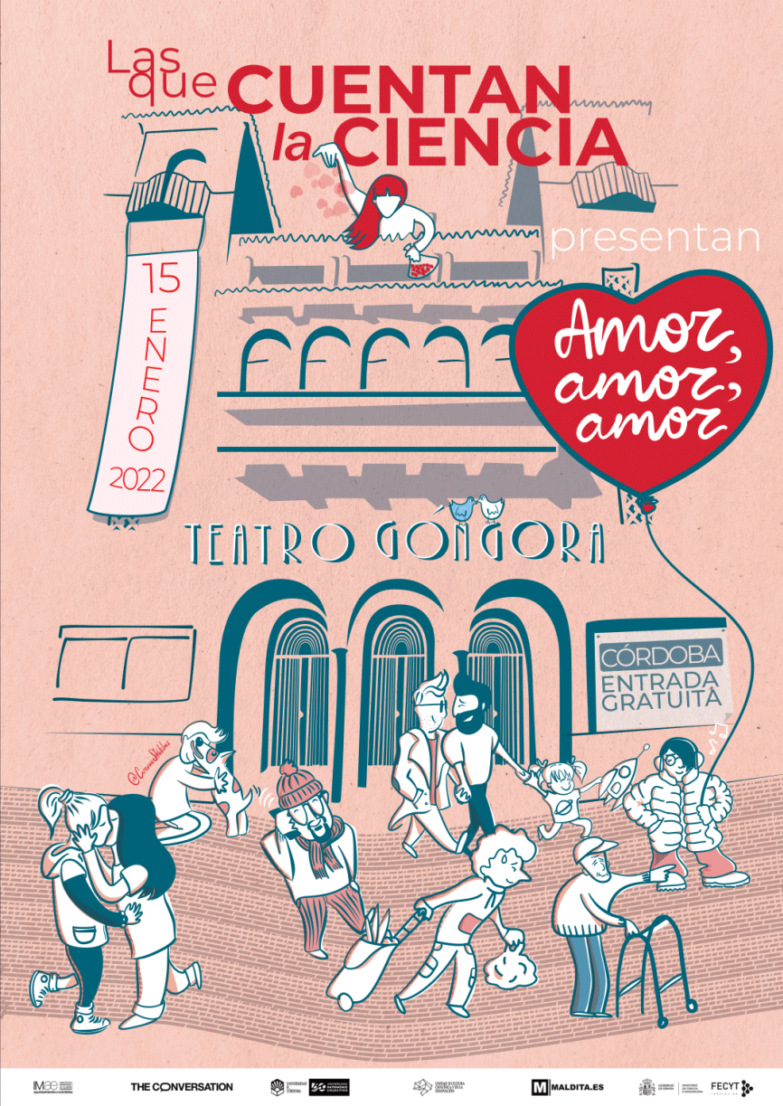
Las Que Cuentan La Ciencia 2022. Animated Poster for Scicomm Event ‘Las Que Cuentan La Ciencia’ (The Women who Explain Science). The 2022 theme was ‘Amor, Amor, Amor’ (Love, Love, Love). In this poster, love is illustrated in diverse forms, while the theatre represents the return to on-site events. This event is organised by the Unit of Scientific Culture and Innovation (UCC+i) of Cordoba (Spain), (University of Córdoba, Maldita.es, The Conversation ES)
What or who are your most important artistic influences?
This is probably one of the toughest questions to answer, since I try to consume as much art/design as I can. From literally everywhere. I’m inspired by movie posters, records covers (I love vinyls), restaurant menus, street signs, street art, graphic novels, etc. However, there are some creators that had made a big impact on the way I see the profession of an illustrator. I really admire the work of many diverse artists like Cristopher Niemann, Malika Favre, Rachel Ignotofsky, Adolfo Arranz, the Etherington Brothers, Andy Riley, Agustina Guerrero, Andry Rasoahaingo (Dedouze), Tom Gauld, Raquel Córcoles (ModernaDePueblo) and many others (I could go on with a never-ending list).
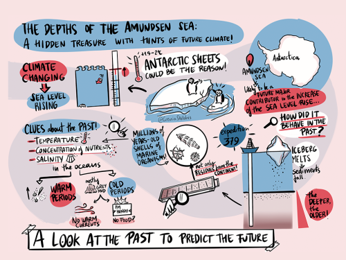
The Depths of the Amundsen Sea. Illustration featured in the article ‘From the depths of the Amundsen Sea’ (Courtillat, M). The image is one of the series of graphic summaries and illustrations created for Horizons Magazine (PAGES- Past Global Changes Project). Horizons highlights paleoscience topics that are of interest for the next generation, and is written in an easy to understand, visual format.
How do you make your art?
Digital tools help me to deliver the work efficiently. It makes the reviewing process easier, and the final artwork can be directly shared in social media, websites, or used for printing or publication. Also, I love the infinite possibilities digital tools provide and learning new resources. Despite all this, I start every project sketching in paper. For my ideas to run wild, I like the feeling of a notebook and a pen, and once I have a plan, I move on to digital fun using both a digital tablet and the computer (depending on the project), hand-drawing apps, vector graphics software, or animation programmes.
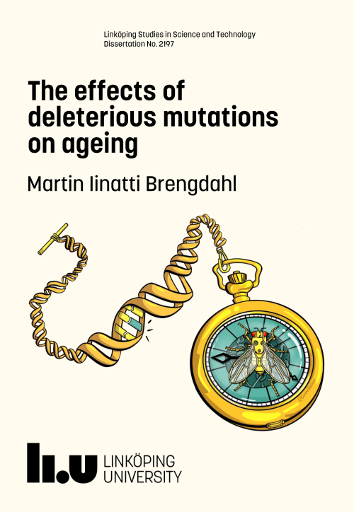
The effects of deleterious mutations on ageing. Cover for Martin Iinatti Brengdahl’s doctoral thesis on evolutionary genetics of ageing and sex differences (Friberg Lab), Linköping University. This illustration reflects Martin’s work investigating the age-specificity of deleterious mutations and their contribution to sex differences in ageing and lifespan in Drosophila melanogaster.
Does your art influence your science at all, or are they separate worlds?
Art has always influenced my science: the way I communicate with my colleagues, planning lab strategies, preparing presentations, posters, figures, giving a chalk-talk, or even thinking about a project. I think creativity and science are completely inseparable. Nowadays, being a freelance illustrator, science is my everyday ingredient in my art, since my job is helping to communicate science efficiently, to both expert and non-expert audiences, and my scientific background plays a key role in allowing me to do so. I also feel very motivated and enthusiastic about having an influence on the way other scientists approach the creation of visuals in their work. In this sense, I really enjoy training other researchers, and give them tips and tools so that they can make better illustrations and have fun instead of struggling!
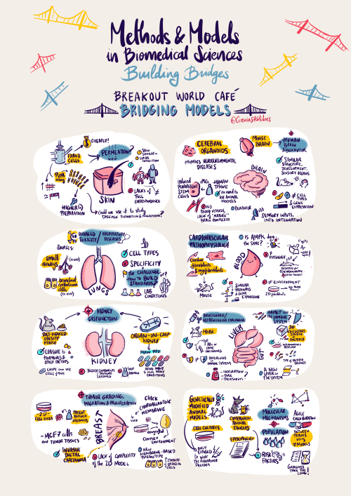
Graphic Summary Poster for Methods and Models in Biomedical Sciences: Building Bridges Meeting, a Champalimaud Foundation workshop co-organised with the European Commission’s Joint Research Centre (JRC), CONGENTO, QuantOCancer and FRESCI. This poster gathers all the ideas and key messages on scientific methods and models used in biomedical research discussed in the session ‘Breakout World Group Exercise’ of the meeting
What are you thinking of working on next?
One of the things I love the most of my job is how exciting it is to work on so many different things, thanks to the diversity of collaborators. In the next few months, I will be working on very cool projects ranging from awareness about vulture conservation, rare diseases, digital gender gap, palaeoecology editorial illustration and producing several infographics, covering topics such as microbiology or climate change, among others. I would like to take advantage of this awesome spot to thank all the amazing people trusting me to help showcase their amazing work. Last but not least, big thanks to The Node for creating the SciArt profile series, it’s great to get to know other colleagues and I am very honoured to be part of it.
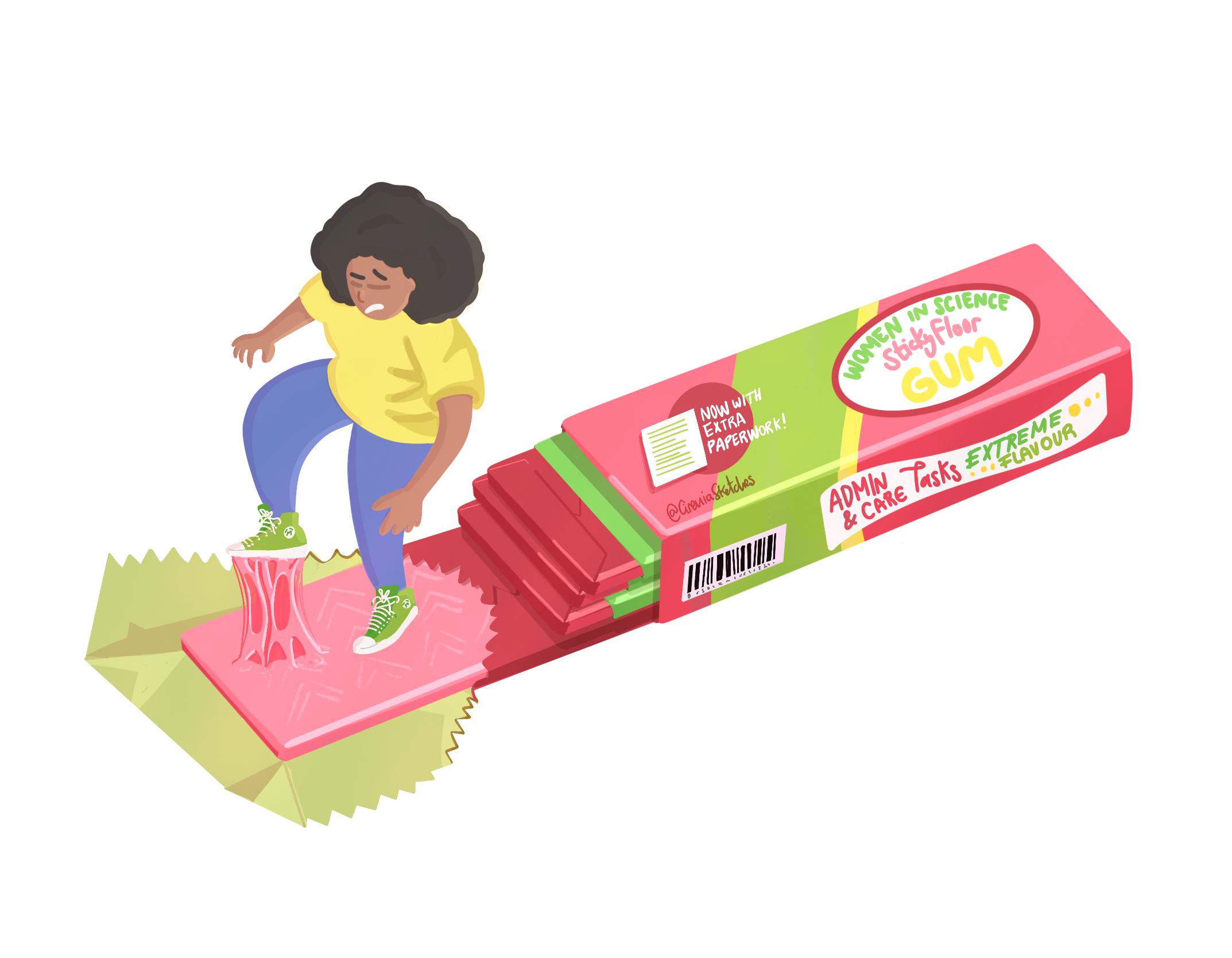
The Sticky Floor. This illustration showcases the ‘sticky floor’ keeping women in the lower ranks of the job scale. Done in collaboration with MAPAS LAB and The Equality Commission of the Spanish Association of Terrestrial Ecology (AEET).
Thanks to Cirenia and all the other SciArtists we have featured so far. You can find the full list here. We’re always on the lookout for new people to feature in this series – whatever kind of art you do, from sculpture to embroidery to music to drawing, if you want to share it with the community just email thenode@biologists.com (nominations are also welcome!)
Read on for our news roundup of the past two weeks, with an emphasis on what has caught our eyes on twitter. We have also include a list of meetings with upcoming deadline, a selection of preLights and finish with our favourite April fools jokes .
Doing better as a community
There have been a few tweets in the last fortnight discussing some of the negative aspects of our research culture with a call to do better in the future. Lets make that happen!
Reviewing papers
Paper Reviewers: STOP asking for nickel and dime, incremental experiments that don’t advance the overall story. This kind of shit is strangling our trainees and accomplishes nothing but making you feel smart and slowing the pace of science. 1/2
Mixed emotions having been back from @Official_BSCB@_BSDB_#BSCBDB22 for 36h. Fantastic to be back in person, meet so many old friends and new faces; faces from screen in real life finally; some amazing and exciting science. And proud of how well the little one did. BUT…
The impact of Brexit on British science continues with notification from the ERC that awardees will need to move to the EU to take up their grants. The UK government is guaranteeing the grants in the UK but details remain sketchy.
And here it is — communication from @ERC_Research to UK-based #ERC starting grant grantees… UK awardees will have two months time to move their grant to the EU proper, otherwise they will lose their STG. Thank you #Brexit#BrexitReality. pic.twitter.com/AnJeCrCQrZ
— Thiemo Fetzer 🇪🇺🇺🇦 – same handle elsewhere (@fetzert) April 8, 2022
All the hard work put in to be awarded an #ERCStG, and we are now in the hands of a political decision. ☹️ https://t.co/YA1f4gezzX
Disaster for U.K. science. U.K. govt knew this was coming and let it happen. Where does the cash come from to replace this invaluable source? @ERC_Research is something we need to be part of. StG support the next generation…..Advanced funds half this old dawg’s group. https://t.co/skYlOv6Vkr
It’s been exciting to see the return of in-person meetings including the joint BSCB and BSDB meeting in the UK (we’ll post our meeting report soon) and #Dros22 in the US. Check out the list below for meetings with upcoming deadline and check our events page for our full listing.
If you are interested in science communication, would like to improve your writing skills and become part of the preLights community, don’t miss the open call for new preLighters.
Just for fun – our favourite April fools
We couldn't believe our eyes at first🤯🤯🤯! We just found the missing link between Cnidaria and Ctenophora. Introducing Pleurocoryna coelenterea, Pleurocorynidae fam. nov., Phylum: Coelenterata. pic.twitter.com/BSegc0B85e
After almost 5 years at the @czbiohub leading an awesome team of computationalists, microscopists and biologists, I have decided to move on and become a marine biologist. 1/n
Happy to announce that we are going to wind down our planarian research program in favor of studying brain regeneration in rodent models. We are all excited that we will finally be doing "real" neuroscience and working in a model relevant for human regeneration.
— Dr. Rachel Roberts-Galbraith (@awormwelcome) April 1, 2022
If you would like to write for the Node, check out our recent list of writing ideas. If you would like to contribute to our ‘Developing news’ blog, please get in touch at thenode@biologists.com
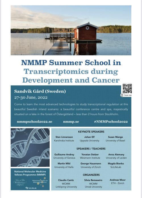
Join us in a Swedish enchanting scenario at the end of June (27-30) when it’s always light in Sweden!
If you are a Student (bachelor/Master’s/PhD), a Postdoc, or a young PI, and are using transcriptomics technologies, or you wish to approach them, this is the right event for you!
Plakoglobin is a mechanoresponsive regulator of naïve pluripotency Timo N. Kohler, Joachim De Jonghe, Anna L. Ellerman, Ayaka Yanagida, Michael Herger, Erin M. Slatery, Katrin Fischer, Carla Mulas, Alex Winkel, Connor Ross, Sophie Bergmann, Kristian Franze, Kevin Chalut, Jennifer Nichols, Thorsten E. Boroviak, Florian Hollfelder
Molecular Signatures and Cellular Diversity During Mouse Habenula Development Lieke L. van de Haar, Danai Riga, Juliska E. Boer, Youri Adolfs, Thomas E. Sieburgh, Roland E. van Dijk, Kyoko Watanabe, Nicky C.H. van Kronenburg, Mark H. Broekhoven, Danielle Posthuma, Frank J. Meye, Onur Basak, R. Jeroen Pasterkamp
TRIM28-dependent SUMOylation protects the adult ovary from activation of the testicular pathway Moïra Rossitto, Stephanie Déjardin, Chris M Rands, Stephanie Le Gras, Roberta Migale, Mahmoud-Reza Rafiee, Yasmine Neirijnck, Alain Pruvost, Anvi Laetitia Nguyen, Guillaume Bossis, Florence Cammas, Lionel Le Gallic, Dagmar Wilhelm, Robin Lovell-Badge, Brigitte Boizet-Bonhoure, Serge Nef, Francis Poulat
LINE-1 retrotransposon activation intrinsic to interneuron development Gabriela O. Bodea, Maria E. Ferreiro, Francisco J. Sanchez-Luque, Juan M. Botto, Jay Rasmussen, Muhammed A. Rahman, Laura R. Fenlon, Carolina Gubert, Patricia Gerdes, Liviu-Gabriel Bodea, Prabha Ajjikuttira, Peter Kozulin, Victor Billon, Santiago Morell, Marie-Jeanne H.C. Kempen, Chloe J. Love, Lucy M. Palmer, Adam D. Ewing, Dhanisha J. Jhaveri, Sandra R. Richardson, Anthony J. Hannan, Geoffrey J. Faulkner
Zfp503/Nlz2 is Required for RPE Differentiation and Optic Fissure Closure Elangovan Boobalan, Amy H. Thompson, Ramakrishna P. Alur, David McGaughey, Lijin Dong, Grace Shih, Emile R. Vieta-Ferrer, Ighovie F. Onojafe, Vijay K. Kalaskar, Gavin Arno, Andrew J. Lotery, Bin Guan, Chelsea Bender, Omar Memon, Lauren Brinster, Clement Soleilhavoup, Lia Panman, Tudor C. Badea, Andrea Minella, Antonio Jacobo Lopez, Sara Thomasy, Ala Moshiri, Genomics England Research Consortium, Delphine Blain, Robert B. Hufnagel, Tiziana Cogliati, Kapil Bharti, Brian P. Brooks
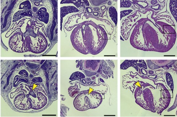
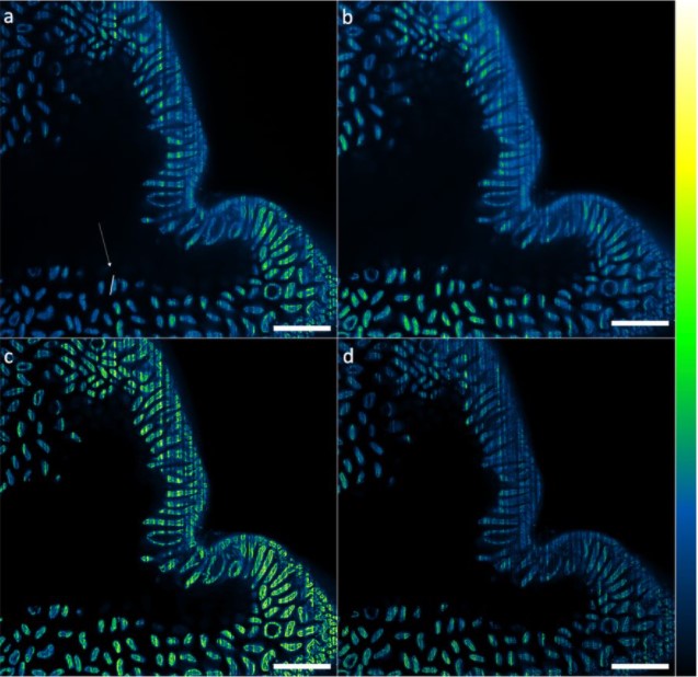
Embryonic mouse hearts stained with hematoxylin and eosin from Chang, et al.
A nuclear receptor facilitates differentiation of human PSCs into more mature hepatocytes Haiting Ma, Esmée de Zwaan, Yang Eric Guo, Paloma Cejas, Prathapan Thiru, Martijn van de Bunt, Jacob F. Jeppesen, Sudeepa Syamala, Alessandra Dall’Agnese, Brian J. Abraham, Dongdong Fu, Carrie Garrett-Engele, Tony Lee, Henry W Long, Linda G. Griffith, Richard A. Young, Rudolf Jaenisch
Stem cells partner with matrix remodeling cells during regeneration Blair W. Benham-Pyle, Frederick G. Mann Jr., Carolyn E. Brewster, Enya R. Dewars, Stephanie H. Nowotarski, Carlos Guerrero-Hernández, Seth Malloy, Kate E. Hall, Lucinda E. Maddera, Shiyuan Chen, Jason A. Morrison, Brian D. Slaughter, Anoja Perera, Alejandro Sánchez Alvarado
SARS-CoV-2 Can Infect Human Embryos Mauricio Montano, Andrea R. Victor, Darren K. Griffin, Tommy Duong, Nathalie Bolduc, Andrew Farmer, Vidur Garg, Anna-Katerina Hadjantonakis, Alison Coates, Frank L. Barnes, Christo G. Zouves, Warner C. Greene, Manuel Viotti
The conserved transcriptional program of metazoan male germ cells uncovers ancient origins of human infertility Rion Brattig Correia, Joana M. Almeida, Margot J. Wyrwoll, Irene Julca, Daniel Sobral, Chandra Shekhar Misra, Leonardo G. Guilgur, Hans-Christian Schuppe, Neide Silva, Pedro Prudêncio, Ana Nóvoa, Ana S. Leocádio, Joana Bom, Moisés Mallo, Sabine Kliesch, Marek Mutwil, Luis M. Rocha, Frank Tüttelmann, Jörg D. Becker, Paulo Navarro-Costa
Cell-intrinsic differences between human airway epithelial cells from children and adults Elizabeth F. Maughan, Robert E. Hynds, Adam Pennycuick, Ersilia Nigro, Kate H.C. Gowers, Celine Denais, Sandra Gómez-López, Kyren A. Lazarus, Jessica C. Orr, David R. Pearce, Sarah E. Clarke, Dani Do Hyang Lee, Maximillian N. J. Woodall, Tereza Masonou, Katie-Marie Case, Vitor H. Teixeira, Benjamin E. Hartley, Richard J. Hewitt, Chadwan Al Yaghchi, Gurpreet S. Sandhu, Martin A. Birchall, Christopher O’Callaghan, Claire M. Smith, Paolo De Coppi, Colin R. Butler, Sam M. Janes
Scalable Generation of Pseudo-Unipolar Sensory Neurons from Human Pluripotent Stem Cells Tao Deng, Carlos A. Tristan, Claire Weber, Pei-Hsuan Chu, Seungmi Ryu, Vukasin M. Jovanovic, Pinar Ormanoglu, Prisca Twumasi, Jaehoon Shim, Selwyn Jayakar, Han-Xiong Bear Zhang, Sooyeon Jo, Ty C. Voss, Anton Simeonov, Bruce P. Bean, Clifford J. Woolf, Ilyas Singeç
A cellular and molecular analysis of SoxB-driven neurogenesis in a cnidarian Eleni Chrysostomou, Hakima Flici, Sebastian G Gornik, Miguel Salinas-Saavedra, James M Gahan, Emma T McMahon, Kerry Thompson, Shirley Hanley, Michelle Kilcoyne, Christine E. Schnitzler, Paul Gonzalez, Andreas D Baxevanis, Uri Frank
Molecular characterization of a flatworm Girardia isolate from Guanajuato, Mexico Elizabeth M. Duncan, Stephanie H. Nowotarski, Carlos Guerrero-Hernández, Eric J. Ross, Julia A. D’Orazio, Clubes de Ciencia México Workshop for Developmental Biology, Sean McKinney, Mark C. McHargue, Longhua Guo, Melainia McClain, Alejandro Sánchez Alvarado
Single-cell atlas of human liver development reveals pathways directing hepatic cell fates Brandon T. Wesley, Alexander D. B. Ross, Daniele Muraro, Zhichao Miao, Sarah Saxton, Rute A. Tomaz, Carola M. Morell, Katherine Ridley, Ekaterini D. Zacharis, Sandra Petrus-Reurer, Judith Kraiczy, Krishnaa T. Mahbubani, Stephanie Brown, Jose Garcia-Bernardo, Clara Alsinet, Daniel Gaffney, Olivia C. Tysoe, Rachel A. Botting, Emily Stephenson, Dorin-Mirel Popescu, Sonya MacParland, Gary Bader, Ian D. McGilvray, Daniel Ortmann, Fotios Sampaziotis, Kourosh Saeb-Parsy, Muzlifah Haniffa, Kelly R. Stevens, Matthias Zilbauer, Sarah A. Teichmann, Ludovic Vallier
Multimodal spatiotemporal phenotyping of human organoid development Philipp Wahle, Giovanna Brancati, Christoph Harmel, Zhisong He, Gabriele Gut, Aline Santos, Qianhui Yu, Pascal Noser, Jonas Simon Fleck, Bruno Gjeta, Dinko Pavlinić, Simone Picelli, Maximilian Hess, Gregor Schmidt, Tom Lummen, Yanyan Hou, Patricia Galliker, Magdalena Renner, Lucas Pelkmans, Barbara Treutlein, J. Gray Camp
An integrated cell atlas of the human lung in health and disease L Sikkema, D Strobl, L Zappia, E Madissoon, NS Markov, L Zaragosi, M Ansari, M Arguel, L Apperloo, C Bécavin, M Berg, E Chichelnitskiy, M Chung, A Collin, ACA Gay, B Hooshiar Kashani, M Jain, T Kapellos, TM Kole, C Mayr, M von Papen, L Peter, C Ramírez-Suástegui, J Schniering, C Taylor, T Walzthoeni, C Xu, LT Bui, C de Donno, L Dony, M Guo, AJ Gutierrez, L Heumos, N Huang, I Ibarra, N Jackson, P Kadur Lakshminarasimha Murthy, M Lotfollahi, T Tabib, C Talavera-Lopez, K Travaglini, A Wilbrey-Clark, KB Worlock, M Yoshida, Lung Biological Network Consortium, T Desai, O Eickelberg, C Falk, N Kaminski, M Krasnow, R Lafyatis, M Nikolíc, J Powell, J Rajagopal, O Rozenblatt-Rosen, MA Seibold, D Sheppard, D Shepherd, SA Teichmann, A Tsankov, J Whitsett, Y Xu, NE Banovich, P Barbry, TE Duong, KB Meyer, JA Kropski, D Pe’er, HB Schiller, PR Tata, JL Schultze, AV Misharin, MC Nawijn, MD Luecken, F Theis
In the latest episode of the Genetics Unzipped podcast, we’re looking at the monkey in the mirror, investigating how flipped genetic switches and long-dead viruses make all the difference between our human faces and those of our closest primate relatives.
“The bits in our genome that encode functional elements like proteins are pretty much identical to a chimp’s. But straight away you can see that a chimp’s face is very different from our own, even though it’s made using the same biological ingredients.”
If you enjoy the show, please do rate and review on Apple podcasts and help to spread the word on social media. And you can always send feedback and suggestions for future episodes and guests to podcast@geneticsunzipped.com Follow us on Twitter – @geneticsunzip
December 3-7, 2022 | Walter E. Washington Convention Center | Washington, DC
Returning in person in 2022—this mid-sized unique joint meeting of the American Society for Cell Biology (ASCB) and the European Molecular Biology Organisation (EMBO) focuses on cell biology as the fundamental basis of biology, while also offering sessions on emerging interdisciplinary topics.
Besides offering attendees opportunities to present their research, the meeting provides an unparalleled forum for attendees to network and initiate collaborations across disciplines. Cell Bio 2022 offers eight different tracks—seven scientific and one covering educational, professional development, diversity, and inclusion.
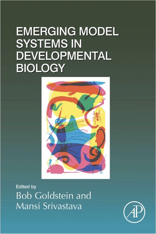
Mansi Srivastava and I have edited a new book that’s packed with scientists’ stories about emerging model organisms. The book presents some interesting additions to the core set of model organisms, with contributions from people who have developed new model systems or advanced tools for emerging models. And it includes personal stories about how and why model systems were developed. The book isn’t comprehensive – we look forward to reading other scientists’ stories about these and other emerging models. But if you’re looking for 700+ pages of fascinating biology, we encourage you to take a look!
A PhD project proposal to be carried out in Patrick Lemaire’s lab at CRBM, Montpellier, France, in tight collaboration with Grégoire Malandain, Morpheme INRIA team, Sophia-Antipolis, France. Funding is conditional upon selection by the CBS2 doctoral school‘s entrance jury.
Why study ascidians?
The embryonic development of ascidians, a group of marine invertebrates, is remarkably conserved, at the single cell level, between individuals of a given species and between species, even if they diverged up to 400 MY ago. Ascidian genomes, however, evolve particularly rapidly. The remarkably simple and transparent ascidian embryos are thus ideal to study developmental systems drift and to identify constraints that could explain the exceptional evolutionary precision and stability of embryonic morphologies.
The proposed project
We propose a computational PhD project, which will use experimental data collected in the team or in public databases to characterise inter-individual and inter-species variability at the geometric, mechanical and transcriptional scales.
This project will involve the development of concepts and computer tools to study and measure, at each scale studied, the variability of different parameters (variability of cell lineages, cell lifetimes, orientation of cell division, pressures, surface and line tensions, gene expression, etc.). These studies will lead to a reflection on the concept of the average embryo and on its computational representation. The tools developed will open the way to the quantitative study of robustness to environmental and genetic perturbations, and to the identification of bridges between scales of analysis (search for co-varying parameters across scales).
What is available to start the project?
The project will benefit from the conceptual and methodological developments made over the last 10 years by the hosting teams and their collaborators (see references below). These breakthroughs (ASTEC, MorphoNet, Aniseed) place them in a unique position to analyse, experimentally and computationally, the variability of animal embryogenesis, whether natural or in response to environmental or experimental perturbations.
Using these advanced tools, we generated high resolution geometric and mechanical descriptions of 7 embryos of the ascidian Phallusia mammillata, over several hours of development and with a 2 minutes time resolution. You can view a short video highlighting this works.
These embryos and tools constitute a solid basis for the proposed PhD project. The embryo collection is currently expanding to include more WT Phallusia embryos as well as embryos cultured in response to environmental (temperature, salinity, pH) or genetic perturbations.
How to apply?
The ideal candidate will have successfully graduated from a Master’s programme in computer science, bioinformatics or physics recognized by France. S/He will have strong computational skills and some knowledge of developmental biology. A working knowledge of English (B2) is needed. There is no prerequisite in French.
To apply to the project, please contact P. Lemaire (patrick.lemaire[at]crbm.cnrs.fr) as soon as possible and by May 11, 2022 at the latest with a motivation letter, a CV and the names and contact details of 2 academic referees including the PhD supervisor.
References
Guignard L. *, Fiuza U.-M. *, Leggio B., Laussu J., Faure E., Michelin G., Biasuz K., Hufnagel L., Malandain G. #, Godin C. #, Lemaire P.# (2020) Contact-area dependent cell communications and the morphological invariance of ascidian embryogenesis. Science, 369 :6500 eaar5663
Dardaillon, J; Dauga, D; …; Dantec, C.#; Lemaire, P#. (2019) ANISEED 2019: 4D exploration of genetic data for an extended range of tunicates. Nucleic Acids Res. 48(D1): D668-D675
Leggio, B; Laussu J; Carlier, A; Godin, C; Lemaire, P and Faure, E (2019) MorphoNet: An interactive online morphological browser to explore complex multi-scale data. Nat Commun. 10(1):2812
The tunicate team at CRBM (Montpellier, France), headed by Patrick Lemaire, is offering a 2-year post-doctoral fellowship (or a 3-year PhD fellowship for an exceptional candidate) to study the robustness of animal embryonic development to genetic and environmental perturbations, using quantitative live imaging of ascidian embryos. A short video describes a recent piece of work of the team relevant to the project.
Why ascidians?
Ascidians are a group of marine invertebrates. Their embryonic cell lineages and early embryonic stage morphologies have remained essentially identical since the group’s emergence about 400 million years ago. This suggests that they are subject to very strong developmental or evolutionary constraints (Lemaire et al. 2011). Ascidian embryogenesis is also very robust to environmental perturbations of temperature and salinity. The extreme evolutionary and environmental robustness of embryonic geometries and cell lineages contrasts with a rapid genetic divergence between species and intra- and inter-specific variability in gene expression.
What is the project?
The proposed experimental project will provide a quantitative assessment of the developmental robustness of a critical morphogenetic process, ascidian gastrulation (1, 3, 5), to two key environmental parameters (water temperature and salinity) and to genetic perturbations of the morphogenetic driving force apparatus (myosin II, Rho kinase, …). The project will study the magnitude of environmental or genetic variations compatible with the production of a viable larva. It will seek to identify the least – and most – robust developmental processes and time points, i. e. those that collapse first, or resist best to the perturbations. Finally, it will characterize the structure of the natural and experimentally-induced variability in the geometry and mechanical properties of embryos. This may lead to the identification of developmental modules.
The project will involve advanced light-sheet imaging of live micro-injected embryos of the ascidian Phallusia mammillata, followed by the computational and statistical analysis of the acquired developmental movies (see 2, 4).
Who funds the project, and how to apply?
The project is funded by an ANR-NSF binational project grant and will be conducted in collaboration with 3 other teams: Prof. Atef Asnacios (MSC, Paris), Prof. Edwin Munro (U. Chicago, USA) and Prof. Madhav Mani (Northwestern University, Evanston, USA).
Expected candidates will have a PhD in cell and developmental biology, an excellent track record of publications and oral communications, strong skills in fluorescent live imaging and some experience in the computational analysis of large datasets. To apply, send Patrick Lemaire (patrick.lemaire[at]crbm.cnrs.fr) by May 11, 2022 at the latest a motivation letter, a CV and the names and contact details of 2 academic referees including the PhD supervisor. A working knowledge of English (B2) is needed, there is no prerequisite in French.
References
Fiuza U.-M. and Lemaire, P. (2021) Mechanical and genetic control of ascidian endoderm invagination during gastrulation, Semin Cell Dev Biol 120:108-118
Guignard L. *, Fiuza U.-M. *, Leggio B., Laussu J., Faure E., Michelin G., Biasuz K., Hufnagel L., Malandain G. #, Godin C. #, Lemaire P.# (2020) Contact-area dependent cell communications and the morphological invariance of ascidian embryogenesis. Science, 369 :6500 eaar5663
Fiuza U-M, Negishi T., Rouan A., Yasuo H.#, Lemaire P. # (2020) Nodal and Eph signalling relay drives the transition between apical constriction and apico-basal shortening during ascidian endoderm invagination. Development 147: dev186965
Leggio, B; Laussu J; Carlier, A; Godin, C; Lemaire, P and Faure, E (2019) MorphoNet: An interactive online morphological browser to explore complex multi-scale data. Nat Commun.10(1):2812
Sherrard, K., Robin, FB, Lemaire, P., and Munro, E. (2010) Sequential activation of apical and basolateral myosin drives endoderm invagination during ascidian gastrulation, Current Biology, 20(17):1499-510.
 (8 votes)
 (No Ratings Yet)