After a year of lockdowns and virtual classes at Bangor University, the opportunity to do a real lab project this summer at the Francis Crick Institute was definitely not one to miss. Under the patient supervision of Adrien Franchet and Sebastian Sorge in Alex Gould’s lab, I set out to explore the roles of some amino acid transporters during the development of the genetic model organism Drosophila.
This two month summer project was my first opportunity to gain hands-on experience doing hypothesis-driven science and to interact with many talented researchers at the Crick. As an undergraduate, my only previous exposure to fruit flies was from reading published papers but, right from day one, I got stuck in to the nitty gritty of Drosophila developmental biology and larval dissections.
The Gould lab are interested in figuring out how the neural stem cells of the developing CNS are so highly protected against environmental stresses such as nutrient restriction (NR) and hypoxia. This process is a key part of brain sparing, which involves sustaining the growth of the CNS at the expense of other organs such as adipose tissue. In mammals, brain sparing is commonly observed in neonates following intrauterine growth restriction. However, the key signalling and metabolic pathways underlying brain sparing are still unclear.
Amino-acids are key signals for growth and they are also critical for protein synthesis. The uptake of amino acids by tissues involves a large number of different amino-acid transporters and I set out to decipher whether two of these transporters (AAT1 and AAT2) are required in the neural stem cell niche (glia in Drosophila) or in adipose tissue (fat body in Drosophila) for CNS and body growth. My project stemmed from Adrien’s and Sebastian’s recent RNA interference (RNAi) screen of amino acid transporter candidates. I followed up two of their screen hits (AAT1 and AAT2) using UAS-RNAi knockdowns lines crossed with Gal4-driver lines specific for glia (repo-Gal4) or fat body (Cg-Gal4). The goal was to measure the phenotypic effects of these cell-type genetic manipulations during standard fed development and also during severe NR on an agar-only diet. Phenotypes were measured for larval and pupal weights using an accurate microbalance. I also quantified CNS phenotypes from confocal microscopy images by measuring CNS area and also neural stem cell (neuroblast) proliferation via the incorporation of a labelled nucleotide analogue (EdU).
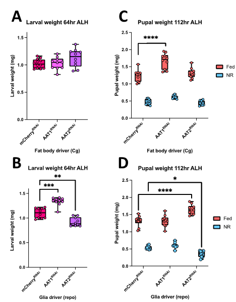
I found that RNAi knockdowns of either AAT1 or AAT2 produced more severe phenotypes in glia compared to fat body (Figure 1). Hence, larval pupal and adult weights were largely normal with the fat body knockdowns (Figure 1A, 1C). However, both glial knockdowns gave modest changes in body weight at the larval stage but, by the pupal stage, these only remained significant for AAT2 (Figure 1B, 1D). I also noticed that glial knockdown of AAT2 eventually resulted in adult lethality, shortly after eclosion, with flies displaying very severe locomotor defects.
Figure 1: Larval and pupal weights of AAT1 or AAT2 knockdowns.Larvae were raised on standard lab diet (Fed) or nutrient restriction (NR). (A,B) Larval weights of fat body (Cg-Gal4) knockdowns (A) or glia (repo-Gal4) knockdowns. (C,D) Pupal weight of fat body (C) or glial (D) knockdowns. mCherryRNAi is a control of RNAi line and times refer to hours after larval hatching (ALH). Statistical significance (asterisks) was determined using Tukey’s Multiple Comparison Test.
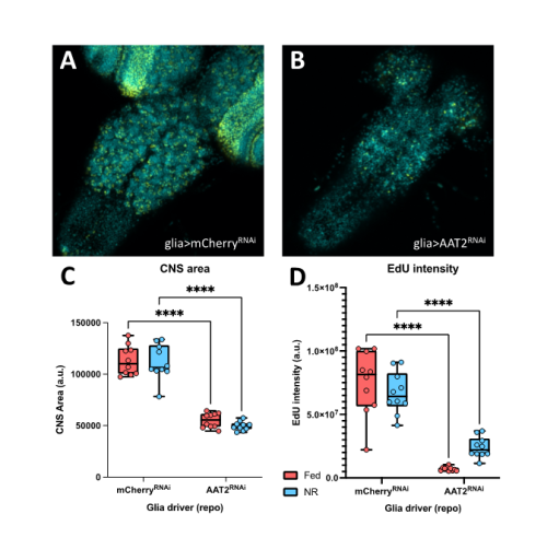
In a parallel set of experiments, I investigated the effects of the AAT2 knockdowns on the growth of the developing CNS and on the proliferation of neural stem cells. To do so, I dissected brains from fed larvae and from larvae exposed to one day of NR. I then performed an in vitro EdU incorporation assay as an indicator for neuroblast progression through S-phase of the cell cycle. I found that the fat body manipulations had no significant effect on CNS size or on neuroblast proliferation. In contrast, the glial manipulations revealed that AAT2 is required in glia for proper growth of the larval brain, as the CNSs of repo-GAL4; AAT2RNAi larvae were strongly reduced in size and likewise the EdU incorporation was much lower than genetic controls (Figure 2A, 2B). This glial requirement for AAT2 for neuroblast proliferation occurred in both fed and NR larvae (Figure 2C, 2D). Thus, in conclusion, my project has revealed a constitutive function in glia for the amino acid transporter AAT2 during both normal CNS growth and brain sparing. It will be important in future to explore whether AAT2 is required in the surface glia of the blood-brain barrier or in the internal cortex glia that surround neuroblasts and their daughter cells. Equally importantly, it will be interesting to identify which specific amino acids are transported by AAT2.
Figure 2: Glial AAT2 is required for neuroblast proliferation in the larval CNS Larvae were raised on standard lab diet (Fed) or nutrient restriction (NR). (A,B) Confocal images of the nuclear marker DAPI (cyan) and the proliferation marker EdU (yellow) for third-instar larvae expressing repo-Gal4 driving AAT2 RNAi and their genotype conrols (mCherryRNAi) (C,D) Quantitation of CNS area (C) and average EdU intensity (D) for control and AAT2 RNAi lines. **** Statistical significance determined suing Tukey’s Multiple Comparison Test.
Overall, this fascinating project has given me a first taste of biological research at the bench and has also allowed me to develop critical thinking and data processing skills. I am indebted to Adrien Franchet and Sebastian Sorge for their fantastic direction, and to Alex Gould and all of his lab for their encouragement throughout. I would also like to thank the Francis Crick Institute for hosting me and the Medical Research Foundation Rosa Beddington Fund for supporting my project and allowing me to contribute to this captivating field of research.
This summer, I was given the opportunity to conduct research at the Francis Crick Institute in the Znamenskiy lab. The aim of the Znamenskiy lab is to understand the relationship between connectivity, gene expression and function of cortical neurons.
The neocortex is a region of the brain integral in performing higher cognitive functions. Neocortical projections can be divided into three broad classes. Corticothalamic (CT) neurons are located mostly within layer 6 and send axons to the thalamus. Pyramidal Tract (PT) neurons are nearly exclusively positioned within layer 5 and project to brainstem and spinal cord. Intratelencephalic (IT) neurons are distributed throughout all six layers and project to distant cortical areas (Kast & Levitt, 2019). The expression of transcription factors during development can affect projection patterns. For example, when Fefz2 is deleted, the cortex no longer sends projections to the brain stem and instead sends projections to the thalamus or contralateral hemisphere (Kast & Levitt, 2019). This shows that genes expressed by a neuron during development play an important role in determining its wiring patterns.
Beyond these broad projection classes, the genetic basis underlying patterns of neocortical connectivity is little understood. The primary visual cortex (V1) is a region of the brain that is important for receiving, segmenting, integrating, and processing visual information relayed from the retinas. Subsequently, the processed information is then sent to other regions of the brain. This is a highly specialised process that allows the brain to recognise patterns quickly and with the absence of a conscious effort. The V1 provides a platform for understanding the neocortex due to its serially homologous structure, and therefore can be used as a model for neocortical projections. The V1 sends projections to several higher visual areas as well as many other areas of the brain such as the lateral geniculate and lateral posterior thalamic nuclei, superior colliculus, striatum, and other subcortical structures but little is known about how these connectivity patterns are established (Kast & Levitt, 2019).
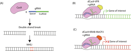
To understand which genes are important for specifying long range connectivity patterns from V1, in vivo genetic manipulations using CRISPR/Cas9 can be used to determine what happens to connectivity patterns when the expression of target genes is altered. CRISPR/Cas9 is a simple, rapid method to modify gene expression which can be pooled together to look at many genes in parallel. As well as knocking-out the gene of interest using the prototypical CRISPR/Cas9 gene editing approach (Figure 1A), methods for modulating gene expression using catalytically inactive Cas9 fused to transcriptional modulators have recently been developed (Figure 1B-C). CRISPR activation (CRISPRa) allows functional analysis of redundant genes through overexpression, whereas CRISPR interference (CRISPRi) allows analysis of gene function by knocking-down gene expression at the transcriptional level and is thought to have fewer off-target effects (Gebre et al., 2018). The aim of my project was to perform preliminary experiments validating whether gRNA constructs designed to be used to examine changes in in vivo V1 connectivity patterns, using CRISPR knockout, CRISPRi or CRISPRa, altered gene expression in vitro. The first part of my project was to clone some of the gRNA CRISPR constructs, and the second part was to test constructs in vitro.
FIGURE 1. Mechanism of CRISPR/Cas9 Genetic Modulation. (A) CRISPR knockout involves co-expressing Cas9 and a gRNA in a cell. The Cas9 protein recognises a specific sequence called the scaffold sequence in the gRNA while another sequence within the gRNA called the spacer region determines the target site within the genome to be modified. The Cas9 protein generates double strand breaks in the gene of interest that are repaired through the non-homologous end joining (NHEJ) pathway that is prone to producing indel mutations (red bases here indicate an insertion) that can render genes non-functional when translated. (B) CRISPR activation (CRISPRa) constructs work via transcriptional activators fused to catalytically dead Cas9 (dCas9) which are targeted near transcriptional start sites of the endogenous gene of interest by the gRNA to induce their overexpression. (C) CRISPR interference (CRISPRi) constructs consist of dCas9 fused to transcriptional repressor domains that are recruited in proximity of the transcription start site of an endogenous gene to repress transcription.
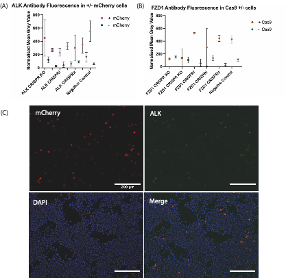
gRNA constructs were tested along with corresponding Cas9s (SP-Cas9, dCas9-KRAB-MeCP2, and dCas9-VPR for CRISPR knockout, CRISPRi, and CRISPRa, respectively) to determine whether a change of expression in our genes of interest occurred within Neuro-2A (N2a) cells. Target genes for validation (Frizzled 1 (FZD1), Androgen Receptor (AR), Polycystic Kidney and Hepatic Disease 1 (PKHD1), and Anaplastic Lymphoma Kinase (ALK)) were identified due their established endogenous gene expression in N2a cells. To determine whether the gRNA constructs worked I co-transfected Cas9’s with the gRNA construct into N2a cells and observed whether this altered expression of target genes by looking at endogenous protein levels through immunostaining. Endogenous protein levels in each condition were compared to a control plasmid without a gRNA insert. The results obtained from the quantification of the transfection and subsequent immunostaining are shown in Figure 2A-C. These results did not reveal expected differences in gene expression between gRNA constructs and further experiments need to be performed using alternative antibodies or staining conditions. However, the project has given me an insight into the molecular basis of developmental biology, and I thoroughly enjoyed learning the techniques and protocols required to complete the cloning process. During my research internship I was able to obtain applied, practical experience within the laboratory which due to the COVID-19 pandemic, has been limited during my undergraduate degree. I also was given a level of independence which I did not expect within the laboratory, completing the transfection of gRNA constructs was an engaging, albeit challenging process as my cells became contaminated during the passaging process. However, I was able to overcome this setback and build resilience. Overall, I really enjoyed my project, and it has encouraged me to pursue a career in scientific research.
FIGURE 2. Quantitative Immunofluorescence after transfection of Cas9 plasmids and mCherry expressing gRNA plasmids targeting either ALK or FZD1. (A) ALK antibody fluorescence in +/- mCherry Cells. (B) FZD1 antibody fluorescence in + mCherry; +/- Cas9 Cells. KO/i/a – KO = Knockout; i= interference, a = activation. Each symbol shows the mean normalized grey value of N2A cells which reflects the level of fluorescence from antibodies targeting the endogenous protein-of-interest after immunostaining. The negative control used was a plasmid without a gRNA insert. SP-Cas9 was used for ALK/FZD1 KO & negative control, whereas dCas9-KRAB-MeCP2 was used for ALK/FZD1 CRISPRi, dCas9-VPR used for ALK/FZD1 CRISPRa. For the FZD1 transfection there was an unexpectedly low number of +Cas9 cells. (C) Immunohistochemistry staining against mCherry and ALK, as well as DAPI staining in N2a cells transfected with SP-Cas9a and a gRNA targeting ALK. mCherry is expressed by the gRNA constructs, staining this protein shows which cells were transfected with our construct of interest, whereas DAPI staining marks the nuclei of all cells. The overlap in ALK and mCherry signals suggests further optimisation of immunostaining and imaging conditions is required to avoid bleed-through.
I would like to take this opportunity to thank the Francis Crick Institute, particularly the Znamenskiy lab for allowing me to undertake research at their facility, alongside my supervisor Benita Turner-Bridger for supporting me in my project. Furthermore, I would like to show my appreciation to the Medical Research Foundation and the Rosa Beddington fund which has provided the financial support for my project. It is an honour to have the opportunity to contribute to The Node and the British Society of Developmental Biology, and I would strongly encourage other undergraduate students to pursue a similar research project during their studies. This experience has been unlike any other.
References:
Gebre, M., Nomburg, J.L and Gewurz, B.E (2018). CRISPR-Cas9 Genetic Analysis of Virus-Host Interactions. Viruses, 10(2), 55.
Kast, R.J and Levitt, P (2019). Precision in the development of neocortical architecture: From progenitors to cortical networks. Progress in Neurobiology, 175, 77-95.
Unique functions for Notch4 in murine embryonic lymphangiogenesis Ajit Muley, Minji Kim Uh, Glicella Salazar-De Simone, Bhairavi Swaminathan, Jennifer M James, Aino Murtomaki, Joseph D McCarron, Chris Kitajewski, Maria Gnarra, Gloria Riitano, Yoh-suke Mukouyama, Jan Kitajewski, Carrie J Shawber
Androglobin, a chimeric mammalian globin, is required for male fertility Anna Keppner, Miguel Correia, Sara Santambrogio, Teng Wei Koay, Darko Maric, Carina Osterhof, Denise V Winter, Angèle Clerc, Michael Stumpe, Frédéric Chalmel, Sylvia Dewilde, Alex Odermatt, Dieter Kressler, Thomas Hankeln, Roland H. Wenger, David Hoogewijs
Retrospective analysis of enhancer activity and transcriptome history Ruben Boers, Joachim Boers, Beatrice Tan, Evelyne Wassenaar, Erlantz Gonzalez Sanchez, Esther Sleddens, Yasha Tenhagen, Marieke E. van Leeuwen, Eskeatnaf Mulugeta, Joop Laven, Menno Creyghton, Willy Baarends, Wilfred F. J. van IJcken, Joost Gribnau
Sex differences and risk factors for bleeding in Alagille syndrome Simona Hankeova, Noemi Van Hul, Jakub Laznovsky, Katrin Mangold, Naomi Hensens, Elvira Verhoef, Tomas Zikmund, Feven Dawit, Michaela Kavkova, Jakub Salplachta, Marika Sjöqvist, Bengt R. Johansson, Mohamed Hassan, Linda Fredriksson, Vitezslav Bryja, Urban Lendahl, Andrew Jheon, Florian Alten, Kristina Teär Fahnehjelm, Björn Fischler, Jozef Kaiser, Emma R. Andersson
Dynamic regulation and requirement for ribosomal RNA transcription during mammalian development Karla T. Falcon, Kristin E.N. Watt, Soma Dash, Annita Achilleos, Ruonan Zhao, Daisuke Sakai, Emma L. Moore, Sharien Fitriasari, Melissa Childers, Mihaela E. Sardiu, Selene Swanson, Dai Tsuchiya, Jay Unruh, George Bugarinovic, Lin Li, Rita Shiang, Jill Dixon, Michael J. Dixon, Paul A. Trainor
Tig1 regulates proximo-distal identity during salamander limb regeneration Catarina R. Oliveira, Dunja Knapp, Ahmed Elewa, Tobias Gerber, Sandra G. Gonzalez Malagon, Phillip B. Gates, Hannah E. Walters, Andreas Petzold, Hernan Arce, Rodrigo C. Cordoba, Elaiyaraja Subramanian, Osvaldo Chara, Elly M. Tanaka, András Simon, Maximina H. Yun
High resolution snRNA-seq analysis of Drosophila Malpighian tubules from Xu, et al.
A cell atlas of the fly kidney Jun Xu, Yifang Liu, Hongjie Li, Alexander J. Tarashansky, Colin H. Kalicki, Ruei-Jiun Hung, Yanhui Hu, Aram Comjean, Sai Saroja Kolluru, Bo Wang, Stephen R Quake, Liqun Luo, Andrew P. McMahon, Julian A.T. Dow, Norbert Perrimon
Bringing TrackMate into the era of machine-learning and deep-learning Dmitry Ershov, Minh-Son Phan, Joanna W. Pylvänäinen, Stéphane U. Rigaud, Laure Le Blanc, Arthur Charles-Orszag, James R. W. Conway, Romain F. Laine, Nathan H. Roy, Daria Bonazzi, Guillaume Duménil, Guillaume Jacquemet, Jean-Yves Tinevez
A reference tissue atlas for the human kidney Jens Hansen, Rachel Sealfon, Rajasree Menon, Michael T. Eadon, Blue B. Lake, Becky Steck, Dejan Dobi, Samir Parikh, Tara K. Sigdel, Guanshi Zhang, Dusan Velickovic, Daria Barwinska, Theodore Alexandrov, Priyanka Rashmi, Edgar A. Otto, Michael P. Rose, Christopher R. Anderton, John P. Shapiro, Annapurna Pamreddy, Seth Winfree, Yongqun He, Ian H. de Boer, Jeffrey B. Hodgin, Laura Barisoni, Abhijit S. Naik, Kumar Sharma, Minnie M. Sarwal, Kun Zhang, Jonathan Himmelfarb, Brad Rovin, Tarek M. El-Achkar, Zoltan Laszik, John Cijiang He, Pierre C. Dagher, M. Todd Valerius, Sanjay Jain, Lisa Satlin, Olga G. Troyanskaya, Matthias Kretzler, Ravi Iyengar, Evren U. Azeloglu, for the Kidney Precision Medicine Project
ShareLoc – an open platform for sharing localization microscopy data Jiachuan Bai, Wei Ouyang, Manish Kumar Singh, Christophe Leterrier, Paul Barthelemy, Samuel F.H. Barnett, Teresa Klein, Markus Sauer, Pakorn Kanchanawong, Nicolas Bourg, Mickael M. Cohen, Benoît Lelandais, Christophe Zimmer
Julia for Biologists Elisabeth Roesch, Joe G. Greener, Adam L. MacLean, Huda Nassar, Christopher Rackauckas, Timothy E. Holy, Michael P.H. Stumpf
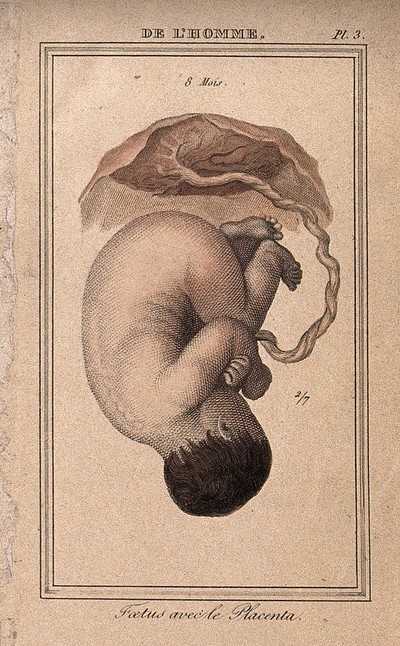
In the latest episode of Genetics Unzipped, Kat Arney explores the science behind one of the most remarkable but often overlooked organs in the mammalian body: the placenta.
To find out more, Kat chats with Ros John, who leads the Pregnancy Research Epigenetics Group or PREGLab at Cardiff University. Ros’s research focuses on understanding maternal mental health, imprinted genes and the role of the placenta during pregnancy and even beyond, with big implications for the health of both mother and child.
Kat also speaks to Sam Behjati, a group leader at the Wellcome Sanger Institute, who recently made the surprising discovery that the placenta is a genetic ‘dumping ground’. The pattern of genetic alterations in the placenta is different to any other human organ and resembles that of a tumour, harbouring many of the same genetic mutations found in childhood cancers.
If you enjoy the show, please do rate and review on Apple podcasts and help to spread the word on social media. And you can always send feedback and suggestions for future episodes and guests to podcast@geneticsunzipped.com Follow us on Twitter – @geneticsunzip
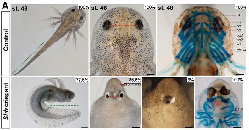
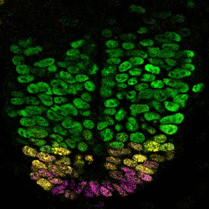
A developing mouse neural tube. Hedgehog signalling is required to produce different types of neuronal progenitor cells, indicated by the different colours. THC inhibits Hedgehog signalling in genetically susceptible mice, leading to developmental defects, including in the neural tube.
A chemical found in cannabis, a common recreational drug, has now been shown to cause birth defects in mice. Scientists from the Icahn School of Medicine at Mount Sinai, USA, show that the psychoactive component of cannabis (THC) can induce face and nervous system defects in genetically susceptible mouse embryos. This study, published in Development, has important implications for cannabis use during pregnancy.
Cannabis, also known as weed or marijuana, is used by hundreds of millions of people worldwide. Although restricted in most countries, the increasing legalisation of cannabis for recreational and medicinal consumption means that cannabis use is rising. Cannabis is also the most common illicit drug used by pregnant women, but the effects of cannabis on embryonic development are not well understood. It is also important to understand the effects of cannabis on individuals with a genetic predisposition, which means they carry genetic mutations that increase the risk of environmental conditions triggering a defect or disease.
A new study from scientists at the Icahn School of Medicine at Mount Sinai, USA, has now revealed that THC, the chemical in cannabis that causes the sensation of feeling ‘high’, can cause birth defects in genetically predisposed mice. In this case, the researchers investigated whether THC could exacerbate a mutation that affects a mechanism that cells use to communicate with each other, called Hedgehog signalling. “Several years ago, it was reported that THC could inhibit Hedgehog signalling in cells grown in a dish,” said the study’s lead author Robert Krauss, PhD, Professor of Cell, Developmental and Regenerative Biology at the Icahn School of Medicine at Mount Sinai. “We reasoned that THC might be an environmental risk factor for birth defects, but that it would require additional risk factors, such as specific mutations in the genes required for Hedgehog signalling, to induce these defects in mice.”
To address this hypothesis, Dr Krauss and colleagues administered a single dose of THC, equivalent to exposures achieved when humans smoke cannabis, to pregnant mice about a week after conception. They then studied the embryonic development of their pups, some of which carried a mutation that meant Hedgehog signalling was not functioning at full efficiency. The scientists found that pups without the mutation developed normally, even when exposed to THC, as did pups that carried the mutation but were not exposed to the drug. However, pups that were exposed to THC and carried the mutation developed a brain and face defect called holoprosencephaly, a common birth defect seen in 1 in 250 human conceptions that includes the failure of the forebrain to divide into two distinct segments.
The researchers showed that the defect occurs because THC can interfere with Hedgehog signalling in the embryo. THC alone is not sufficient to disrupt Hedgehog signalling and cause defects but, in animals where Hedgehog signalling is already weakened through genetic mutation, it has significant effects. “THC directly inhibits Hedgehog signalling in mice, but it is not a very powerful inhibitor; this is presumably why a genetic predisposition is required for it to cause holoprosencephaly in mice,” explained Dr Krauss.
These first studies in mice have important implications for human health, highlighting the need for more research into the effects of cannabis use during pregnancy in humans. “The THC concentration in cannabis is now very high, so it is important to perform epidemiology studies looking at whether cannabis consumption is associated with developmental defects. Women are already advised not to consume cannabis while pregnant, but our results show that embryos are sensitive at a very early period, before many women know they are pregnant. Cannabis consumption may therefore be inadvisable even when women are trying to get pregnant,” Dr Krauss warned.
Although this study focussed on one chemical in cannabis and one genetic mutation, further research could reveal other combinations that cause similar effects. “Many of the mutations found in human holoprosencephaly patients could conceivably synergise with THC,” said Dr Krauss. “We would also like to test the related chemical CBD in genetically predisposed mice. Like THC, CBD inhibits Hedgehog signalling in cells grown in a dish, but CBD appears to work differently. As CBD is widely available and often viewed as beneficial – or at least innocuous – it would be worth investigating this as well,” he added.
There is no doubt we are in the information age, and this is especially true in our world of biological research. We are ever more reliant on finding and accessing data, from genetic and genomic to epigenetic, transcriptomic and expression data. Searching and sorting through published data can be time consuming and difficult. Another challenge can be navigating resources. With the wealth of reagents, it can be difficult to figure out what is available and best suited for your system. This is especially true when our work involves an organism that is less widely used than rodents. Enter the individual model organism databases (MODs). These are total game changers for our research. By curating data and providing a “one stop shop” for resources specifically related to your model organism of choice we can easily find what we need to move our research forward. But these databases don’t run themselves. They require expertise to keep them current and functional and this expertise requires funding. MODs are currently supported by grants from the NIH and this support is absolutely essential to the continuation of this resource. But continued funding isn’t guaranteed. What can we as individuals due to help keep this funding going? We can let the NIH know just how vital MODs are to us and our research. The NIH is currently soliciting feedback about use of scientific data sources and tools, including MODs. If your MOD is important to you, let the NIH know. A great response from the community could be the difference in securing that next funding cycle. Below is a collection of voices from around the community sharing their stories of what their MOD means to them and their research. We hope that these testimonials inspire you to share your own stories. Please help our community and provide feedback to the NIH through the survey link by October 15. https://www.surveymonkey.com/r/5NGLNNJ
Heather Ray, PhD
Assistant Professor, Idaho State University Department of Biological Sciences
I began working with Xenopus laevis for the first time at the start of my postdoc. Besides beginning a new research project, I needed to get up to speed quickly on the specifics of working with a new organism. I spent many hours scouring Xenbase and it was all of the resources available in this database that helped me get my research up and running quickly. I continue to access Xenbase almost every day for a variety of purposes but most notably to access genomic and related gene expression data. The genetics of Xenopus laevis are complex as this species is a pseudo-tetraploid with differing alloalleles. With Xenbase, I can easily access the genetic sequences for each alloallele to identify the similarities and differences for primer design, gene targeting strategies, etc. I can also easily visualize gene expression data to compare expression patterns of each alloallele over developmental time.
I have recently started my independent lab and Xenbase is now an essential resource for training and education. It has great visuals to teach about embryonic cell fate, morphogenesis, and identifying developmental staging which I use in my undergraduate developmental biology course. It helps the students in my lab find the resources they need to conduct their research and aid in their independence. Without Xenbase as a resource, I would lose countless hours of valuable time. Instead of having a single resource with easily accessed species-specific information, I would have to search through an endless amount of primary literature and synthesize information that is reported in separate places. The time and effort that has gone into curating the interfaces on Xenbase should not have to be repeated a million times over by every individual investigator.
Joaquin Navajas Acedo, PhD
Postdoc, Schier Lab, Biozentrum, University of Basel
“Where is this gene expressed?”, “I wonder if there is an mCherry version of this transgene”, “Oh, lab X is looking for a Technician!”. I have been using Zebrafish as a model organism for my research for around 8 years now, and I don’t think I have spent more than a week without using any of ZFIN’s varied features.
Model organism databases like ZFIN are a central pillar of our work and must be protected at all cost. I cannot imagine doing my job without such a centralized and well curated database. This is thanks to the fantastic job that the folks at ZFIN do, but we should strive for giving them all the space and resources they need, and keep their funding. It would be amazing that some day ZFIN’s features expand to a point where it becomes an Alexandria’s Library where people come, interact and brainstorm together, present their work and leave with new ideas and expertise. A ZFINFish Virtual Forum, where this and other models can be integrated.
Money is a little price to pay to have such wonderful resources that ultimately benefit everyone.
PhD candidate, University of Alabama Birmingham Department of Cell, Developmental and Integrative Biology
As a graduate student who entered the Xenopus research field with minimal knowledge of Xenopus, either conceptual or technical, the wealth of information about all things Xenopus on the Xenbase website has been (and continues to be) a cornerstone for my early career development. My lack of knowledge and skill was glaringly obvious during the first few months in my thesis lab and I desperately wanted to seek resources that could help me. I distinctly remember the moment when a senior lab mate showed me the Xenbase website. I was amazed, and quite frankly, I was relieved to have a seemingly overwhelming amount of information curated in an easily navigable fashion. I initially spent much of my time on Xenbase searching genes of interest, easily comparing their RNA (and now, protein) expression levels, and reading linked references to know more about these genes. I watched beautiful videos of Xenopus embryo development, learned how to time embryo development between Stages, and read numerous protocols designed and time-tested by others in the field. Over the past 4 years, I have continued to rely on Xenbase for pertinent scientific information as well as community updates and conference information. Xenbase has been magnificently and carefully designed to not only support those just beginning their research with Xenopus but continues providing excellent scientific support over a career.
Recently in the literature is the surge of large datasets generated via -omic experiments and analyses. These datasets can be extremely challenging to navigate and identify helpful or relevant information to one’s research. Xenbase compiles and presents these large datasets in a more accessible, meaningful way for researchers. One can easily obtain information to help design experiments, such as using RNA-seq datasets to identify highly expressed RNA isoforms for a gene, or finding the gene structures for L and S alloalleles of a gene to decide whether translational or splicing-blocking morpholinos would be best to achieve gene knockdown. Furthermore, Xenbase is also continuously improving to better serve our froggy community – they frequently add new features such as protein expression information that I previously mentioned and most recently, a new phenotype search function.
I am extremely grateful that Xenbase was funded by the NIH and established prior to my starting graduate school. I know that should I not have had the critical information that Xenbase organizes about Xenopus and associated techniques, it would have immensely slowed my progress as a Xenopus researcher. During graduate school, we are taught to be reliable, honest, independent researchers but scientific information does not need to be a constantly frustrating, convoluted process of diving into deep “rabbit holes”. Xenbase does a fantastic job of organizing data and placing it at a researcher’s finger tips. I truly hope that our community can rally behind Xenbase to keep it running for the generations of scientists to come after us, as I know how important it has been and continues to be for me.
Andrew Plygawko, PhD
Postdoctoral Fellow, Campbell lab, University of Sheffield, UK
My current project uses single-cell RNA sequencing data to identify genes required for Drosophila embryonic midgut development, and I don’t think it’s an exaggeration to say this would be almost impossible without FlyBase. Being able to browse the collective findings from a century’s research streamlines this analysis by distilling the immense amounts of information available into a single webpage that tells me about a gene’s structure, phenotypes, existing tools and more. I’m hopeful that the further identification of gene expression patterns using single-cell techniques will lead to a greater integration of these datasets into FlyBase in the future to build on existing knowledge. This would make it easier for researchers to identify orthologues which are expressed in homologous tissues in other species, accelerating the process through which flies can be established as a model system for understanding development and disease.
Ann Miller, PhD
Associate Professor, University of Michigan Department of Molecular, Cellular, and Developmental Biology
Xenbase is essential for my group to successfully carry out our research! Lab members use Xenbase at least 1x/week. When I polled my lab members about their most used features of Xenbase, they responded:
• We all use Xenbase for up-to-date information on genes of interest: getting sequences, finding alloalleles, looking for related papers using Xenopus, using RNAseq/proteomics data to get a sense of expression levels during relevant stages, looking at spatially localized expression levels at different developmental stages using in situ hybridization data.
• We all use Xenbase when looking up potential morpholinos for proteins of interest and antibodies that have been verified experimentally in Xenopus.
• We all use Xenbase to look up Xenopus protocols.
• We all use Xenbase to consult the Nieuwkoop and Faber Xenopus developmental staging series – and download images of different stages for presentations.
• We use Xenbase to access the lineage map information to do lineage-specific microinjections.
• In addition to the above, I have used Xenbase to access useful material for teaching, writing animal care protocols and grant proposals, and gain support for human disease modeling in Xenopus.
• One of my newer PhD students says: “Xenbase provides an excellent starting point for many of the processes, genes, and small molecule treatments that I’m interested in using for my projects. I also find the educational/background material (such as developmental videos) highly useful while building background for presentations”
• One of my senior lab members says: “I use this as a resource for my current research and also as a resource for undergraduates who join the lab and are not familiar with the model system. The images and movies of Xenopus development are excellent for helping new students learn the basic developmental biology. It is especially helpful that this information is gathered together in a single location so that researchers and students can quickly and easily find the information they need.”
• Another senior lab member says: “I often reference the data on mRNA expression during development as a way to determine whether a gene is of interest for my research. This type of data is not something that we could generate in our own lab, and it is helpful that the Xenopus community openly shares that information on Xenbase. Referencing gene expression data on Xenbase saves time and helps focus my efforts on genes that are most important for the developmental stage I am interested in.”
It would be a major blow to the community and to my group’s research if Xenbase did not exist. For example, it would be difficult to design primers/morpholinos/guide RNAs, time spent searching for background source material and supporting papers would be significantly longer, it would be harder to find trusted protocols and materials when trying out new experiments. Xenbase is a reliable resource that my group uses to quickly and easily find information about the Xenopus model system. It provides foundational knowledge for researchers who are new to working with Xenopus and catalogs up-to-date genomic information that is essential for our research. Finally, the staff members at Xenbase are knowledgeable, supportive, and flexible. They are always happy to explain new Xenbase features and very receptive to new ideas to make Xenbase more useful to the community.
Sumantra Chatterjee, PhD
Research Assistant Professor, NYU Grossman School of Medicine
I have been trained both as a developmental biologist as human geneticist working on deciphering the functional consequences of genetic mutations leading to congenital diseases like Hirschsprung disease (HSCR; congenital enteric aganglionosis). This research has necessitated the extensive use of mouse models as access to human fetal tissues is limited. I have extensively used MGI in the last 15 years to utilize its curated data of various mutant mouse models and their expression patterns to complement our transcriptomics studies of the developing mouse enteric nervous system (ENS).
The MGI data has been extremely useful to us to validate many of our finding without the need for generating multiple mouse lines. Without this it would have been extremely difficult to fully flesh out the downstream effect of many genes we observe disrupted in the ENS and build a comprehensive gene regulatory network, which is disrupted in HSCR. Now that we have started our forays in looking ta human fetal tissue, the MGIs vertebrate homology dataset has been very crucial in trying to integrate our mouse data with the human data in a logical comprehensive manner rather than a piecemeal gene name by name comparison. I cannot imagine the progress I have made in my career studying rare congenital diseases would have been possible without MGI. Finally, it would be good if we can start adding some of the multiple single cell genomics data that is been generated in various mouse tissues at different developmental stages. The cell state changes die to specific genetic mutations would be a great addition to the phenotypic panel.
John Wallingford, PhD
Professor and Mr. and Mrs. Robert P. Doherty, Jr. Regents Chair in Molecular Biology, University of Texas College of Natural Sciences
We use Xenbase every day. In fact, for our new proteomics analysis, we may sometimes query Xenbase hundreds of times per day. We literally could not continue the more cutting-edge omics approaches without this crucial resource.
Grégoire Michaux, PhD
Principle Investigator IGDR, France
As a C. elegans lab we use Wormbase on a daily basis, for two main reasons. First, examine various gene features (sequence, genetic localization, expression pattern, homology, phenotypes, relevant literature, etc). The other frequent use is the Blast tool allowing the fast identification of homologs and paralogs of genes from other organisms in the C. elegans genome. Without Wormbase it would take ages to find these data and I used it so frequently that I cannot imagine my work without it.
Ideally, I would love to see Wormbase converging with Flybase and other similar online resources to make it easier to explore other databases.
Ben Steventon, PhD
University Lecturer, University of Cambridge
Our lab uses ZFIN on a more-or-less continuous basis. We are constantly using it to look up gene expression patterns of interest- something that is becoming of increasing importance now that we are discovering new genes of interest while combing through transcriptomic datasets. How would we ever assign cell types to clouds of scRNAseq data without such an easy access to gene expression patterns? In addition to this, we use the database to look for available mutants and transgenic lines. A huge amount of effort has gone into building resources such as these, including continual engagement with the community’s needs. For these reasons MODs are essential resources for all and need to be maintained. An exciting direction for such databases would be to link in with single cell datasets, so that one could click on a dot and go straight to a gene expression and back. This would allow for a much more rapid exploration and validation of cell type assignment.
Richard Behringer, PhD
Professor, University of Texas MD Anderson Cancer Center Department of Genetics
Xenbase has some very useful information and tools for education. In the Marine Biological Laboratory Embryology course, the Frog Module exploits Xenbase for that information. We also use it here in our first-year graduate course during Developmental Biology week.
For examples, the Anatomy and Development section is very useful for embryo staging. In addition, the fate maps tool is really useful for students. The Movies section has numerous time-lapse movies of different periods of X. laevis and X. tropicalis development that lets students see the morphological changes that occur over time.
Dan Bergstralh, PhD
Assistant Professor, University of Rochester
I use Flybase multiple times a week, and I’ve been doing that for over a decade now. I love it. It’s an invaluable resource for our research and we use it many ways, including: to investigate and to track down alleles, to examine patterns of gene expression, and to help identify homologs in other species. I can’t imagine working without it!
Adult Neural Stem Cells (NSCs) have a remarkable capacity to produce new neurons and glia cells that integrate into pre-existing neural networks. Adult NSCs are found in all mammals, including humans, giving us hope of using the pool of adult NSCs to replace damaged/diseased cells affected by neurodegenerative disorders and brain trauma.
NSCs can be found in two states, quiescent (resting) and active. Activated NSCs eventually divide to generate new cells. However, while in rodents NSCs are activated and produce neuronal progenitors, in humans they are found mostly in the quiescent state. Given this, the promise of using the endogenous capacity of NSCs to repair brain damage cannot be fully realized without understanding the mechanisms that regulate NSC quiescence and activation. Indeed, the uncontrolled proliferation of NSCs may result in the formation of tumours, whereas the enhancement of NSC quiescence may restrict their regenerative capacities. It is thus essential to understand the mechanisms that control the transition between the quiescent and active states in order to develop strategies to fine-tune NSC activity.
We recently published a paper (Gengatharan et al., Cell, 2021) that shed light on the mechanisms that regulate the transition of NSCs from the quiescent to the activated state in freely behaving animals. This paper addresses several questions related to NSC physiology.
Do animal behaviour and environmental factors influence the transition of NSCs from the quiescent to the active state?
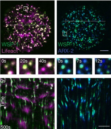
Although we have a growing understanding of various pro-proliferative and pro-quiescent molecular cues, it is unclear how NSC proliferation can be reliably and cyclically regulated throughout every day of an animal’s life and how the environment in which animals live affects NSC activation and division. What happens in the life of an animal that occurs every single day independently of anything else? The day/night cycle! We used advanced imaging techniques to record NSC division in freely behaving animals and showed that ~70% of divisions occur during the day and only ~30% at night. The “shining of light” induces NSCs activation, while the lack of light during the night switches some of the cells to the quiescent state.
Melatonin is front of mind when talking about the day/night cycle. Melatonin levels increase in our body at night as they do in nocturnal animals such as rodents. Such a big systemic change appears to regulate the delicate balance between proliferation and quiescence every day of an animal’s life, preventing excessive NSC proliferation.
However, it is undoubtably not the only an environmental change and a signaling molecule that affect the state of NSCs. Imaging of freely behaving animals will allow us to correlate changes in proliferation with animal behaviour and the surrounding environment. It can also be used to uncover other systemic factors that increase or decrease NSC proliferation and to decipher the molecular mechanisms underlying these changes.
What are the intracellular pathways in NSCs on which various extracellular signals impinge to modulate affect cell proliferation or quiescence?
How NSCs can decode and integrate pro-proliferative and pro-quiescent signals to decide whether to become active and divide or to remain quiescent? Although signals can be very different in abundance, time scale, signaling cascade, etc., would it be possible to combine all these signals into one overarching mechanism?
We used in vivo and ex vivo imaging to show that calcium dynamics serve to translate micro-environmental changes and various inputs into changes in the state of NSCs. Calcium is a versatile cellular messenger and many of the parameters of its dynamics can be influenced. We showed that quiescent and active cells display different frequencies and amplitudes of calcium events and have different intracellular loads of calcium ions. Most cells are quiescent and display a high frequency of calcium events and low intracellular calcium levels. This calcium signature is maintained by pro-quiescent signals. In response to pro-proliferative factors, the frequency of calcium events decreases and intracellular calcium levels increase, triggering NSC activation.
Can calcium alone trigger a change in the state of NSCs or is it merely reflection of the state of NSCs?
We used optogenetic tools to mimic the calcium pattern of quiescent cells in animals. Constant daylight was used to stimulate NSC activation. Interestingly, this optogenetic stimulation decreased NSC proliferation even below the baseline level. This fascinating result showed that the transition from one state to another can be manipulated and that increased proliferation can be reversed using the universal nature of calcium signals.
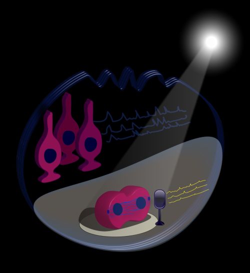
To summarize, we designed an illustration (Figure 1) featuring a stem cell on a performance stage with a light shining on it. It sings solo with low frequency calcium traces and divides. It is accompanied by a backstage chorus of quiescent cells (which are the majority of stem cells) that sing with high frequency calcium traces. “Shining a light on stem cells” is thus also a figurative expression of NSC division in live animals, where they play a role on stage and are watched by an audience of researchers.
–
Figure 1. Simplified representation of NSC states.
Gengatharan, A., Malvaut, S., Marymonchyk, A., Ghareghani, M., Snapyan, M., Fischer-Sternjak, J., Ninkovic, J., Götz, M. and Saghatelyan, A. (2021). Adult neural stem cell activation in mice is regulated by the day/night cycle and intracellular calcium dynamics. Cell 184, 709-722.e713. doi:10.1016/j.cell.2020.12.026
Daisy Vinter (PhD Student, Ashe lab, University of Manchester) ‘Dynamics of hunchback translation in real time and at single mRNA resolution in the Drosophila embryo’
Ping Wu (Associate Professor, Chuong lab, University of Southern California) ‘Cyclic growth of dermal papilla and regeneration of follicular mesenchymal components during feather cycling’
Yan Gong and Dominique Bergmann (Bergmann lab, Stanford University) ‘The Arabidopsis stomatal polarity protein BASL mediates distinct processes before and after cell division to coordinate cell size and fate asymmetries’
The webinar will be held in Remo, our browser-based conferencing platform. After the talks you’ll have the chance to meet the speakers and other participants at virtual conference tables. If you can’t make it on the day, talks will be available to watch after the event on the Node. You can also sign up to our mailing list for email alerts.
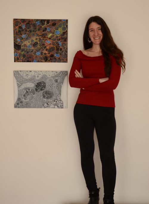
Our eleventh SciArt profile of the series features Ayelén Valko, a postdoc in Sebastian Schuck’s lab at Heidelberg University
Ayelén with two of her artworks that describe the autophagy process from different perspectives and magnifications
Where are you originally from and what do you work on now?
I am originally from Argentina, where I studied Biology at the University of Buenos Aires (UBA). During my PhD I explored the molecular mechanisms that trigger starvation-induced macro-autophagy in the fruit fly, Drosophilamelanogaster. This is a physiological process by which cells digest their own cellular material in an attempt to compensate for nutrient deprivation. Later on, I became interested in studying a different kind of autophagic degradation, called micro-ER-phagy, by which the endoplasmic reticulum (ER) is eliminated during ER stress, and for which the underlying mechanisms are not completely understood. With this aim I joined Dr Sebastian Schuck’s laboratory at Heidelberg University one year ago, as a postdoctoral fellow, where I am using budding yeast as a model organism to tackle this issue.
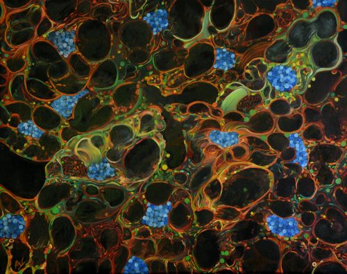
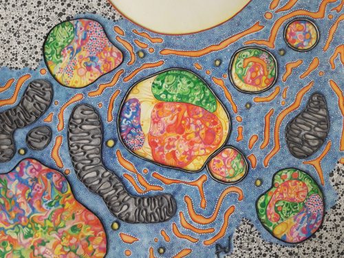
Oil on canvas depicting the autophagy process induced by starvation in fat body cells from Drosophila. The painting, made during my PhD, was inspired by fluorescence confocal microscopy images. Red and green colours are used to represent fluorescent autophagic markers. The black areas within each cell represent lipid droplets, and the nuclei appear in blue as if they were DAPI-stained. This artwork was selected as a cover image in the Autophagy Journal.
Were you always going to be a scientist?
Definitely yes! According to my family, when I was 5 or 6 years old I used to spend hours in the garden observing insects and plants, and drawing them on notebooks. At around 10, my dad gave me a small microscope, an old and very simple device that belonged to him, which I used to look at water samples and fragments of leaves. I remember my excitement seeing for the first time this amazing tiny world through the lens of this enigmatic artefact. That was when I knew that I wanted to hold to that feeling and keep exploring that microscopic universe.
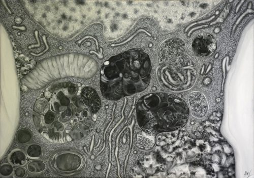
Oil on canvas that represents the autophagy process with a higher level of magnification than the previous painting. The painting was made using a grayscale of tones as if the tissue was seen through a transmission electron microscope.
And what about art – have you always enjoyed it?
For me art has always been as important as science. It was also during my childhood that I became interested in depicting human faces, as I thought that by doing so I would be able to catch the essence of each of my family members on paper. Later on, as an adult, I started to see art as a useful way of conveying my appreciation of life and nature, and share with the general audience the beauty hidden inside a cell, which my work as a scientist allows me to grab. To do so, in parallel to my scientific training, I took several art courses at the National University of Art in Argentina. After having been trained in different art techniques, I specialized in scientific and naturalistic illustration. My works have been exhibited in several galleries and museums back in Argentina, among them the Quinquela Martín Museum and the Rómulo Raggio Foundation in Buenos Aires. I have also described two of these scientific paintings in a commentary article for the Autophagy journal.
Artistic interpretation of autophagy. The vesicles represented here are inspired by autophagosomes (double line delimiting the structure) and autolysosomes (single line) of fat body cells of Drosophila. I made this painting using a mixed technique that combines watercolours, acrylics, Indian inks, and collage. Cover image for Volume 16, Issue 1 of Autophagy
What or who are your most important artistic influences?
The main inspiration for my scientific artwork comes from my own photographs of cells and tissues that I need to take for my scientific work. The complexity of these subcellular worlds and their biological processes are constantly feeding my imagination. In fact, I am motivated not only by their mystery and beauty but also by the fact that this magnificent cellular universe remains veiled to the general public. Thus, my main aim is to create attractive paintings that can appeal to the general public, as an instrument of enjoyment and popularisation of science. But, obviously, not only cells inspire me. There are a large number of artists, that belong to very different artistic traditions, whose work has affected me deeply. I feel very moved by the incredible representation of hell and heaven by Hieronymus Bosch, the monochromatic landscapes depicting alien universes by H. R. Giger, Remedios Varo’s surrealist paintings with alchemic creatures, and many other exponents of the Surrealist movement such as Salvador Dali and Joan Miró, just to name a few.
“My main aim is to create attractive paintings that can appeal to the general public, as an instrument of enjoyment and popularisation of science.”
‘Imaginary autolysosome’. The painting shows an artistic interpretation of a chimerical autolysosome, summarising different selective and non-selective autophagic processes in the same vesicle. Elements inside this vesicle could come from virophagy, reticulophagy, mitophagy, lipophagy, and general non-selective autophagy. The artwork was made employing a mixed technique that involves watercolours, Indian inks, markers, and digital integration. Cover image for Volume 16, Issue 12 of Autophagy
How do you make your art?
The challenge of making art based on scientific images lays in the need to represent the elements of study using artistic resources without completely losing the rigor of scientific observations. For me, this means that subcellular elements should be recognisable in the final artwork. However, my paintings are not in any way copies of photographs, but instead are the product of a creative process inspired by my observations and planned carefully. The first step of this process is sketching the original idea, ordering the elements of the composition harmoniously and attractively. It is only then that I decide the best technique for the work, as well as the kind of support in which I am going to make the artwork. I usually employ oil colours, acrylics, watercolours, Indian ink, collage, or a combination of them. As for the support, I usually employ canvas, a particular type of paper or cardboard.
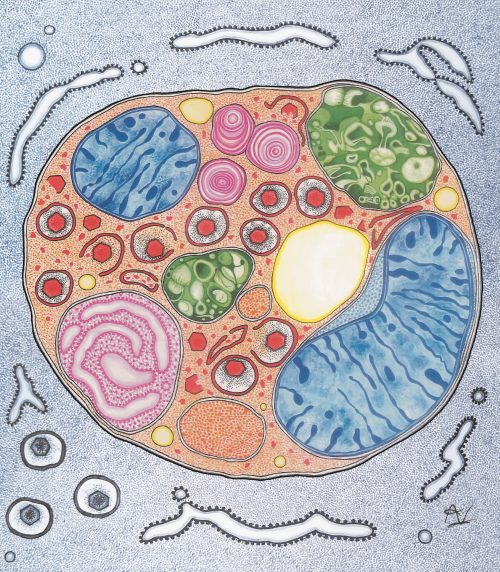
The painting illustrates the micro-ER-phagy process, which I am currently studying. The main structure represents ER whorls that are being incorporated by the vacuole. The artwork was made using a mixed technique that combines watercolours, acrylics, Indian inks, and collage
Does your art influence your science at all, or are they separate worlds?
Art can influence science for sure! I think both activities are connected in deeply synergistic ways. In general, to understand a new idea or fact, normally we have the intuitive tendency to draw it. It is often considered that our understanding of a particular subject can increase by the act of trying to put what we are learning on paper, and for this, of course, science is no exception. To represent a scientific phenomenon, either artistically or schematically, one needs to develop a high level of comprehension of the fact. I have found that by drawing outlines of the experimental designs and the expected results, I usually can visualize better the biological questions that I am trying to answer. And this practice has often led me to new ideas for future paintings. Thus, my artistic and scientific lives nurture each other, giving me a unique point of view that ended up being essential for my scientific and artistic projects.
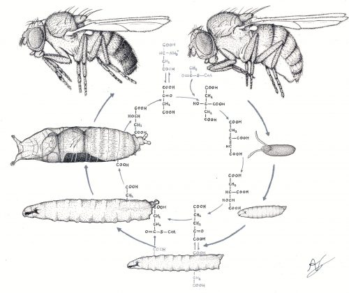
Parallelism between two different biological cycles: the Drosophila life cycle and the Krebs cycle. I made this painting with Indian inks and digital integration of individual drawings. It was used to illustrate a Doctoral Thesis
What are you thinking of working on next?
Apart from two cover images for different scientific journals on which I am currently working, and some other art projects, I am planning to study which are the elements or features that generate a stimulating and intriguing scientific artwork. My feeling is that the general audience is not as enthused by scientific art as it is by the rest of figurative art. To address this issue, I plan to systematically analyse people’s responses to a collection of slightly different cellular paintings, in order to identify which elements can maximise the identification of the viewer with the artwork. In line with this, I also want to explore interdisciplinary approaches to divulge science. I would like to work with professionals from other disciplines, such as psychologists, graphic designers, and physicians, to study different ways to convey to the general public scientific knowledge through art.
Artistic interpretation of the Drosophila lymph gland, the larval hematopoietic organ. In the background, a whole field of larvae is depicted, using a mixed technique involving watercolours, Indian inks, markers, and digital integration. A version of this painting was selected as a cover image for Volume 462, Issue 1 of Developmental Biology
We’re looking for new people to feature in this series – whatever kind of art you do, from sculpture to embroidery to music to drawing, if you want to share it with the community just email thenode@biologists.com (nominations are also welcome!)
Sophie Karolczak, Dowling Lab, Genetics and Genome Biology, Hospital for Sick Children, Canada
As a graduate student who started my program during the Covid-19 pandemic, I have never attended an in-person conference. I have heard stories of the serendipitous connections during social hours that turn into amazing future collaborations, and the opportunities to visit new places, both domestic and abroad. However, this has not been my reality in the conferences I have attended thus far. I have been sitting in my living room in Toronto, often in a different time zone than the conference, hoping that my Wifi will stay connected long enough to hear a talk!
While being in an actual conference hall with other people sounds quite appealing, I had a really enjoyable time overall attending the Developmental Disorders: From Mechanism to Treatment conference virtually. Allowing speakers to pre-record their talks made the flow really seamless, and for the most part prevented the technological hiccups that we have grown so accustomed to in this day and age. I appreciated the opportunity to chat with the speakers after each round of talks, and the Remo platform allowed for easy hopping from table to table if there were a few people I hoped to reach. During one brief session, I got to meet scientists at varying career stages, from multiple different countries, employing different model organisms, and asking vastly different scientific questions. At times I would just sit there listening to conversations taking place between experts in fields totally different than mine, and I was enjoying every minute of it!
One thing I really appreciated about this conference was the emphasis on the connection between basic science and translational research. We got to hear from researchers doing amazing work at all points along this continuum, including some who managed to follow projects from the discovery of a mutation to implementation of new treatments in the clinic. As someone who is working on the more basic characterization of a disease phenotype but hoping to see treatments head towards the clinic someday, it was fascinating to see how this process can work in real life. I also absolutely loved the patient testimonials, which help remind us that the diseases we study, while sometimes abstract in our minds, are things real people are struggling with every day. I hope to keep seeing this type of session implemented in disease-focused conferences.
Overall, this conference was a fantastic opportunity to hear about cutting-edge science in the disease modelling and developmental biology field, and I look forward to attending it again in the future!
Nicole Edwards, Postdoctoral Fellow, Cincinnati Children’s Hospital, USA
Virtual scientific conferences have made it possible for early career researchers to share our work and interact with colleagues during the COVID-19 pandemic. Having run very successful virtual seminars (namely “Development presents…”) in the past year, I looked forward to a wonderful experience at the online meeting run jointly by the journals Development and Disease Models & Mechanisms, Developmental Disorders: From Mechanism to Treatment. The meeting struck an excellent balance between invited talks, short talks, and flash talks – a series of 3-minute presentations given by participants. Flash talks were a great opportunity to learn about the breadth of science being conducted by meeting participants and gave talking points for discussion time after the talks. Presenting posters at online meetings are challenging, so 3-minute flash talks were a great alternative to get maximum engagement at this virtual meeting.
I greatly appreciated that this meeting also incorporated views and collaborations between physicians, clinical geneticists, and basic scientists working on a variety of disease models. These collaborations are where the rare disease field is headed in order to make the most impact clinically, and to help understand the underlying biology of these developmental disorders. With this, one of the most impactful session included a series of patient and family interviews, reminding us of the real implications our research on rare diseases has.
I had the opportunity to give a ten-minute short talk which I pre-recorded on Zoom, making it overall less stressful and lessened the chances of technical glitches giving a live talk. Having dedicated “Meet the Speaker” networking time afterwards was a plus, and the online platform Remo was easy to navigate and to facilitate discussion. If you couldn’t catch a particular participant you wanted to talk with, it was easy to use the chat feature in Remo to make connections.
Finally, I encourage any early-career researcher to attend a Workshop and/or Journal Meeting run by The Company of Biologists, either virtually or hopefully soon, in-person. Having been to both their in-person and online meetings, I have experienced how much The Company of Biologists strives to support early-career researchers. The Company of Biologists meetings truly are unparalleled opportunities to be involved with the future of your scientific field.
 (2 votes)
 (2 votes)


