The programme offers generous four-year DPhil studentships which cover full fees, pay a tax-free, enhanced stipend of ~£17,609 pa, and provide £5,300 pa for research and travel costs.
Student on this programme start research on their main projects immediately and so they have a full four years to work on their research project. If their work is delayed by the pandemic students will be given a fully funded extension.
Individuals of all nationalities are welcome to apply.
Applications for entry in October 2022 must be submitted before 12 noon, 3rd December 2021.
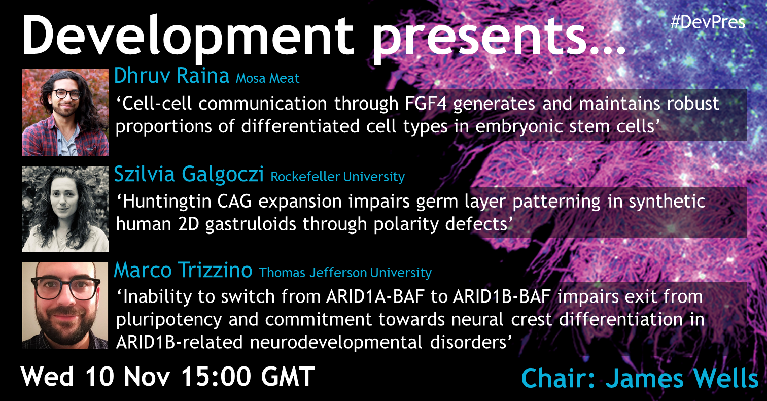
Dhruv Raina (Senior Scientist, Mosa Meat, previously a PhD Student in Christian Schröter‘s lab at the Max Planck Institute for Molecular Physiology) ‘Cell-cell communication through FGF4 generates and maintains robust proportions of differentiated cell types in embryonic stem cells’
Szilvia Galgoczi (Research Specialist/Visiting PhD student in Ali Brivanlou‘s lab at Rockefeller University) ‘Huntingtin CAG expansion impairs germ layer patterning in synthetic human 2D gastruloids through polarity defects’
Marco Trizzino (Assistant Professor at Thomas Jefferson University) ‘Inability to switch from ARID1A-BAF to ARID1B-BAF impairs exit from pluripotency and commitment towards neural crest differentiation in ARID1B-related neurodevelopmental disorders’
The webinar will be held in Remo, our browser-based conferencing platform. After the talks you’ll have the chance to meet the speakers and other participants at virtual conference tables. If you can’t make it on the day, talks will be available to watch after the event on the Node. You can also sign up to our mailing list for email alerts.
The current NIH director, Francis Collins, is retiring at end of the year. He urged that a woman should succeed him, but what would your list of eligibility requirement be? Is a medical degree an essential? The ability to address the inbuilt inequity in science?
Articles in Science and Nature address these and many other issues with the appointment.
You are not alone. Even Nobel laureates fail to get all grants funded
I received another disappointing un-fundable score for my @NIH grant today. I am privileged, and I realize no-one wants to hear me complain. Just sharing to make the point that everyone experiences this kind of feedback, and that it never stops from stinging! 1/3
American Association for Antomy scholar program and the story behind it.
I am so proud of the American Association for Anatomy for supporting this program. This is a great example of "putting your money where your mouth is". https://t.co/h7tFlmdPPG
So, I am breaking tradition and I'll tell you a little bit about how the idea for the ASP program came to be. I hope it will help explain why I think it will be a game changer.
Thanks to the #DevBio community for sharing their thoughts, especially on twitter. If you have some news that you think we should share on our blog, please get in touch at thenode@biologists.com. If you are interested in getting involved with writing preLights you can find out more here.
Register yourinterest by October 31st 2021 for the Edinburgh Gallus Genome and Embryonic Development (EGGED) Workshop in 2022. EGGED 2022 will run on the 12-15th of July 2022 at The Roslin Institute and R(D)SVS, Easter Bush Campus, The University of Edinburgh. Full registration will open in due course and spaces will be limited due to the nature of the workshop.
The EGGED 2022 Workshop will provide hands-on training for developmental biologists that use or would like to use the chicken embryo in their research. Instruction will include fundamental techniques, ex ovo culture, and imaging to advances in transgenics, gene editing, and genomics and using the chicken embryo to teach developmental biology. This practical workshop is open to researchers with a range of experience; from students and early career researchers to group leaders and principal investigators. The workshop will also provide an opportunity for scientists to share, learn and develop embryological techniques that use chicken embryos.
The UKRI-BBSRC funded Roslin Institute and the National Avian Research Facility (NARF) have developed globally unique chicken resources, including a range of transgenic fluorescent reporter chicken lines. EGGED will bring together the world’s embryology experts to share their skills and showcase these exceptional resources. To date, speakers for EGGED 2022 include; Prof Marian Ros, Prof Claudio Stern, Prof Tatjana Sauka-Spengler, Prof Neil Vargesson, Dr Raman Das, Dr Hervé Acloque, Dr Ben Steventon, Dr Jérôme Gros, Dr Mike McGrew, Dr Jacqueline Smith, Dr Joe Rainger, Dr Adam Balic, and Dr Denis Headon.
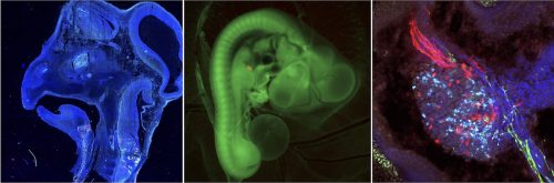
From left to right; 1) Whole head cross-section (x10) of a Chameleon transgenic chicken embryo, with cells either labelled in blue, red, green or cyan. 2) GFP chicken embryo with a ‘red’ graft placed into the limb bud with micro-surgery. The graft is about 50-100uM. 3) Dorsal Root Ganglion of the nervous system of a Chameleon transgenic chicken embryo. Nerves going into the dorsal root ganglion are red, and nerves coming out are green.
A Royal Society of Edinburgh Saltire Facilitation Network Award has been awarded to The Roslin Institute, R(D)SVS and the NARF, both based at The University of Edinburgh Easter Bush Campus, to hold these practical workshops in both 2022 and 2023. EGGED is also supported by The Company of Biologists, including support towards making the meeting sustainable.
We also aim to document the specialist skills demonstrated at the EGGED workshops and make them available online as an important developmental biology community resource.
EGGED is held in memory of Dr Donald Ede, a graduate of the University of Edinburgh, a talented chicken embryologist and member of the RSE, who passed away in 2018.
Event organisers are Dr Megan G Davey (The Roslin Institute, R(D)SVS) and Dr Lindsay Henderson (The Roslin Institute, NARF). Read more on the EGGED website and register your interest here.
In the latest episode of Genetics Unzipped, presenter Kat Arney is squelching through the Californian mud, swimming with platypuses, bearing witness to daylight robbery and even finding time to catch an episode of Star Trek as she looks back on some of the most mind-blowing stories from the world of genetics in 2021.
We meet the Borgs – huge genetic elements in archaea that can assimilate genes from their neighbours – and discover how whitefly pulled off a genetic theft that enabled them to become one of the world’s most destructive agricultural pests.
We hear how researchers are developing mirror-image DNA polymerases that can make mirror-image DNA – perfect for long-term, stable data storage. Then there’s the strange discovery that hundreds of viruses use a DNA base called 2-aminoadenine, known as Z, instead of the usual adenine (A), with big implications for our understanding of the genetic code as we know it.
And finally, we take a dive into the duck-billed platypus genome, to discover what these mysterious monotremes can teach us about mammalian evolution.
If you enjoy the show, please do rate and review on Apple podcasts and help to spread the word on social media. And you can always send feedback and suggestions for future episodes and guests to podcast@geneticsunzipped.com Follow us on Twitter – @geneticsunzip
The 8th Edition of the Annual Portuguese Drosophila Meeting (#DrosTuga2021), aims at bringing together national and international members of the Portuguese Drosophila community. Along with them, Portuguese Drosophila scientists abroad and any participant from other country interested in Drosophila and developmental research are invited. https://igc.idloom.events/drostuga2021
–
The purpose of this Drosophilameeting is to promote open sharing of data and ideas, as well as to provide arich forum for discussion of new research findings and conceptual breakthroughs in an informal environment.
–
Due to the ongoing uncertainty caused by the COVID-19 pandemic, this edition will be held entirely ONLINEon the afternoons of the 29th and 30th of November 2021.
The event will include selected short and long talks presented at the Plenary Sessions via Zoom. Thanks to our sponsors, the best talks and posters will receive a prize. In addition to the presentations, there will be time for discussion and mixing between researchers at all career stages during the two interactive Poster Sessions (held in the Hopin platform). You can take a look at the programme for more details.
In this DrosTuga 2021 edition, we will have the pleasure of listening to two great Keynote Speakers. Their exciting work spans a broad range of topics of interest to our community: Isabel Palacios and Nicolas Gompel. Besides our speakers research, we will have the opportunity of talking about outreach and the importance of Drosophila studies in science.
Attendance is FREE, but registration is compulsory.
NEW Abstract submission deadline for posters only: 7th November 2021 (23:59h GMT+1). Click here to submit your abstract. The deadline for short and long talks is now closed (24th October 2021).
Registration deadline: 24th November 2021 (23:59h GMT+1). Click here for registration.
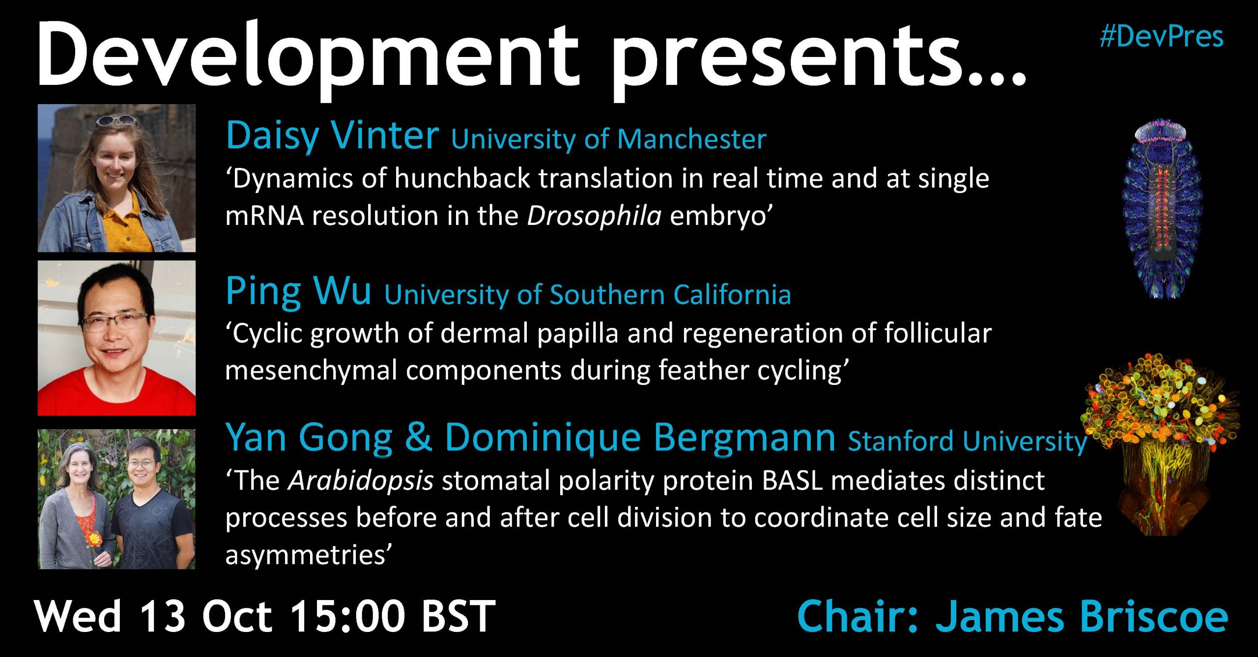
Below you’ll find each of the talks, plus a Q&A chaired by Development Editor-in-Chief James Briscoe. The next #DevPres webinar will be held on 10 November 2021, and chaired by James Wells – subscribe to our mailing list for updates.
Daisy Vinter (University of Manchester) – Dynamics of hunchback translation in real time and at single mRNA resolution in the Drosophila embryo
Ping Wu (University of Southern California) – Cyclic growth of dermal papilla and regeneration of follicular mesenchymal components during feather cycling
Yan Gong & Dominique Bergmann (Stanford University) – The Arabidopsis stomatal polarity protein BASL mediates distinct processes before and after cell division to coordinate cell size and fate asymmetries
In April 2020, I should have attended The Company of Biologists Workshop “The Cytoskeletal Road to Neuronal Function”. If only there was not the beginning of the SARS-CoV-2 pandemic. The Workshop was ultimately canceled, however the organizers suggested to us (the enthusiastic and disappointed participants) to initiate a Webinar as a virtual platform for the neuronal cytoskeleton research community. I and four other participants (Satish Bodakuntla, Meng-meng Fu, Oliver Glomb and Lisa Landskron) volunteered as organizers, and this is how “The Cytoskeleton of Neurons and Glia” Webinar was established in April 2021. Since then, we hosted more than twenty diverse and inspiring speakers. On October 7th our efforts reached a milestone – the twelfth Webinar marked six months of its existence!
Actin monomers as cellular cobblestones
I wrote this Node inspired by one of our speakers – Dr. Eric Vitriol. He presented a work from his Lab focusing on how actin dynamics are affected by actin monomers in neuronal cells. But first, let us take one step back and borrow the cytoskeletal road metaphor from The Company of Biologists workshop. Imagine a trail made of cobblestones, assembling underneath your feet and in front of you while you are walking. This trail will fall apart at the back once it is no longer needed. Next, imagine the cobblestones are self-aware of their numbers, and they will never start making the trail unless they “know” there is a sufficient number of them to start the construction (“critical concentration”). Additional factors like a solid ground to build on, stabilizing mortar, continuous supply of high quality cobblestones, all facilitate the trail assembly. While a self-aware and self-building trail is still far from our reality, practically all cells in our bodies have plenty of “cobblestones” called actin monomers. The monomers are present way above the critical concentration and can assemble into trails and many different cellular structures. As a matter of fact, with such a good supply of actin monomers why not building all the time?!? Well, cells have figured a way to protect themselves from an energy-costly unproductive actin assembly. This is achieved by sequestering actin monomers via another molecule called Profilin (PFN), that will release the “cobblestones” once there is a “construction permit”.
Profilin, actin monomers and neurons
In cells with neuronal origins, the team of Dr. Vitriol studied how different actin structures assemble from a common monomer pool and how Profilin 1 (PFN1) is influencing this process. They varied the concentration of PFN1 and induced cellular shortage, normal levels, or excess of PFN1. In those three conditions, they analyzed two established actin assemblies (branched Arp2/3-mediated and linear Mena/VASP based) at a highly dynamic part of the cell called leading edge. The cells responded with downsizing actin assembly throughout the cell when there is a shortage of PFN1. In addition, the lack of PFN1 repositioned the Arp2/3 nucleator complex towards the center of the circular cells, and reduced Mena/VASP function, ultimately disrupting the architecture of the leading edge. At low concentration of PFN1 cells seem to employ a mechanism to be resourceful and favor linear networks constructions. Abundance of PFN1 signals that both linear and dendritic actin networks can be reestablish. You can find more details in the paper published in 2020 in Current Biology, with Dr. Kirsten Skruber as a first author (1). This study provides us with a glimpse on how mammalian cells reshape actin assemblies when they face challenging situations when the concentrations of a major “guardian” and nucleators of the actin building blocks are changed. These disturbances must come at high costs for cell fitness, especially in long-lived, specialized cells like neurons. And while short term each cell has certain capacity to cope with different intra- and extracellular challenges, long-term exposure to the same “stretch” will eventually lead to neuronal dysfunction.
Mutations in PFN1 are direct cause of a late onset, incurable neurological disorder called amyotrophic lateral sclerosis (very recent review from another speaker in the Webinar series Dr. Kai Murk (2)). In addition, decreased levels of PFN2 were detected in cells from patients with Charcot Marie Tooth disease – genetically heterogeneous disorder affecting the peripheral nerves, as found by the team led by one of my PhD supervisors – Dr. Vincent Timmerman (3). Thus, the building blocks of the actin cytoskeleton are getting the closer attention they deserve, as there are plenty missing pieces in the puzzle that costs humans their health.
Skruber et al., 2020, Current Biology 30, 2651-2664;
Murk et al., 2021, Front. Cell Dev. Biol. 9, 681122;
Juneja et al., 2018, J Neurol Neurosurg Psychiatry 89, 870-878.
Our twelfth SciArt profile of the series features Giacomo Moggioli, a PhD student at Queen Mary University of London studying genomics of deep sea worms
Where are you originally from and what do you work on now?
I am from Milan, Italy. I did both my bachelor’s and master’s degrees at University of Milan-Bicocca. During my years as undergraduate student I started to feel fascinated by deep sea environments, so I decided to apply for the Erasmus project and spend one year working on my master’s thesis at Heinrich-Heine University in Düsseldorf. My thesis was focused on the role of iron-sulphur clusters in the origin of life, which, accordingly to the most robust hypothesis, happened in hydrothermal vents environments. My curiosity for this kind of deep-sea environments motivated me to apply for a PhD at Queen Mary University of London. Now, as a PhD student, I work on a fascinating clade of Annelid worms, Siboglinidae, which thrive in hydrothermal vents. I love to be able to learn more and more about these creatures. They possess such unique features such as the lack of a digestive system in favour of a symbiotic lifestyle that allows them to harness the energy contained in sulphur compounds in order to keep their metabolism going.
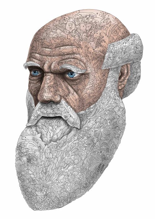
Darwin of Life I made this for an Art and Science contest organized by the university of Milan-Bicocca and it has been exhibited at the Natural History Museum of Milan. I have tried to shape the tree of life as the portrait of the famous scientist. The origin of life is at the tip of the beard and from there you can follow the evolution organism by organism upward. Plants, animals, fungi, invertebrates, I have simply tried to put as many different organisms as possible. Can you spot the Koala?
Were you always going to be a scientist?
Not at all. I believe that what happens in our lives keep on shaping our desires, aspirations and interests, so I don’t feel I ever had a clear path ahead of me. Nevertheless, when I was a little kid I was fascinated about being a marine biologist, and here I am today! Over the years I had many different part-time jobs, for example I was working in a flower shop for a couple of years during University, I worked at a stand during design week promoting some very nice lamps and I have also worked in a motorbike customisation shop, where I was mainly painting on helmets and bikes. So I definitely had the opportunity to try different paths, but overall the part time job I loved the most during my time in Milan was being a freelance illustrator. As an illustrator I was taking part in art fairs, art markets and exhibitions all over Italy showing my art and selling my prints. I am happy where I am now, working with deep sea animals in London, but I definitely considered making a living out of my illustrations.
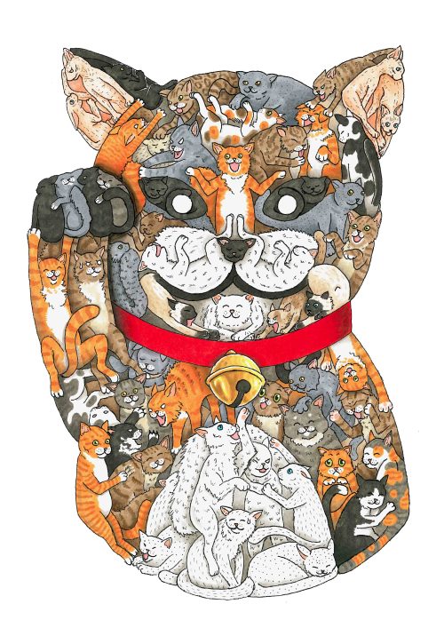
Neko-no Kami, the spirit of cats. This is part of a project on Japanese spirits, my idea is to make a sort of an encyclopedia about the many different “Kami” or spirits that are coming from the Shintoism. This ancient animistic religion believes every natural and even artificial entity has its own soul, its own spirit. I have expanded this concept and started to imagine how these spirits might look.
And what about art – have you always enjoyed it?
Yes, as far as I can remember. When I was a kid my mother was buying me awesome illustrated books about sea animals. I was spending hours looking at those beautiful drawings of all the amazing life forms we can find in our oceans! I always tell my friends that those books made me who I am today, having marine biology and illustration as the main passions in my life. I am very grateful to have had the opportunity to study art history at high school. In those classes I learnt a lot about artists and understand how they made their art and what they wanted to say with what they were doing. I really feel there is a world behind every single art piece we see; a world made by the experiences of the artists and their opinions, ideas and way of looking at the world. This feeling really enhanced my curiosity about Art. Now I really enjoy going to art museums and exhibitions and when I am walking around the city, I always love to search for street art, murals and graffiti, trying to imagine the world that might be behind them.
“I really feel there is a world behind every single art piece we see; a world made by the experiences of the artists and their opinions, ideas and way of looking at the world.”
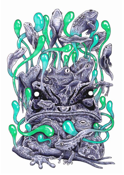
Henshin-no Kami, the spirit of metamorphosis Another artwork from my Kami series.
What or who are your most important artistic influences?
There were many amazing artists that impacted me and there will always be new ones as well. To name just a few of them: Salvador Dalí, who showed me that the only limit in art is our imagination, an Italian street artist named “Blu”, his rich and meaningful works taught me to always keep an eye open for hidden beauty that always surround us, Masashi Kishimoto, the author of the manga “Naruto” from which I understood the artistic potentiality of a black outline, and finally another Italian street artist “Hitnes”, from which I have learnt the artistic potentiality of not using a black outline.
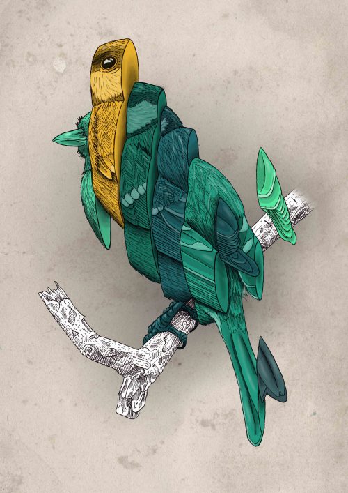
Disassembled tit bird. I am really fascinated by birds and I love to play with their shapes and their colours and see the final result!
How do you make your art?
When I was living in Milan I had a studio and this allowed me to try many different techniques. After these experiments, I managed to find a technique which I felt comfortable with. I was first drawing with pencil on paper, then inking the lines with a black marker, erase the pencil away and finally color the drawing with alcohol-based markers which allowed a very good control of the shades. Then I move to London, and I couldn’t carry my studio with me. Therefore, I decided to switch to digital techniques and now I am mainly working using my tablet and a drawing app. After a steep learning curve, I now feel comfortable drawing with my tablet and I love to be able to draw from my sofa instead of sitting on a desk fully covered in ink and all sort of different drawing tools.
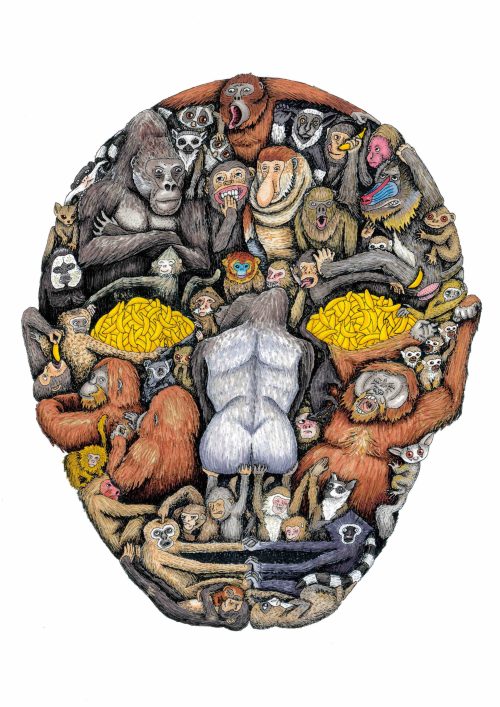
Primate’s face Primates all together are forming a human face. When making this composition I always like to depict different interactions between the characters. Can you spot the monkey trying to steal the baboon’s banana?
Does your art influence your science at all, or are they separate worlds?
I would say that science is influencing my art more than the other way around. I love to make science-inspired drawing, but I have never really done art-inspired science before. Nonetheless, I think that art may indirectly influence my science. While doing my art I have learnt how to transform big, complicated and very detailed subjects into some way simpler yet still very descriptive drawings. I find this “simplification process” I use in my art to be very useful in science as well. As a scientist a big part of my work is handling huge amounts of genomic data and identity the key features in order to be able to simplify the information and make a good description of the organism I am studying.
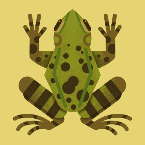
Common frog This is part of my icon-like animal series, heavily inspired by the British illustrator Owen Davey. I have started to make these icons and share them with my colleagues so that they could include them in their slides and hopefully have a nice simple representation of the organisms there are studying.
What are you thinking of working on next?
At the moment, my priority is completing my PhD and publish my first paper as first author on deep sea worm genomics. After that I would like to keep on working with marine organism genomics in a Postdoc. As a side artistic project, I have started to work on a card game during the long lockdown evenings and I would like to finish the last details and try to release it to the public. Little spoiler: there will not be deep sea creatures but there will be some dinosaurs in my game 😊.
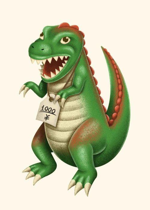
T-rex toy This vintage-like dinosaur toy will be part of my upcoming card game. Stay tuned!
If you want to have a look at my other works and be updated on my new projects you can follow me on Instagram: https://www.instagram.com/kelp_art/
We’re looking for new people to feature in this series – whatever kind of art you do, from sculpture to embroidery to music to drawing, if you want to share it with the community just email thenode@biologists.com (nominations are also welcome!)
We needed an afternoon snack in the Morris Lab today, so I conducted a blind taste test. With a sample size of 7 (everyone loitering around lab this afternoon), I investigated whether people preferred @AldiUSA energy bars or Clif bars. Here are my results: pic.twitter.com/jZkYXJBSaJ
Instead of using the word "interestingly" every five sentences, what if we start incorporating exclamation points in the scientific literature? Why is this taboo?
Our Production Editors at Development have responded that they are happy to accept exclamation marks in the text, but only if they follow the instructions below: Authors must indicate their level of excitement/surprise using a scheme similar to the use of asterisks for significance level: !, interesting finding; !!, surprised by this result; !!!, gobsmacked/couldn’t believe it; !!!!, that can’t be right, we should repeat that experiment
Thanks to the #DevBio community for sharing their thoughts, especially on twitter. If you have some news that you think we should share on our blog, please get in touch at thenode@biologists.com. If you are interested in getting involved with writing preLights you can find out more here.
 (No Ratings Yet)