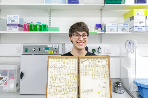
In this SciArt profile, we meet Petra Korlević, a scientist at the Wellcome Sanger Institute, interested in retrieving DNA from historic mosquito collections.
Can you tell us about your background and what you work on now?
I work on human malaria transmitting mosquitoes; population genetics, insecticide resistance, and currently trying to marry all that up with landscape genomics. My background is on getting DNA from difficult samples, for my PhD I worked on method development for ancient DNA extraction from bones and teeth, for my postdoc I developed a method for minimally morphologically destructive DNA extraction from museum pinned insects.
I was going to be way too many things, a veterinarian, a cartoon artist, an astronomer, a physicist… and also a nature researcher, so being a biologist fits at least that box. Though thinking of plans “for retirement” having a rescue for all the abandoned cats and dogs back home in Croatia wouldn’t be bad either.
And what about art – have you always enjoyed it?
My first ever recorded and preserved doodle was crayons on my dad’s arm at 11 months old, so I had a pretty quick start. Next was the living room wall. Then paper. I was lucky I grew up in an environment where art was encouraged together with, not against, a scientific pursuit. Then it got a bit tricky in school because I would still doodle things in my notebooks which not all teachers appreciated. I had a pretty harsh art-block during my undergrads and masters, but in the end art persevered!
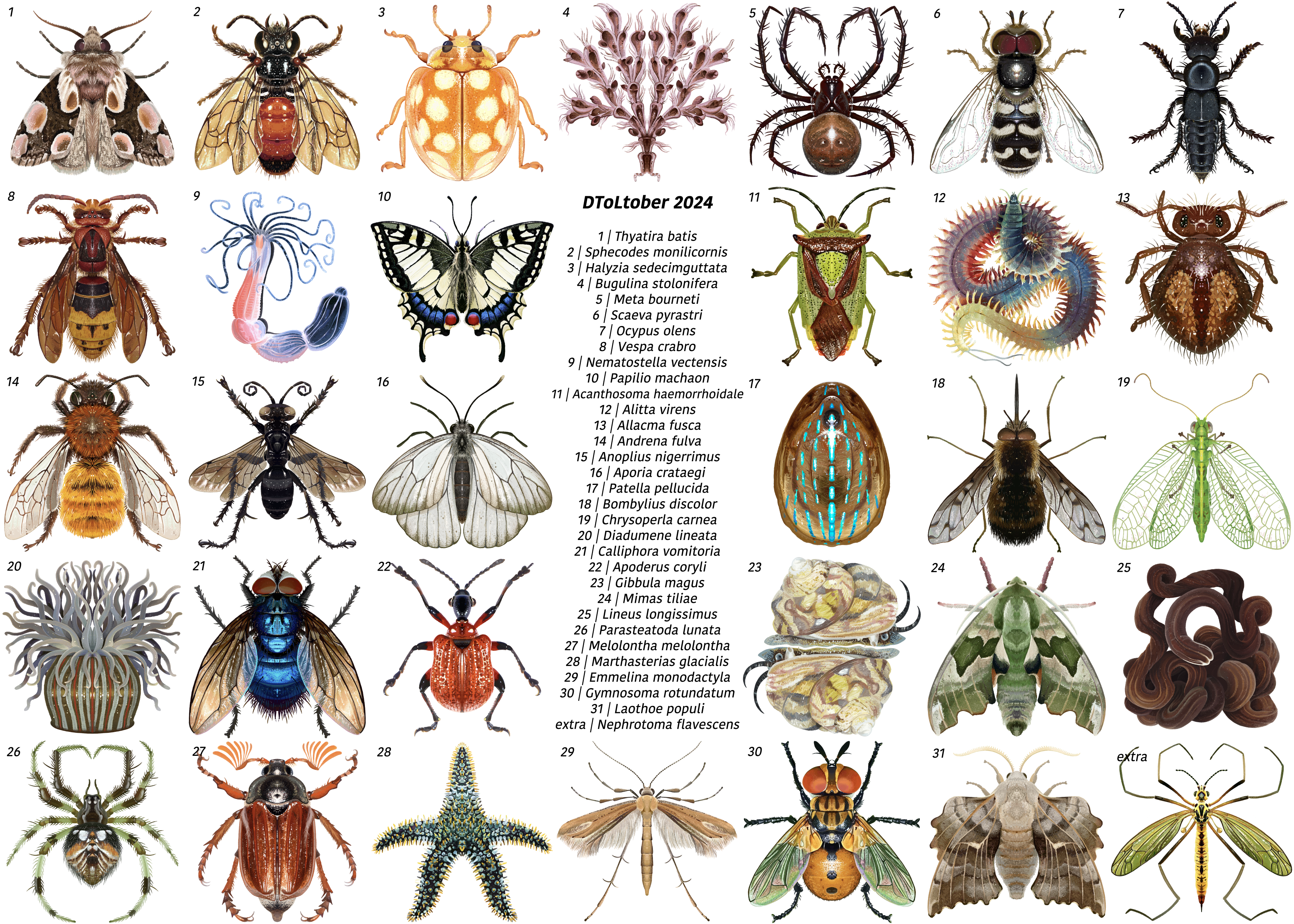
Huevember (drawing of one Croatian animal per day/hue)
What or who are your most important artistic influences?
I grew up on Franco-Belgian and Italian Topolino comics. Back then my main influence was my dad since he, my sister and I would sit down and paint/draw together. There are just too many artists whose work I admire to list here, but a few: Vincent van Gogh (grew up with the starry night on the wall), Michael Whelan (his sci fi and fantasy realism is breathtaking, we used to sit down and make stories from his paintings without knowing the books attached to them), Joshua from False Knees (the ways he keeps animal drawings mostly realistic but with such beautifully woven personalities in nothing but simple pencil strokes is such an inspiration), Chen Zha (chentomology) and Nicole (fossilforager) (the two of them have some of the cutest invertebrate art out there, and invertebrates always need more love) and so so many more I am so sorry I can’t put you all in here!
While I do still enjoy the occasional traditional media project (and do embroidery as a hobby), I primarily use Procreate on my iPad, and Inkscape on my laptop. One thing I wish is to have more time to actually learn new things, I would love to learn animation and other new software.
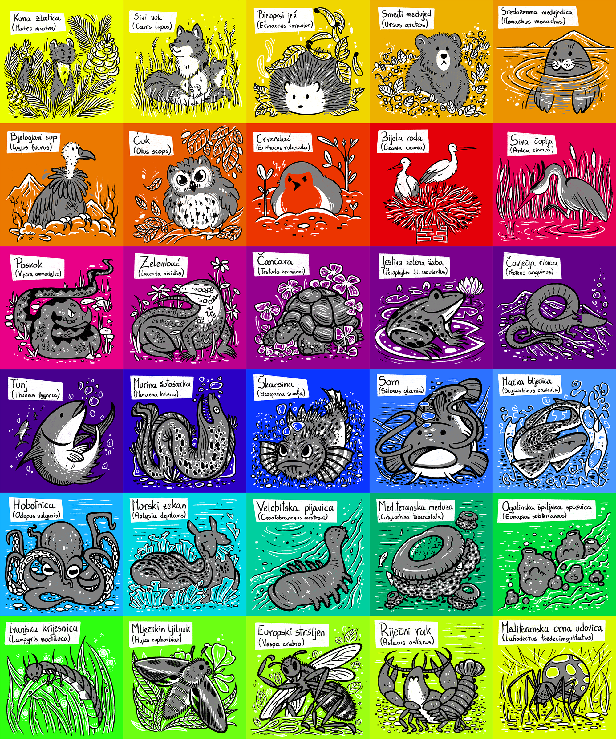
Fig2-v1 (schematic from the paper in the above Trends in Genetics issue, “Future of DNA-based insect monitoring”; really I just love the little snail getting pampered by vacuuming and bathing)
Does your science influence your art at all, or vice versa, or are they separate worlds?
It is a big symbiotic organism, both are amazing outlets for my creative process. And it definitely helps with creating engaging talks (for scientists or the general public) and having pretty paper figures.
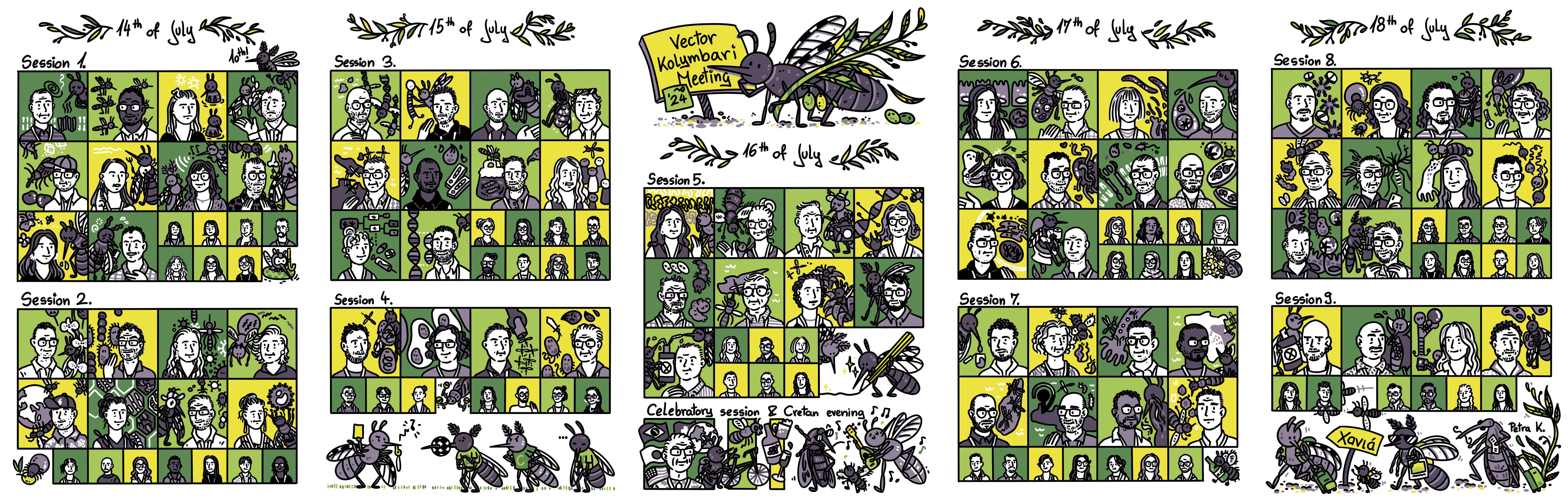
Vector Kolymbari Meeting 2024 (live sketching of the event, with some little embellishments like a tick eating an olive and the EURO 2024 final recreated with mosquitoes)
What are you thinking of working on next?
Science-wise I need to get a wishlist of new Anopheles reference genome and get to mapping a bunch of understudied malaria vectors, then putting them into their habitat/climate context.
This year, we proudly celebrate the 30th anniversary of the Spanish Society for Developmental Biology (SEBD)—a milestone that marks three decades of passion, dedication, and collaboration among Spanish scientists worldwide, committed to uncovering the mechanisms that dictate the development of living organisms. Since its founding the 5th of May of 1994 by the visionary Antonio García-Bellido, SEBD has evolved into a vibrant and inclusive community, uniting established researchers with a growing number of emerging scientists eager to contribute to this dynamic and ever-evolving field. The integrative nature of developmental biology remains essential for understanding processes ranging from regeneration to evolution, and the influx of young talent continues to enrich our Society with fresh ideas and perspectives, ensuring that Spanish science remains at the forefront of innovation.
To commemorate this special occasion, we have gathered interviews with SEBD members who, over the years, have significantly contributed to the Society’s growth and success. These conversations reflect on personal journeys, celebrate key scientific advancements, and emphasize what makes SEBD unique: a combination of scientific excellence, creativity, and a collaborative spirit. They also underscore the Society’s role as a supportive environment where established scientists and early-career researchers work together to expand the boundaries of our understanding.
Through these interviews, we honour not only our rich history but also the exciting future that lies ahead. As we celebrate this milestone, we continue to foster connections, inspire creativity, and build upon the legacy of this extraordinary community. We hope you enjoy these reflections, which celebrate the individuals and the collective achievements that have shaped SEBD over the past 30 years.
We are confident that these qualities, which have made SEBD so special, will continue to drive its success in the future.
Eloisa Herrera is the current SEBD president. Eloisa’s research focuses on axon guidance and the formation of the nervous system. She investigates the mechanisms by which neurons establish precise connections, shedding light on the processes that shape brain wiring and their implications for developmental disorders. By combining molecular, genetic, and imaging techniques, Eloisa’s work aims to unravel the complexity of neuronal connectivity, providing critical insights into how proper brain architecture is established and maintained during development.
Sofia J. Araújo is the current SEBD secretary. Her research focuses on the genetic and cellular mechanisms that control branching morphogenesis and cell migration. Her work uses Drosophila as a model to investigate the processes involved in the development of organs like the trachea and nervous system, particularly how single-cell branching mechanisms contribute to organ formation. In addition to her research, Sofia is a keen science communicator and SciArt enthusiast. She is committed to exploring creative ways to make science more accessible, helping to bring complex biological concepts to a wider audience in an engaging and understandable manner.
Cristina Pujades specializes in vertebrate neural development, using zebrafish as a model organism to investigate the genetic and cellular processes that govern brain and spinal cord patterning. Her research centers on key processes such as cell migration, differentiation, and the establishment of neural circuits, offering fundamental insights into nervous system development and its evolutionary adaptations. Through her work, she contributes to a deeper understanding of how the nervous system forms and functions across species.
Former SEBD president Fernando Casares is a leading researcher in developmental genetics, with a particular focus on eye development and evolution. His work explores the genetic networks that regulate eye size and shape, primarily using Drosophila as a model organism. Through his research, Fernando has deepened our understanding of how alterations in developmental pathways contribute to morphological diversity, providing valuable insights into the fundamental processes of organogenesis.
Former SEBD president Miguel Torres’s research centers on the mechanisms underlying embryonic morphogenesis and tissue regeneration. He has made significant contributions to our understanding of cardiac development, employing cutting-edge tools to construct a dynamic 3D atlas of heart formation. This work has offered valuable insights into the origins of congenital heart defects and holds potential implications for regenerative medicine. Additionally, he investigates endogenous regeneration pathways in the heart, drawing inspiration from highly regenerative species such as amphibians and zebrafish.
Former SEBD treasurer Paola Bovolenta focuses on key signaling pathways, including Wnt and Shh, that regulate brain development. Her research investigates the genetic and cellular mechanisms involved in brain patterning, with a particular emphasis on congenital diseases and brain malformations. Paola’s work has been instrumental in advancing our understanding of the molecular basis of developmental disorders, providing crucial insights into how disruptions in these pathways contribute to neurological conditions.
Former SEBD secretary Miguel Manzanares investigates transcriptional regulation during early embryogenesis, focusing on how molecular mechanisms drive lineage specification and organogenesis. His research aims to uncover the regulatory networks that govern the differentiation of cells and the formation of organs, providing valuable insights into fundamental processes of early development. Additionally, his work has important implications for regenerative medicine, as understanding these processes may contribute to strategies for tissue regeneration and repair.
Former SEBD treasurer Pilar Cubas is an expert in plant developmental biology, with a focus on the regulation of axillary bud dormancy and shoot branching. Her research delves into the genetic and molecular networks, including the role of TCP genes and strigolactone signaling, that control these processes. By employing model systems such as Arabidopsis thaliana, tomato, and potato, her team investigates how these regulatory mechanisms influence plant adaptation and productivity, providing insights that could enhance crop yield and agricultural sustainability.
Former SEBD president Ángela Nieto is widely recognized for her pioneering work on epithelial-to-mesenchymal transitions (EMT), processes that are crucial in development, fibrosis, and cancer metastasis. Her research has significantly advanced our understanding of how these cellular transitions regulate embryonic development and the pathological consequences when they are dysregulated. Ángela has received numerous prestigious national and international awards in recognition of her transformative contributions to the field of developmental biology.
This year’s SEBD Science Communication prize winner Luisma Escudero is a researcher best known for his discovery of the scutoid, a geometric shape important for understanding tissue formation and organ development. His work explores how geometry influences cellular packing and tissue organization, contributing to advancements in developmental biology and tissue engineering. Alongside his research, Luisma is an enthusiastic science communicator, and has recently published a children’s book “Papá, ¿cómo se enroscan las caracolas?”, which explains complex scientific concepts like scutoids in an accessible way. Through his research and outreach efforts, he aims to make science more accessible and engaging for the public.
Luisma Escudero, winner of the 2024 SEBD Science Communication Award
Former SEBD board member Isabel Fariñas is a leading expert in neural stem cell biology, investigating their role in brain development and adult neurogenesis. Her research aims to understand how neural stem cells maintain their potential for regeneration and differentiation throughout life. Isabel’s work has implications for understanding brain plasticity, repair mechanisms, and neurodegenerative diseases. Isabel has received this year’s Ramon y Cajal National Science Award from the Spanish Science Ministry.
Is there such thing as a standard career path for a scientist? Some people find fulfilment in pursuing a PhD, postdoc, then starting their own research group; others may have second thoughts or may be drawn by other interests and aspirations.
In this new series, we chatted to several developmental biologists who have had vastly different career trajectories. This interview series was inspired by this 2013 post from Kara Cerveny, who went back into academia as an assistant professor after a stint as a scientific editor.
Below is a sneak peak of the career stories.
One common thread throughout all the interviews is that there is no right or wrong when choosing a career path. Every decision is profoundly personal.
We hope that whichever career stage you are at, and whatever career journey you are embarking on, these stories can help demonstrate that many valid career paths exist. It is okay to take breaks, make U-turns and go on tangents – you might end up doing something you’ve never dreamed of!
I’d like to thank everyone featured in this series so far, who’ve kindly taken time out of their busy schedules to chat to me. I appreciate everyone’s honesty and willingness to share their personal stories.
I hope you enjoy reading these interviews.
Joyce Community Manager, the Node
Do you have an unconventional career path and want to share your story? Or do you know someone who does? Get in touch at thenode@biologists.com
Sneak peak of the career stories
Eve Seuntjens is currently the Principal Investigator of the Developmental Neurobiology Lab at KU Leuven, Belgium. Originally trained as a pharmacist, Eve decided to embark on an academic career but spent almost 16 years as a postdoc before landing an independent position. What is her advice to people currently in the endless postdoc period? Find out more from our interview with Eve.
Originally trained as an architect, Karen Liu made an active decision in her late 20s to pursue a scientific career, starting from washing the dishes as a lab technician. How was Karen’s journey towards becoming a professor in developmental biology? Read the interview to find out.
“I don’t think I would’ve been better off being a biologist to begin with”
Bill Hinchen has always wanted to go down the academia route, but an unfortunate series of events led him to pivot his career to science writing. When he was given a second chance to do a PhD, what made him decide to turn down the offer? How is he finding life as a freelancer? Read our conversation with Bill.
“I never wanted to leave academia, but now I love being a freelance science writer”
Before becoming the Reviews Editor of Journal of Cell Science, Sara Morais da Silva has dipped her toes in many different careers – from teaching, to pharma, and even a brief stint in the police! What has she learned along the way? Check out the interview with Sara.
“Choose what fits with your personality”
To Maria Rostovskaya, science and dance have always been equally important in her life. Between her undergraduate and PhD, Maria was a dancer and taught dancing. What made her switch careers and follow an academic path? How did her experience in dancing shape her subsequent career? Read the interview.
“I made a conscious choice to switch from dancing to research, and I don’t regret it at all“
Instead of doing a postdoc right after his PhD, Christos Kyprianou joined The Company of Biologists as FocalPlane’s Community Manager. After setting up the community site for microscopy, what made Christos decide to return to academia as a postdoc? We interviewed Christos to find out more.
“There’s no one particular path of doing science – explore different options”
No such thing as a standard career path – an interview with Karen Liu
Karen Liu is Professor of Genetics and Development at King’s College London, UK. She did English and architecture as an undergrad and worked in an architectural firm for a couple of years before deciding to pursue a scientific career. We had a chat with Karen to find out more about her switch from architecture to science, and how her own experience has influenced how she guides early career scientists through their career journeys today.
Can you briefly tell us about your career path so far?
I’m currently Professor of Genetics and Development at King’s College London, and I’ve been here my entire independent career. I started here as a lecturer in 2007 and before that, I was at Stanford for three years in the labs of Michael Longaker and Gerald Crabtree as a joint postdoc. Prior to that, I did a PhD at UC Berkeley with Richard Harland. Before my PhD, I was a technician at Columbia for two years, in a mouse lab with Argiris Efstratiadis and that was where I started in science. I was the only tech in the lab, so my duties included washing the dishes, lab management and a lot of mouse work. I barely knew what DNA was! I’m still astounded that I now have this job as a biology professor, because as an undergraduate, I went to Columbia University and studied architecture and English.
Karen talks about making an active decision to switch from architecture to biology.
Let’s wind back the clock: you started out training as an architect. How was that experience?
The architecture degree I did was very theoretical. I knew during the degree that I didn’t want to be an architect. Like science, architecture is a creative field but it’s very much about the funding pressures. To be a famous architect, you need a lot of resources backing you up, and the field comes and goes with the vagaries of the economic situation. I was at university in the early 90s, when there was a major economic crash. When I got out of university, I managed to get a job at I. M. Pei. This was a very exciting period in architecture – they had just done the Louvre Pyramid, the Rock and Roll Hall of Fame and other amazing buildings. But I didn’t work as an architect. I had an English degree as well, so I worked in the office where we put out press releases and wrote grant applications. I did a lot of architectural writing and putting together proposals.
What influenced your decision to pursue a scientific career?
Because of the economic situation, it was not a stable job. I didn’t want to stay in architecture, but I had to think hard about what to do next. I had a fair amount of science background in high school. When I was working at I.M. Pei, I was ill for a while. I thought I needed to do something useful for humans, but I also want to do something fun. With biology, there’s an infinite number of questions out there. I thought if I could become a biologist, there will always be something I could be excited about. I didn’t know anyone who was a research scientist; it just seemed like a fun thing to go to grad school for.
Your entry into the research world was a lab technician job. How did that come about?
I didn’t take any biology in college, so initially I was looking around for night school. Then I found out that at Columbia, where I’d done my undergrad, offered employees two courses a term for free. So, I decided to find a job at Columbia. I was talking to the HR person at Pei, and she mentioned she had a friend who ran the sequencing facility at Columbia. After talking to them, they in turn mentioned that the lab of Argiris Efstratiadis was looking for a technician. I went to talk to Argiris, and amazingly, he gave me a job based on nothing. I think he liked interesting people who had done different things and who genuinely wanted to be in biology.
I started out washing pipettes and bottles and making media in the lab. In the meantime, I took some undergraduate classes, like genetics, developmental biology, and biochemistry. Biochemistry included organic chemistry, which totally kicked my ass! By the second year I worked there, they said I could take the first year PhD seminar course in the genetics department. It was during this course that I read about all these famous and wonderful genetics and development papers. The tutors included Gary Struhl and Iva Greenwald, and Ginny Papaioannou taught us mouse genetics. They made a developmental biologist out of me!
You’re currently a Professor of Genetics and Development at the Centre for Craniofacial and Regenerative Biology, King’s College London. What is your research about?
My lab works on the neural crest. It’s a vertebrate-specific population that arises in the embryo. The neural crest is multipotent in the embryo. The neural crest cells are born at the neural crest border, and then migrates. We are interested in the migratory properties of these cells and how they interact with their surrounding environment as the embryo develops. Because the embryo is changing dramatically during those stages, the neural crest cells need to crawl through tight spaces and navigate complicated environments. We’re interested in the molecules and the proteins that are important for human development and the phosphorylation events that control the interactions between cells. That’s the fundamental basis of the lab. On the other end, we’re identifying new gene variants from human patients who have congenital neural crest anomalies. Our focus is on head structures because the crest forms most of the skeleton in the head and some cardiac tissues. We collaborate with people who work on other neural crest derivatives, like the enteric nervous system. From a migration perspective, we’re also interested in a neural crest cancer called neuroblastoma, which arises from the sympathoadrenal system. We try to keep our work rooted within human anomalies, and that’s an active decision that I made when I started my lab. I wanted to make sure the things I do are relevant to the human condition.
Are you involved in other research initiatives?
I lead the Congenital Anomalies Cluster of the MRC National Mouse Genetics Network. Our overarching goal is to improve the way we model novel gene variants identified from patients with congenital anomalies. There is an infinite number of gene variants, but if you can’t attribute functional changes to them, then it’s meaningless data. The initiative is aimed at modelling these variants in mice. We are loosely linked with the Rare Diseases UK network, because a lot of the neural crest anomalies are rare diseases. We’ve just finished a Horizon 2020 training network called NEUcrest, which is a clinical to basic science PhD training programme spanning 15 students in 10 countries across Europe. That was exciting and rewarding to have a network of people focused on modelling neural crest anomalies. For me, at this stage of my career, I enjoy enabling these kinds of scientific interactions to have a broader impact. I’m also the academic lead for the Crick. I have oversight over all the Crick PhD students who are registered at King’s. I’m stepping down now after seven years. I run a PhD programme at King’s called Multiscale Models for Life, which is a new multidisciplinary programme where the two supervisors are from different disciplines. I also have a role at Kings as a postgraduate coordinator. In general, I think a lot about the different aspects of being a PhD student.
How did your previous roles help you get to where you are now? Do you see any similarities in architecture and science?
I think everyone winds up being the person they are like based on their route. From the architectural perspective, a lot of the themes still go through the research that we’re interested in now – how things are built, how they work together, how they function and what happens when things go wrong. I think the aesthetics of architecture really made me intolerant of bad figures or posters. I like schematics to be clean and figures to be aligned. I’ve not done any bench science myself in a long time, but I still love sketching out ideas and making schematics of our hypotheses. We’re also interested in 3D modelling now, like AlphaFold modelling of variants. Computational modelling in biology has matured over the years. A lot of that arose from architectural drawing and 3D renderings. I appreciate architecture from a distance much greater now than from within.
There’s also a similarity in terms of the collaborative nature of the work. The architectural field is very much about group work. People have their own expertise, but they have to bring all that together to make a building. The same is true in biology. I have people in my lab who love biochemistry and will only run western blots, and people who specialise in dissecting embryos. We need people to come together to build a magnificent story. I like seeing how things fit together and I like helping my people see that.
Karen compares architecture and biology.
Anything you’ve learned from switching career paths? Looking back, would you have done anything differently?
It’s a big question! I don’t think I would’ve been better off being a biologist to begin with. I don’t regret the long way to where I am now. For me, being a biologist is an active decision I made when I was in my 20s. I’m really fortunate that I was able to choose that and then move into it and wind up being a professor now. I was pretty terrified when I went into the PhD – I was 28 years old, and I knew zero biology going in. I think I did not take enough advantage of my undergraduate education. I was a really bad student, but one thing I would say to students now is, even if you’re a really bad student, you can come out of that and figure out what you really like, and then slowly work your way there. I don’t know if I could switch careers like I did now. I probably won’t be brave enough. It’s also helpful if you have a safety net. I think a lot of people couldn’t do that. Back then, perhaps I was a bit naïve. I figured if I could pay my rent, I’d be okay. If grad school didn’t work out, I could always get another job.
Do you have any advice to someone thinking of switching career paths? Has your own experience influenced how you give career advice?
I think people who haven’t switched careers can’t appreciate how difficult it is. Some people think that because they’ve just finished a PhD, switching feels like they’re failing. It’s not. You just did this PhD where you became the world’s expert on your topic! I think the hardest part is to get over that feeling that it’s a failure. Then you need to do a bit of self-reflection. What are you good at? Some people think, I’m not good at anything other than running Western blots. That’s so not true. You just wrote a 200-page thesis. You’ve presented your data again and again, coordinated with many people, and put together an entire project with many moving parts. You can ask yourself, am I good at the people part? Or the writing part? Would I prefer to be by myself all day, or would I prefer to spend a lot of time with others? Those simple questions are a really good start.
Then you can talk to all kinds of people and find people who are supportive. This can be difficult, but people are more supportive than one thinks. Looking back, I found that people really made an effort to point out particular opportunities and help me move to the next phase. Like that woman from the architectural firm, she didn’t have to tell me about her friend at Columbia’s sequencing facility, but she was helpful and was excited about my career prospects (I should really write to her and thank her!). So now in my own lab, I always try to point out opportunities to students.
I did work hard to get to where I am now, and I think people should own that. Of course you have to work hard, but it’s easy to work hard if you’re in a nice environment. But certain work environments are not so great, and if you could tell it isn’t the right place for you, then it’s time to take a step back and try to figure out how to get yourself out of that situation. We read these articles about changing careers and how you have to be brave etc. I think people in more senior positions need to appreciate that being able to change is a privilege, and I feel strongly about this. You cannot change if you don’t have a safety net, so we need to be providing that safety net, within the university and within the PhD programme.
Karen stresses the importance of providing career support for PhD students.
Finally, what do you enjoy doing outside of research?
I like good food and I like traveling. I like to do long-distance walking. My husband, who is also a scientist, and I have been trying to do things that are a little bit outside of our comfort zone. We took a pottery class recently. I’m terrible at it, but it’s fun!
No such thing as a standard career path – an interview with Bill Hinchen
Bill Hinchen is currently a freelance science writer, but he never wanted to leave academia in the first place. Following the devastating loss of his PhD data while thesis writing, he ended up falling into the world of science writing. A few years ago, he was offered a second chance to do a PhD. What made him decide to turn away from his dream of an academic career? What advice does he have for early career researchers? We chatted to Bill to find out more.
Let’s start from the beginning. Have you always been interested in science?
I watched lots of David Attenborough and wildlife programmes growing up. I love the natural world and I wanted to be a vet. At secondary school, I chose biology, chemistry and art as my A levels subjects. Then I started to learn about research and decided I would do either toxicology, marine biology or biochemistry at university. At the last possible minute, I decided I wanted to do fine art, but my art teacher told me I should do science, because there’s no money in art. That swayed me, so I chose to do marine biology in Plymouth. (I want to go back and tell my art teacher that there’s no money in science either…)
How did you end up doing a PhD at the University of Cambridge?
After my undergrad, I did a Master of Research in marine biology at Plymouth, focussing on ecotoxicology – specifically I looked at the neurotoxic effects of dietary copper on catfish. On the side, I was also reading a lot about ecophysiology work, looking at how environmental conditions affect early embryogenesis. And in no time at all I just fell in love with research. As I was wrapping up my Master’s, I applied for PhDs at several places. An opportunity at Cambridge popped up. The project was on the early development of the marine crustacean Parhyale hawaiensis. That sounded interesting, but I didn’t know how I could end up getting a PhD in Cambridge. I grew up in a rough part of East London and went to secondary school and college in Tower Hamlets. I applied anyway, and I got the interview. At the time, I had a clean-shaven head, so I tried to grow my hair as fast as I could and took my piercings out. Superficial, I know, but the thought of Cambridge made me panic! At the panel interview, they fired all kinds of questions at me. My molecular biology knowledge was very lacking, but my biochemistry knowledge was good. One of the questions was “What’s your favourite experiment?” I immediately went back to my time in Plymouth at the Marine Biological Association, where Hodgkin and Huxley conducted the experiments on ion channels in giant squid axons. You can still see the giant squid axons that they used to throw up to the ceiling after an experiment. I told the interviewers that was my favourite experiment and one of them said, “That’s my favourite too!” To this day, I still think that’s the only reason I got the PhD. Then, Cassandra Extavour, my supervisor, told me she’s got an associate professorship at Harvard and asked if I’d like to go there for a year. I was a bit shell shocked – I just landed a PhD at Cambridge and Harvard! It was great, but hard. It was a big transition, going from Plymouth, feeling like a big fish in a small pond, to Cambridge, where I felt like a very small fish in a very big, scary pond.
What did you work on during your PhD?
I was using 4D microscopy to figure out cell migrations patterns during the very first few cleavage stages before gastrulation. Once we’d modelled how the cells migrate, we did single cell ablation of individual blastomeres to see if their neighbours carried on going where they need to go, or whether cells needed each other to figure out where they were in the embryo. Interestingly, it turns out that there’s plasticity and compensation at the early stages of development, where you can just do the horrible things the embryo, and it just carries on developing.
What happened towards the end of your PhD as you were thesis writing?
My PhD was based on 4D microscopy, where I did multiple z-plane time-lapse recordings. I accumulated about 300GB of data. This is a long time ago, now. The computer was rubbish, and there was no cloud storage. I went to Harvard for a year and came back to the UK to write my thesis. Things were going really well. I was writing at home one day, and I went out to do a few errands. When I came back to my house, the window was ajar. I looked in and it was just ransacked – the TV, DVD player, my laptop, external hard drives and USB sticks – literally everything on my desk had been swiped off. So that was all my PhD data gone. The only bits of my PhD left were the few drafts I’d emailed to my supervisor and advisor. I needed at least six months to re-do all my key experiments, but there was no funding available. I was already working part time to cover rent. I rewrote the thesis but there was no data to support anything I wrote. Cambridge told me they simply couldn’t examine me on data I didn’t have. I was told there wasn’t enough to submit as a PhD, so I had to submit it as an MPhil. I breezed through the viva examination, but I didn’t bother going to the graduation at the end of it all. I didn’t want anything to do with it.
Following the devastating loss of your PhD data, how did you end up getting a job in medical writing?
At that time, I never wanted to leave academia. I loved being at the bench. After the MPhil, we had to leave Cambridge because it was too expensive to live there. My wife got a job in Peterborough, and so we moved there. Work was hard to find because I was constantly told I was over-qualified. So, I worked as a cleaner in a dental surgery and as an Apple reseller at a local shop. Whatever it took. Finally, I was hired as a Research Associate in a biophysics lab at King College London. They were developing a technique to do controlled light exposure microscopy in C. elegans embryos. They were all physicists and needed someone who knew how to work with embryos. I was there for four months until they ran out of funding. It was possibly the most fun four months I’ve ever had. After that, someone mentioned medical writing to me. I ended up getting a job in an agency as a medical writer, where I produced publications and marketing materials for pharma companies. I didn’t really like it. It was very much about whatever the client says, you do. After working at a few agencies as a medical writer, I moved to Abcam as a science writer, and it was a complete change of pace. I was writing marketing materials, blogs, and guides on how to run IHC or ELISA. I eventually became the senior writer. I moved away from writing lengthy technical stuff, to doing more creative writing, like a lovely full-page advert in Nature about ELISA kits. But I decided I wanted to do something more wholesome, so I joined the charity Kidney Research UK as their Research Communications Manager. My job was to take the research they fund and turn it into something the public can understand. That was good fun but hectic.
You were offered another chance to do a PhD in 2020. How did that come about, and how did you make the difficult decision of turning down the offer?
I was getting tired of writing for people and I longed to be back at the bench doing experiments. A friend of mine mentioned that her old boss, Grant Wheeler at the University of East Anglia, was looking for a PhD student in ecotoxicology. I thought this could my opportunity to get back into academia. At the interview, they were lovely. They offered me the PhD and it became very real. I spoke to a lot of people in academia, and they all mentioned the lack of funding, lack of job security, and the huge competition – stuff I knew deep down, but needed to hear. It would’ve meant moving my wife and son to Norwich, taking a massive pay cut, and working all the hours required for a PhD. After a lot of consideration, I decided to turn down the offer. I felt bad for messing Grant around, but I think it was the right thing to do. Part of me will always long for the lab, but I think at that point, having had the chance to go back, I could close that chapter, and move on.
Bill recounts when he was given a second chance to do a PhD.
You’re currently a freelance science writer, specialising in copywriting. What does your job entail?
Copywriting used to mean writing short copy, but it’s since come to mean anyone who writes anything salesy. I call myself a science copywriter, as I focus on short ads, but I also still write long copy like blogs, web pages and white papers. I do creative directing and consulting as well. I made a copywriting 101 video a while ago. That was for my sister initially, who runs her own business, but I thought I should make it available online for anyone who doesn’t know where to start. I’ve also run some training workshops. And I use LinkedIn a lot as a platform to not only attract clients but put the word out there that science writing doesn’t have to and shouldn’t be boring and dry – it can be interesting and fun!
How are you finding freelancing compared to working for an organisation?
I went freelance nearly five years ago. Going freelance has been the best thing ever and the scariest thing all at once. On one hand you have complete freedom. I take off whatever days I like. I set my own breaks. And I’m lucky enough to be established now and so I can choose my clients. Freelancing works out very well for me, because I get to push back a lot more on my clients. I attract a certain type of clients who don’t just want a word monkey. Quite often they want me to show them how to do things differently, so sometimes they become more like collaborators. But the other hand, I have absolutely zero security. If I take a day off, I earn no money. If I get sick, I earn no money. If my clients decide I’m off, I’m unemployed. But the freedom and the control of being freelance is something I don’t think I could give up.
How did your experience in all the different organisations you’ve worked in help you as a freelancer?
My previous experiences have been instrumental. There was no way I could have hit the ground running quite as fast as I did, without all that experience. I’m now acutely aware of what companies need from a content marketing perspective having worked with a variety of clients. People tend to want the similar sort of things, even if they’re from different industries. Because I’ve seen such a broad spectrum of content types and strategies, I can easily come up with ways to make something stand out.
I really thought I’d like the public sector, but it wasn’t that different from working in a big private company, except the budgets are smaller and people’s roles are less defined. In the public sector, we’re communicating this wonderful work and funding researchers who’re doing brilliant science. But the reality is, most of this research will never be heard about. It’s more interesting to us (with a science background) than it will be to most people, but the goal is to get more donations for the charity. At the end of the day, whether it’s a company or a charity, there is always the drive to make more money.
Do you have any advice for people interested in getting into science writing?
The first thing is to think about the types of writing you like. Do you like writing short copy, or long, detailed pieces? There are so many styles of writing. Don’t feel like you need to pick one branch. Be open to trying different things, and then you’ll figure out which types of writing you like. I never expected I’d end up writing short copy.
Back to my point about private versus public sector: if you’re considering a job in science writing, I would think less about the overall cause of the organisation and think more about whether the science you’re writing about is interesting, because that’s what will help you write engaging pieces. I would also tell people to steer clear of recruiting agencies, if possible. Some of them are fine, but most of the ones I’ve encountered are trying to get you into almost any position. I think you’ll be more rewarded by finding people who write the things you’re interested in, and then either look for jobs with them or just message them. People are way more receptive to random questions than you think, especially on LinkedIn.
One last point I want to make is that your ambition doesn’t always need to mean becoming senior writer, creative director, or head of copy. Ambition doesn’t have to mean moving up the corporate ladder. Your ambition can be to become really good at writing this one thing. You don’t always need to be climbing some corporate ladder.
Bill’s advice to people thinking of going into science writing.
Do you think you’ll stay in science writing?
I’ve been doing science copywriting for 13 years. I don’t know if anyone would pay me to do anything else. I would like to move away from writing words for people and move more towards the consulting side, like helping people figure out the tone of voice, coming up with a marketing or communications plan, and helping people get better at writing.
Apart from science writing, you said you almost did fine art at university. Did you ever consider any other careers?
All the time! I did some steel engineering work at building sites. I thought maybe I could do an apprenticeship. I also thought about doing a graphic design course. I love drawing. My wife and I started a small T-Shirt company called Old and Board, because we like to skate and surf and we’re in our 40s now. If I could somehow turn that into a job, that would be lovely.
What do you like to do in your spare time?
I surf a lot. We moved from the middle of England down to Cornwall, so we can live by the sea and surf on demand. If I’m not swimming, surfing, or writing, I’m probably in the gym.
Thank you for sharing your story! Do you have one final piece of career advice for the Node readers?
Do not ever think you need to have everything figured out. No one has it figured out. Everyone is winging it. If you feel like you don’t know what you’re doing, or you’ve got imposter syndrome, that’s totally normal. Everyone feels like that. Don’t worry about it. Just keep doing what you’re doing!
The 20 November 2024 Development presents… webinar was chaired by BSDB Chair Marysia Placzek (University of Sheffield) and featured four talks from the prize winners of the 2024 BSDB Spring meeting. Catch up on the talks below.
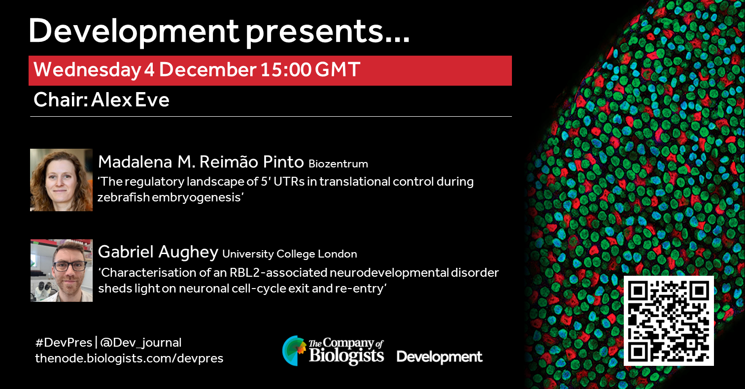
The final webinar of 2024 features two early-career researchers working on gene regulation and will be chaired by Development’s Senior Editor, Alex Eve.
Wednesday 4 December – 15:00 GMT
Madalena M. Reimão Pinto (Biozentrum) ‘The regulatory landscape of 5′ UTRs in translational control during zebrafish embryogenesis’
Gabriel Aughey (University College London) ‘Characterisation of an RBL2-associated neurodevelopmental disorder sheds light on neuronal cell-cycle exit and re-entry’
At the speakers’ discretion, the webinar will be recorded for viewing on demand. To see the other webinars scheduled in our series, and to catch up on previous talks, please visit: thenode.biologists.com/devpres
Queer people, trans individuals in particular, remain significantly underrepresented in STEM and academia. In August 2023, Nick wrote an Honest Conversations post on the Node entitled ‘Coming Out of my Cage and I’ve Been Doing Really, Really Good’, discussing his experience of being a transgender scientist, the importance of the support he received from his lab, and the freedom he found in living authentically.
In this Voices piece, we hear from two PhD students who identify as trans. They discuss their experiences navigating academia, issues prevalent in academia that trans people still face, and the support systems they have found that have empowered them on their journeys.
Of being a trans immigrant – an uphill journey through science
by Aflah Hanafiah
As an 8th year PhD student, my journey to climb this career ladder consisted of numerous hurdles, setbacks, and emotional and mental rollercoasters. I was lucky enough to navigate myself out of one of the most queerphobic countries in the world. The UCLA Williams Institute ranks Malaysia 115th of 175 countries based on social acceptance. The Human Rights Watch (HRW) reported on certain Malaysian laws criminalizing same-sex sexual acts and transgender people’s gender expression and sentencing those who found guilty with jail time, fines, and whipping. These laws and the generally conservative culture that is hegemonic in the country easily subject queer people to discriminations and violence, especially towards trans femme people. I grew up learning very early on that expressing myself outside of the heteronormative traits would make my life difficult, so I learned to conform the best I could.
I spent my high school years throwing myself into schoolwork and getting my grades up so I could qualify for a government’s scholarship that would later take me to the USA where I eventually completed my bachelor’s degree in molecular biology. This opportunity allowed me to not only pursue my career goals, but it had also provided me with the space to start addressing my queer identity that I had long suppressed. During my time at Rochester Institute of Technology (RIT), I met many people, student body and faculty included, who were accepting and affirming of queer identities in general. They essentially created a safe space for people like me to feel comfortable in my own skin. However, there was still lingering stigma behind being openly queer and pursuing post-graduate education. I was reminded of this as I applied to several graduate programs. During my interviews, I pivoted strongly to presenting as the gender that appeared on my legal documents so I could avoid the possible awkwardness and questions that could arise from my queer identity.
As I went through the process and started my PhD, I felt like I was given another opportunity to further explore my queer identity now that I was in a totally new environment. This was where I started to feel comfortable to use my preferred pronouns, dressed the way that affirmed my gender identity, and explored gender affirming care. By this time, I was already 27 years old and well past my junior PhD years. My decision to undergo hormone replacement therapy (HRT) that depends on my school’s health insurance while at the same time working on my PhD was a difficult one to make. The PhD already came with its own sets of challenges, and I was unsure if I should add to this chaos by physiologically jumbling my hormonal level. However, the persistent gender dysphoria that is always occupying my mind would later drive me to embark on this next phase of my transition. I am fortunate because my student’s health insurance covers gender-affirming care, so I knew that this was an opportunity that I could not pass on.
Though I have not faced much explicit transphobia in my graduate school, which I largely attribute to me being passable enough, my other trans friends on the other hand, have a much different experience. Despite universities improving the workplace environment for queer people and other minorities; prejudice, micro- and macro-aggressions are still prevalent. I have no doubt that if I did not pass as what society deems as a woman, I would have had a very different experience going through life and specifically, working in academia. I am not “out” per se, in graduate school because it never became a question. I do make it a point that I am transgender around other queer colleagues and in queer spaces because I think that it’s important to let people know that they are not alone and that they are safe with me. As I am at the tail end of my PhD, I can’t help but be worried for my future. A slew of anti-trans laws is being passed in almost every state in the country and more companies are reportedly moving away from investing in LGBTQ+ oriented diversity, equity, and inclusion (DEI) efforts. Coupled with my F1 visa status, the career choice that is viable to me are ever narrowing. However, it is times like these that I rely on my chosen family, loved ones, and community in navigating these challenges. I believe that with a supporting community behind you, you can overcome any obstacles.
Searching for others like me: navigating academia as a trans person
By James Lythall
As a queer and trans person in STEM, I’m acutely aware of how few LGBTQIA+ people there are in STEM. There are plenty of heterosexual and/ or cisgender people who I enjoyed working and socializing with, the vast majority of whom have been very supportive. At the same time, I can count the number of queer academics I know in the field of life sciences without running out of fingers, and the number of trans academics in any scientific field on one hand. There are probably many more out there, but the number of visible, openly trans and/or queer academics is vanishingly small. Of course, representation will not solve all the problems facing queer and trans researchers, but it can help us fight the feelings of isolation that are often common amongst under-represented groups. To know that someone like you has managed to succeed, despite the odds, can both be comforting and motivating. On a more practical level, it also means there are people you can ask for advice who are familiar with the problems you may be facing and may have already found solutions to them.
I can’t help but feel that I am having to forge my own path all the time, and whilst that is sometimes exciting, it is often exhausting. There are many people who I feel I can ask for scientific guidance, but almost none who I feel comfortable asking for support on issues I face from being trans. This is not because I believe those around me to be trans- or homophobic, but simply because they are often unaware of specific problems that trans and queer researchers face.
As a researcher, I am acutely aware that my personal experience is not necessarily representative of others- an n of 1 is not much! Frustratingly, there is precious little data available on the experiences of the queer – and particularly trans – people in academia. Much of the current data focuses on the experiences of undergraduate students, often in the US, and often not stratified beyond science and humanities. Both this data and surveys conducted by scientific organizations rarely collect data on the trans status of participants, and group everyone who doesn’t identify as a woman or a man as ‘other’. Nonetheless, the little data that there is suggests that queer and trans people are often underrepresented in STEM compared to in the general population, with one US survey finding LGBTQIA+ people were ~20% less prevalent in STEM fields compared to the general population (1). The same study also reported 70% of academics felt uncomfortable being out at work. Focusing on undergraduates, another study found that LGBTQIA+ undergraduates were 9.4% less likely to remain in a STEM major (2), with this rising to 10% for trans undergraduates (3). Another study reported 45.67% of natural sciences students compared to 14.96% of social sciences students reported being misgendered (4). This is particularly alarming in the light of a new study that has found an association of higher levels of microaggressions (including misgendering) and worse mental health outcomes in trans adults in the UK (5).
Despite these gloomy statistics, I am optimistic. I do believe science is slowly becoming a more welcoming place for queer and trans people. I have had some very positive experiences and moments of connection as a trans person in STEM, and in academia. I have had lecturers who have gone above and beyond to create a welcoming teaching and learning environment for trans and queer students, as well as offering considerable personal support to me and other queer students. I have also been encouraged by the acceptance and support for trans and queer people that other students have offered, such as using and advocating for more inclusive language to challenging the heteronormative and cisnormative narratives that often pervade medical sciences. More recently when I was applying for PhDs, one potential supervisor went out of their way to ensure that only my preferred name would be used throughout the application process, and another offered to correct a colleague when they noticed they had got my pronouns wrong.
I would like to finish by suggesting a few things that I think allies can do to help improve LGBTQIA+ experiences in the academic world. Firstly, if you have teaching responsibilities alongside your research, include queer, trans and intersex people whenever possible. On a more individual level, ask and listen to what your LGBTQIA+ students and colleagues need and try to avoid making assumptions. Another important thing you can do is look into how your institution collects data on students and staff and whether this includes appropriate gender and sexuality choices, including options to not disclose. This makes institutional data much more helpful for researchers trying to understand the experiences of queer and trans people in STEM.
These are just a few suggestions, but there are numerous excellent articles out there on how to support queer and trans students and colleagues that I would encourage you to read. Equally, I would also encourage you to think about what you can do outside of the academic sphere to support LGBTQIA+ people. Visible and meaningful support for LGBTQIA+ people has never been more important, particularly in the UK where hate crime rates continue to rise and transphobic rhetoric has become increasingly commonplace in media and politics. Building a better academia also means building a better society in large.
References:
1: Freeman, J. B. (2020). Measuring and Resolving LGBTQ Disparities in STEM. Policy Insights from the Behavioral and Brain Sciences, 7(2), 141-148. doi:10.1177/2372732220943232
2: Hughes, B. E. (2018). Coming out in STEM: Factors affecting retention of sexual minority STEM students. Science Advances, 4(3), eaao6373. doi:doi:10.1126/sciadv.aao6373
3: Maloy, J., Kwapisz, M. B., & Hughes, B. E. (2022). Factors Influencing Retention of Transgender and Gender Nonconforming Students in Undergraduate STEM Majors. CBE—Life Sciences Education, 21(1), ar13. doi:10.1187/cbe.21-05-0136
4: Whitley, C.T., Nordmarken, S., Kolysh, S. and Goldstein-Kral, J. (2022), I’ve Been Misgendered So Many Times: Comparing the Experiences of Chronic Misgendering among Transgender Graduate Students in the Social and Natural Sciences. Sociol Inq, 92: 1001-1028. https://doi.org/10.1111/soin.12482
5: Wright, T., Lewis, G., Greene, T. et al. The association between microaggressions and mental health among UK trans people: a cross-sectional study. Soc Psychiatry Psychiatr Epidemiol (2024). https://doi.org/10.1007/s00127-024-02775-2
from Irene Karapidaki, Béryl Laplace-Builhé and Michalis Averof
What is this?
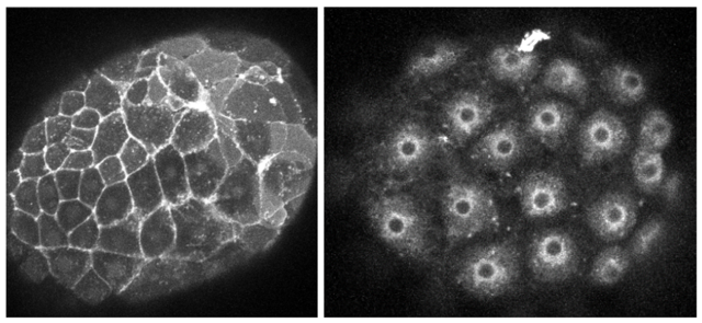
These are crustacean embryos injected with mRNA encoding a red fluorescent protein bound to membranes. On the left, the fluorescent protein is localised on the plasma membrane, on the right it is trapped in the endoplasmic reticulum and/or Golgi.
How was this image made?
These images were made while testing different membrane localisation tags in the marine crustacean Parhyale hawaiensis. One-cell stage embryos were injected with mRNAs encoding mScarlet3 fused to a Lyn tag, directing protein myristoylation and palmitoylation (on the left), or a signal peptide and the CD8 transmembrane domain (on the right). The embryos were allowed to grow for a day and then imaged live on a confocal microscope.
Why should people care about this?
Targeting fluorescent proteins to cell membranes allows us to visualise the shape and behaviours of cells in living embryos, as they build the body. See for example this amazing movie, showing the choreography of cells as they build sensory organs in the fish embryo: https://thenode.biologists.com/ready-steady-cooooooonga/research/
Can I do this in my favourite research organism?
The problem is that existing membrane-localising tags do not work equally well in all species. The SP-CD8 tag for example (on the right) gives good plasma membrane localisation in Drosophila, but gets stuck in the secretory pathway in our crustacean (Parhyale) embryos. In non-conventional model organisms, one needs to test several tags to find a good one.
To facilitate this process we generated a toolkit of 11 membrane-localising tags, which can be screened rapidly by microinjecting mRNA in your species of interest. Comparing results obtained in different species will help to identify tags that work well in a wide range of eukaryotes. If you are interested in trying the toolkit and joining our comparative screen, please get in touch.
My name is Katie Pickup and I wanted to introduce myself as I’m a new Reviews Editor at The Company of Biologists. I am going to be working with Development and the Node, as well as with three of the Company’s other journals (Journal of Cell Science, Disease Models & Mechanisms and Journal of Experimental Biology). I’m really looking forward to being exposed to a broad range of bioscience topics in this role.
My research background is mainly in stem cell biology. I did my PhD at the MRC Human Genetics Unit at the University of Edinburgh investigating the role of DNA methylation in pluripotency and differentiation of mouse embryonic stem cells in Richard Meehan’s lab. I used 3D models of differentiation including gastruloids and embryoid bodies to understand the impact of DNA hypomethylation on cell lineage trajectories and exit from pluripotency. I’m particularly interested to see where stem cell-based embryo model research goes over the next few years, both scientifically and from the regulatory side. I’m also excited for the opportunity to branch out in my new role as an editor and learn more about other areas of developmental biology across the spectrum of model organisms.
I’m really looking forward to getting to know the wider community better through working with authors on Review articles and other front-section journal content like interviews and poster articles. I also can’t wait to travel to lots of different conferences and workshops across the world, so hopefully I’ll get to meet some of you there! In the meantime, feel free to get in touch via email, LinkedIn or X.

 (1 votes)
 (No Ratings Yet)