X-rays and frog embryos: new features of gastrulation revealed
Posted by Gary McDowell, on 19 August 2013
For decades, the development of the early embryo and patterning of tissues has been studied with the help of a workhorse of developmental biology, the frog embryo. Xenopus embryos are large and undergo clear morphological changes throughout their development that make them very quick and easy to work with in answering questions surrounding the formation of early germ layers and processes such as gastrulation.
However, a major disadvantage to working with Xenopus embryos is the amount of yolk contained within them. Unlike in organisms such as zebrafish embryos, which have a separate yolk cell from which to draw their nutrition, the fertilised Xenopus embryo contains large yolk platelets, predominantly in the lower (vegetal) half of the embryo. This yolk is primarily made up of the protein vitellogenin and can account for half of the protein content of the cell – a problem that has contributed to the somewhat lagging development of Xenopus mass spectrometry proteomics. But the yolk also results in an opaque embryo that limits the options for imaging live embryos with light microscopy. Mouse embryos are also opaque and so a method of imaging these embryos would be of great interest to many fields of developmental biology.
One approach to overcome this problem – published recently in Nature – has been developed at the Karlsruhe Institute of Technology by Jubin Kashev and Ralf Hofmann, with contributions from Xenopus embryos from the lab of Carole LaBonne. This technique is phase-contrast X-ray tomography, using synchrotron radiation.
That sounds more complex than it is! Regular X-ray imaging is typically carried out by firing X-rays at a subject and building up an image of tissues depending on how much they absorb X-rays. However this is not at a good enough resolution for small samples such as Xenopus embryos, and requires a high blast of radiation and additionally injection of some sort of reagent to add contrast to the image. This is where the synchrotron comes in. This provides a coherent wave of radiation that is slowed through different tissues in a distinctive pattern at high resolution. A series of 2D images is produced whilst the embryo is rotated and these 2D images are used to build up a 3D picture – this is the tomography part.
The researchers were able to control timing and duration of X-ray blasts to minimize damage to the embryo whilst capturing as much of the process of gastrulation as possible between stages 11.5 and 12.5 (the authors comment that a 2 hour window of development is reasonable to capture before damage to the embryo becomes a significant problem). They found further evidence for the idea that the archenteron expands by uptake of external water; and also suggest previously unseen adhesive interactions occur between mesoendodermal cells, forming a ridge of contracted ectoderm at the point where dorsal and ventral mesendoderm meet and ectodermal cells begin to spread over the surface of the gastrula. They suggest this structure may be destroyed in the traditional process of taking explants for further study, hence why it has not been observed prior to these studies.
The obvious limitation in this approach is the current availability of synchrotrons (in the Research Highlight in Nature, it is suggested that there are only 8 facilities in the world!). But it is exactly this sort of innovation, at the frontiers of developmental biology and biophysics, which can help us overcome the traditional limitations to studies in developmental biology.
References:
Moosmann, J. et al. X-ray phase-contrast in vivo microtomography probes new aspects of Xenopus gastrulation. Nature 497, 374–377 (2013).
Nawy, T. Embryos under the X-ray. Nature Methods 10, 603



 Innervation of the mammalian pancreas is crucial for endocrine and exocrine function. Neural crest cells that give rise to neural progenitor cells are responsible for intrinsic pancreatic innervation, but the specific cues that guide this process in the developing embryo are largely unknown. Now, on
  Transcriptional elongation via RNA polymerase II (Pol II) is an essential component of gene expression. It has long been thought that a necessary step of elongation is the serine 2 phosphorylation (Ser2-P) of Pol II, which is catalysed by a CDK-9/cyclin T complex called P-TEFb. Although this dogma holds true in the soma, Elizabeth Bowman, William Kelly and colleagues now present evidence (see
  The blastocoel of mammalian embryos is a fluid-filled cavity surrounded by a monolayer of trophectoderm, and its formation marks one of the earliest stages of embryonic development. During this process, the trophectoderm undergoes a metabolic shift toward oxidative phosphorylation, which increases the level of reactive oxygen species (ROS) and places the blastocyst under oxidative stress. In this issue (
 The Drosophila wing primordium is a simple epithelial structure with significant regenerative capacity. As in any regenerative system, it is essential to maintain correct tissue size and shape during the repair process, yet little is known about the mechanisms that enable such control. In this issue (
 The proper formation of somatic muscle depends on morphological and molecular events that orchestrate the specification, maturation and terminal differentiation of muscle precursor cells during development. Many of the transcriptional regulatory networks that regulate these processes have been defined, but little is known about the possible post-transcriptional mechanisms that might also be involved. In this issue (
 The ubiquitin proteasome system regulates protein expression at the post-translational level by tagging certain proteins with ubiquitin and thereby marking them for degradation by the proteasome complex. This system is highly conserved and is used by almost every eukaryotic cell to degrade and recycle proteins. In this issue (
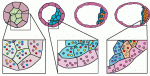 Early mammalian embryos exhibit remarkable plasticity, and recent studies of mouse embryos implicate a network of transcription factors in governing this plasticity and the establishment of embryonic lineages. Here, Alfonso Martinez Arias and colleagues summarise this information, link it to classical embryology and propose a molecular framework for the establishment and regulation of developmental plasticity. See the Hypothesis article on p.
  François Guillemot heads the Division of Molecular Neurobiology at the MRC National Institute of Medical Research in London, where his research focuses on the transcriptional control of neurogenesis and the epigenetic regulation of gene expression in neural development. François recently became an editor for Development, and we asked him about his research and career, as well as his lab’s future move to the Crick Institute. See the Spotlight article on p.