The community site for and by
developmental and stem cell biologists

In Development this week (Vol. 138, Issue 3)

Posted by , on 4 January 2011

Here are the research highlights from the new issue of Development:

Wnt/PCP signalling, microtubules and gastrulation

During vertebrate gastrulation, convergence and extension (C&E) movements shape the germ layers to form the anterioposteriorly elongated body axis of vertebrate embryos. Non-canonical Wnt/planar cell polarity (Wnt/PCP) signalling regulates C&E by polarising the morphology and behaviour of cells, which suggests that the Wnt/PCP pathway might influence the microtubule cytoskeleton. Here, Lila Solnica-Krezel and co-workers investigate this possibility by assessing the position of the centrosome/microtubule organising centre (MTOC) relative to the cell nucleus and the body axes during zebrafish gastrulation (see p. 543). They report that MTOCs occupy a polarised position within the plane of the ectoderm and mesoderm, becoming biased to the posterior and dorsal/medial side of the cell between mid and late gastrulation. This polarisation, they report, depends on intact Wnt/PCP signalling. Conversely, microtubule disruption experiments show that microtubules are required to initiate the anterior localisation of Prickle, a core PCP signalling component. These and other results suggest that reciprocal interactions between Wnt/PCP signalling and the microtubule cytoskeleton are required during C&E gastrulation movements.

Skin deep: Adam10 regulates Notch signalling

Notch signalling plays a crucial role in the development and maintenance of the epidermis: the stratified epithelium that forms the skin’s outer layer and protects organisms from dehydration, mechanical trauma and microbial invasion. Now, on p. 495, Carien Niessen, Paul Saftig and colleagues reveal that the disintegrin/metalloproteinase Adam10, a `sheddase’ involved in Notch processing, is essential for epidermal integrity and Notch-mediated epidermal signalling in mice. The researchers show that epidermal-specific deletion of Adam10 in mouse embryos leads to perinatal death, impairment of the skin’s barrier function and an absence of sebaceous glands. Moreover, deletion of Adam10 in adult mice causes hair loss, epidermal hyperproliferation and cyst formation. These phenotypes closely resemble those produced by epidermal inactivation of Notch signalling. Indeed, the researchers report that epidermal loss of Adam10 severely impairs Notch processing and signalling in the epidermis. Together, these data identify Adam10 as the major Notch processing enzyme in the epidermis in vivo and as a central regulator of skin development and maintenance.

SAD (kinase) tales of neural-specific glycans

Several aspects of neural development and function rely on the regulated expression of specific glycans, but what are the mechanisms that govern neural-specific glycosylation during embryogenesis? On p. 553, Michael Tiemeyer and colleagues report that Sugar-free frosting (Sff) – the Drosophila homologue of SAD kinase, which regulates synaptic vesicle tethering and neuronal polarity in nematodes and vertebrates – drives neural-specific glycan expression in the Drosophila embryo prior to synaptogenesis. They performed a genetic screen for mutations that affect the expression of neural-specific N-linked glycans known as HRP-epitopes; neural expression of HRP-epitopes requires ectodermal expression of Tollo, a Drosophila Toll-like receptor. Analysis of the sff mutant recovered from this screen reveals that Sff modulates glycan complexity by altering Golgi dynamics in neurons that respond to Tollo transcellular signals. The researchers propose that multiple protein kinases facilitate flux through divergent Golgi processing pathways, thereby sculpting tissue-specific glycan expression patterns during development.

nanos1: novel structure-based translational regulation

During development, translational control of mRNAs regulates gene expression. Translational control is usually achieved through binding of trans-acting factors to mRNA untranslated regions but, on p. 589, Mary Lou King and co-workers reveal a novel, structure-based mechanism for translational repression of Xenopus germline nanos1. Nanos translational repressors maintain primordial germ cell identity during development. nanos1 RNA is transcribed during early oogenesis and stored in germinal granules. Surprisingly, the researchers report that, unlike other mRNAs, nanos1 RNA translates poorly after injection into Xenopus oocytes. Thus, sequestration within germinal granules cannot explain translational control of nanos1 mRNA. Instead, they report, a secondary structural element immediately downstream of the mRNA start site is necessary and sufficient to repress the initiation of nanos1 translation through steric hindrance of ribosome scanning; insertion of 15 nucleotides between the start codon and this element relieves repression. Although structure-based translational regulation is common in prokaryotes it has not been observed before in eukaryotes and represents a new, developmentally important mode of nanos1 regulation.

Fast Nodal/Lefty movements set LR asymmetry

Nodal and its feedback inhibitor Lefty instruct left-right (LR) asymmetry in vertebrates, but what controls the spatial distribution of these ligands in the embryo? On p. 475, Lindsay Marjoram and Christopher Wright address this question by expressing functional epitope-tagged Nodal and Lefty from grafts implanted into tailbud Xenopus embryos. Both ligands move long distances along the extracellular matrix (ECM), they report, with Lefty moving faster than Nodal. Moreover, sulphated proteoglycans in the ECM seem to facilitate Nodal movement. Thus, the researchers propose, Nodal autoregulation aided by rapid ligand transport underlies the anteriorward shift of Nodal expression along the left lateral plate mesoderm (LPM), with higher levels of chondroitin-sulphate proteoglycan in more mature anterior regions providing directional transport cues. Finally, they report, Lefty moves from the left to the right LPM, a result that strengthens LR patterning models that involve active blocking of right-sided Nodal expression. Future molecular studies into how Nodal and Lefty interact with sulphated proteoglycan-rich ECM should provide additional insights into the establishment of LR asymmetry.

Moved to radial intercalation by PDGF-A

Radial intercalation – a common morphogenetic process in which cells from germ layers deep in developing embryos interdigitate into more superficial layers – is essential for the tissue rearrangements that occur during gastrulation. Here (p. 565), Erich Damm and Rudolf Winklbauer use scanning electron microscopy and time-lapse recordings to analyse radial intercalation in the prechordal mesoderm (PCM) during Xenopus gastrulation. They show that this process involves cell reorientation in response to a long-range platelet-derived growth factor A (PDGF-A) signal and directional intercellular migration towards the ectoderm, the source of this signal. The PCM, they report, fails to spread during gastrulation when endogenous PDGF-A signalling is inhibited. However, expression of a short-splicing isoform of PDGF-A, but not of a long-splicing form that binds to the extracellular matrix, rescues PCM radial intercalation. These results provide the first insights into the molecular basis of radial intercalation movements in the vertebrate gastrula and identify distinct roles for PDGF-A isoforms during gastrulation.

Also…

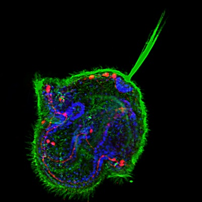
As part of the Evolutionary crossroads in developmental biology series, Pauline Schaap introduces Dictyostelium discoideum, a social amoeboid that exists as both uni- and multicellular life forms, studies of which have provided key insights into the evolution of multicellularity.
See the Primer article on p. 387.

Thumbs up (No Ratings Yet)
Loading...

Tags: , , , , , , , , , , , , , , , ,
Categories: Research

Navigate the archive

Use our Advanced Search tool to search and filter posts by date, category, tags and authors.

Celebrating NYE with Bioluminescence

Posted by , on 31 December 2010

Let me take you on a Bioluminescent journey across many kingdoms.

If you’re not well acquainted with the term, it’s the ability of living things to chemically produce light. It’s also a natural widespread feature to many organisms, from jellyfish to algae, fireflies to fungi. In recent years, it’s become a standard molecular biological tool for visualizing gene expression.
ResearchBlogging.org

(Blue Jellyfish by Maaco. CC. link here.)

On some coasts (and even lakes), if you disturb the water you might notice the sudden appearance of hundreds of tiny bright lights in the water. Sometimes it’s observable in the crest of a powerful wave.

(bioluminescent wave, Phil Hart, CC, Ask Nature)

The light comes from the thousands of tiny unicellular algae, or dinoflagellates. The same species that cause deadly red tides (toxins released from the algae, which can cause paralysis in concentrated amounts. hence, never eat shellfish in red tide areas)

On land in fireflies, the luciferin pigments in their bodies can react with oxygen, to emit radiant light energy. This is catalyzed (or sped up) by the luciferase enzyme. Many biologists are probably familiar with Luciferase reporter genes, as markers for gene expression.

For instance, this reporter system can be used to track circadian rhythms in plants, by fusing the firefly luciferase gene to a plant one. The light signals are controlled by the plant genes, and are switched on by the plant itself during different parts of the day.

(Image: Firefly, by qmnonic)

The artificial lights cue researchers when the plant genes are switched on an off according to it’s circadian clock. Of course, the plants need a water with luciferin pigments, which they don’t naturally produce.

The idea of using bioluminescent genes & proteins (at least in plants) originated in tobacco in the late 80s. The moment was captured in Science & Time Magazine, it was so groundbreaking and breathtaking. Link here (Google Books) for the comparison of tobacco before and after “switching on” the luciferase action.

(CC from the Harmer Lab. Luciferase activity in a transgenic Arabidopsis plant)

Firefly luciferase is slightly different from GFP, a photoprotein. Photoproteins don’t require any special pigments, enzymes or chemicals to set it off. Once expressed, it simply needs mineral substrates to emit fluorescence. GFP was originally recruited from jellyfish. It has the same purpose as the luciferase enzyme in molecular biology, to mark the expression of select genes. At least in the lab, it GFP also requires you to shine blue light (or sometimes UV) for it to emit the green fluorescence. It’s such a standard tool now, in mice, plants, flies, fish studies. And it garnered it’s inventors the Nobel Prize for Chemistry in 2008.

In RNAi industry, it’s becoming a convenient diagnostic tool for tracking the efficiency of RNAi drugs. Previously, to gauge how well a target gene could be silenced by RNAi, substantial amounts of tissues needed to be extracted and ground up to conduct a quantitative RNA assay. this gave a numeric reading of how much was silenced compared to untreated tissues or model animals. However, this was a rather intensive method. Reporter genes, such a luciferase one, offer a non-invasive way of perceiving how strong the silencing is occurring.

Some fungi also naturally have a green glow at night. Their traditional names include jack-o-latern fungi, ghost fungi and foxfire (image on left, CC from the Cornell Mushroom Blog). Perhaps these guys were the original fairy rings 18th century cottagers thought they saw.

Thanks for reading & hope you enjoyed the show

(Image, CC from Hither & Thither).

References & Interesting Reading:

Luciferase in Tobacco:

OW, D., DE WET, J., HELINSKI, D., HOWELL, S., WOOD, K., & DELUCA, M. (1986). Transient and Stable Expression of the Firefly Luciferase Gene in Plant Cells and Transgenic Plants Science, 234 (4778), 856-859 DOI: 10.1126/science.234.4778.856

Luciferase in Arabidopsis for tracing circadian rhythms:
Harmer SL, Hogenesch JB, Straume M, Chang HS, Han B, Zhu T, Wang X, Kreps JA, & Kay SA (2000). Orchestrated transcription of key pathways in Arabidopsis by the circadian clock. Science (New York, N.Y.), 290 (5499), 2110-3 PMID: 11118138

New Developments with Luciferase in the Pink Tentacle Blog

Review on Fluorescence in Molecular Biology:

Mavrakis M, Pourquié O, & Lecuit T (2010). Lighting up developmental mechanisms: how fluorescence imaging heralded a new era. Development (Cambridge, England), 137 (3), 373-87 PMID: 20081186

Luciferase and RNAi Diagnostics:
McCaffrey, A., Meuse, L., Pham, T., Conklin, D., Hannon, G., & Kay, M. (2002). Gene expression: RNA interference in adult mice Nature, 418 (6893), 38-39 DOI: 10.1038/418038a

Just noticed that Wiki has a list of proposed uses of bioluminescence here (just have to scroll down a bit). Some original ones include using luciferase trees to line highways and save on electricity. Crops that light up when they’re thirsty. Glow in the dark pets. etc.

Thumbs up (1 votes)
Loading...

Tags: , , ,
Categories: Research

HHMI Biointeractive

Posted by , on 30 December 2010

Each year in early December, the Howard Hughes Medical Institute hosts a series of educational seminars, called the Holiday Lectures, in which researchers explain the very basic concepts of their work. The lectures make a great introduction to a topic, and all past lectures are available on the HHMI Biointeractive site or as DVDs for teachers to use in the classroom. This year’s Holiday Lecture was on viral outbreaks, but a few past lectures have been on topics more closely related to developmental biology.

The Biointeractive site also features short videos and animations related to each year’s lectures, and the 2006 Holiday Lecture series on “Potent Biology: Stem Cells, Cloning, and Regeneration” offers many interesting clips for use in teaching developmental biology or stem cell science. For example, there’s an 11 minute mini documentary in which Alejandro Sanchez Alvarado explains planarian regeneration.

On the animation section of the Biointeractive site you can find, among other things, a short explanation about creating embryonic stem cell lines, also from the 2006 Holiday Lectures.

Have a look the lists of videos and animations on the site. There are too many to all watch, but it’s worth looking around just to see what’s there, especially if you’re teaching introductory courses. There’s even an interactive transgenic fly lab on the site, and a museum!

(Screencaps used with permission.)

Thumbs up (No Ratings Yet)
Loading...

Tags: ,
Categories: Outreach, Resources, Video

Steering a changing course

Posted by , on 29 December 2010

(This is the editorial from Development’s first issue of 2011. It first appeared on the Development site on December 7, 2010.)

As I wrote in last year’s editorial, developmental biology is facing a major revolution with the emergence of the stem cell field, to which many of our best scientists are drawn. Thus, one of my main priorities for 2010 was to raise the profile of Development in the stem cell community and to try to make the journal a premier forum for publishing the best stem cell work. To achieve this goal, we have created a new section of the journal called ‘Development and stem cells’, which groups together papers of interest for the stem cell field. As you might already have noticed, we take a broad perspective on stem cell biology in this section, and have published papers that range from embryonic to adult stem cells, from both animals and plants. Over the course of 2010, we featured at least 75 papers in this section.

We have also expanded our team of editors by recruiting stem cell specialists to increase the visibility of Development in the stem cell field. In 2009, we recruited Shin-Ichi Nishikiwa to join our other stem cell experts, Austin Smith and Ben Scheres, on Development‘s Editorial board. We are also very pleased to announce that Development‘s editorial stem cell expertise is to be further strengthened by Professor Gordon Keller, currently the director of the McEwen Centre for Regenerative Medicine in Toronto (Canada), who will be joining Development‘s Editorial board from January 2011. As many of you will already know, Gordon is a world renowned specialist in the field of human and mouse embryonic stem cell differentiation.

(more…)

Thumbs up (4 votes)
Loading...

Tags:
Categories: News

Keep up and blog on – my route to the Node

Posted by , on 23 December 2010

This is the first in a series of posts about careers outside of academic research. See here for more information.

To start the series on alternative careers, I’m going to share the story of how I ended up as community manager of the Node. But first I need to bring up a pet peeve: I don’t like the phrase “alternative career”. For me, leaving the bench was never an “alternative” – it was my goal.

I studied chemistry at VU University in Amsterdam – first with the intention of becoming an environmental scientist and saving the planet, but once I was in the program I found that there were things I enjoyed much more than research itself. By the time I finished, I had done research in three biochemistry and pharmacology labs, but I’d also organized a study tour to Scandinavia, assembled the chemistry students’ year book, ran two career days and organized a number of other activities for science students. I really liked connecting scientists and talking about science. However, I wanted a few years of additional research experience, so I did a PhD in the Biochemistry program at the University of Toronto, where I researched pathways involved in mouse skin pigmentation.

A visit to AkzoNobel in Malmö, Sweden, during the study tour I organized in 1999. I’m in the brown striped scarf, near the middle.

During my PhD, I started writing a science blog in my spare time. I wanted to create a site where I could write about the kind of science-related things that were not directly relevant to my work in the lab, just for fun, in the evenings after work. Starting the blog led to a number of other science writing opportunities, ranging from a blog on Nature Network to writing fact sheets for season four of ReGenesis – a Canadian TV drama about scientists. I started doing more and more writing on the side, as well as other science-related things: I was on the editorial board of Hypothesis (a small journal based out of the University of Toronto), I visited classrooms to talk to kids about science for outreach organization Let’s Talk Science, and I instigated an “unconference” (a meeting without a program) for scientists called SciBarCamp.

Connecting people at SciBarCamp: programmers, cognitive scientists, and biologists learning about engineering students’ solar car.

I really liked doing all my side projects, and wanted to do more of that. When I came close to the end of my PhD, I made myself a promise: I would try a freelance career for a year, and if at the end of that year it looked like it was not sustainable, I would find a fulltime job. My backup plan, in case nothing worked out, was to do a postdoc, but I really didn’t want to. I felt that that would take me too far down the track I didn’t want to be at. Doing a postdoc was, for me, the absolute last-choice alternative.

I defended my PhD thesis in December 2008. Unfortunately, with the plummeted economy, 2009 was not the best year to start a freelance career, and, when I earned little enough that I qualified for employment insurance, I started looking for full-time work. I sent out job applications here and there, mostly for editorial positions, because scientific publishers are key players in connecting scientists and in communicating science.

The job search was frustrating, not just because there weren’t very many jobs at the times, but also because I didn’t quite have the right experience for anything. Eventually, I applied for a reviews editor position at Development, even though I knew I didn’t qualify for that either, and got a reply asking me if I wanted to interview for the Online Editor position instead – the job that involved setting up and running the Node. As you know, I got that job, and I get to blog during work hours now.

I like that running the Node gives me a chance to keep in touch with academic research, through conferences, lab visits, and reading papers, but without having to do any experiments myself. Even though I now mainly work with developmental biologists, I’m more broadly interested in the practice of science in general, and my dream job would be to think about general questions related to the vague concept of “the scientific community”: Why did postdocs become a requirement for academic jobs in some fields but not others? How does the public think about scientists, and why? Is there a better way to do peer review than the current system? (A current side project of mine is to find out why so many scientists are also musicians. It has leaked over to the Node on a few occasions…) To be able to think about these things, about the process of science and the life of scientists, I really needed the experience I’ve had doing research myself, as part of my PhD. So while I didn’t use my PhD in the traditional way of a sort of training program for a specific field of research, it has definitely been indispensable and worthwhile.

The main reason I got my current job at the Node, and not any of the others I applied for, was that all the things I’d been doing in the previous years formed the kind of experience that the job required. I had done research, but I’d also done science writing and community building – mostly as a hobby! Everyone is going to ask for experience, no matter what job you apply for, but it’s possible to get experience in things you think you’re just doing for fun, and that might just be the best job yet.

For a career involving science writing, blogging is a good way to start. You can set up your own, but we could also always use new people to write for the Node. In the wise words of this beer coaster spotted at a London pub, “Keep up and blog on!”

Thumbs up (7 votes)
Loading...

Tags:
Categories: Careers

GFP, YFP and RFP = Christmas Themes!

Posted by , on 22 December 2010

The other beauty of Fluorescent Reporter Gene Systems is its undeniable likeness to holiday colours. So then, will we eventually see Christmas trees with genes hooked up to GFP, YFP and RFP?
ResearchBlogging.org

The purposeful beauty of the system is in our ability to view artificial fluorescence (and hence the genes they report) throughout development in living tissue. There’s no need to freeze tissues in lethal chemicals to obtain results. Furthermore, we can observe fluorescence from teeny organelles under the confocal microscope to whole plants under a UV lamp. (Fantastic, now how do you switch it off?)

(Image: Ethereal looking Arabidopsis seed with glowing red nuclei, Development Cover Oct. 2009. Read more about it in the original article by Fitz Gerald et al., 2009)

Since the advent of RNAi, they’re now being used as molecular sensors for the occurrence of gene silencing in vivo. Recently, RNAi signal transport in tissues was uncovered using GFP. Researchers constructed a small RNA directed against the GFP sequences. What they found was quite surprising. If they injected their small RNAs in one leaf of a tobacco plant, which is widely expressing GFP, over time the GFP diminished first in the veins and then in tissues. Eventually, the fluorescence was quenched (summarized in Voinnet 2002).

Here in Australia, Peter Waterhouse (a Grand-daddy of RNAi in plants) is working on making live action movies of RNAi in progress. One new aim in his lab is to produce the “Zebrafish” of plants…a chlorophyll-less Arabidopsis plants. Weird or genius?

(Image: Tobacco plant afflicted with a virus can cause an unusual green glow similar to GFP. The red is autofluorescence from chlorophyll. Viral transport can overlap with the transport of RNAi signals, which also serves as a plant defense mechanism against viruses & other foreign nucleic acids. Flikr CC, by Xmort)

While beautiful, it’s a not perfect system in plants for a variety of reasons. The main one is autofluorescence, or the bright red grinch that inadvertently contrasts with the merry green. A microscope tech described it as nothing short of a total nightmare. Plants naturally fluoresce because of the pigments they contain ~ chlorophyll. The huge heap of noise it makes can block the signal from the reporter

gene. However, it’s getting easier to get around this using a range of light filters. Some filters only allow a narrow wavelength of light to go through. Usually they’re specific to just the wavelength of the GFP/RFP/YFP signal, and this causes non-fluorescing tissue to appear black.

(Image: my own hideous encounter with autofluorescence :(, note that chlorophyll is absent in the pollen & petals).

At any rate, I won’t keep my fingers crossed for that fluorescent Christmas tree. But it could be potential electricity saver. Instead of purchasing a bunch of energy consuming twinkle lights, simply purchase a fluorescent bulb (doesn’t have to be UV) and switch it on in place of your regular house lights. And if you get tired of the fluorescence, spray or inject some RNAi on a branch.

On a side note, while looking for possible videos on reporter genes, I came across the following video on Youtube:

Growing Nerve Cells from Hair Follicle Stem Cells in Mice?

So…stem cells in hair follicles…highly related to nerve & brain stem cells? Exciting new trend..or weird?

Thanks for reading!

YFP in the Heart Stage embryo of  Arabidopsis thaliana (Gifford 2003)

References:
Gerald, J., Hui, P., & Berger, F. (2009). Polycomb group-dependent imprinting of the actin regulator AtFH5 regulates morphogenesis in Arabidopsis thaliana Development, 136 (20), 3399-3404 DOI: 10.1242/dev.036921

Gifford, M. (2003). The Arabidopsis ACR4 gene plays a role in cell layer organisation during ovule integument and sepal margin development Development, 130 (18), 4249-4258 DOI: 10.1242/dev.00634

Voinnet, O. (2002). Molecular Analysis of Post transcriptonal Gene Silencing. Science. [Accessed 21 Dec 2010] link
Thumbs up (3 votes)
Loading...

Tags: , ,
Categories: Images, Research

An interview with Ben Scheres

Posted by , on 22 December 2010

(This interview by Kathryn Senior originally appeared in Development on December 21, 2010)

Ben Scheres is an expert in plant development. He has been investigating development in Arabidopsis at Utrecht University, The Netherlands, since 1990, where his group uses the root tip as an easily accessible supply of plant stem cells. Ben agreed to be interviewed by Development to talk about his interest in stem cells and the beauty of self-organisation in plants.

When did you first realise you were going to follow a science career?

When I was 14, we visited a fancy science museum called ‘Evoluon’ and I bumped into a very lucid explanation of how DNA ‘encoded life’. I still remember the big spiral staircase model and the impression it made on me. Even as a small kid I had often wondered how life works – and here was a model that seemed to make it possible to start to understand some of that!

What first made you interested in stem cell research in plants?

For my PhD, I chose a research project in the field of plant-microbe interactions. At that time, breakthrough papers in fly and worm development were appearing thick and fast and I was fascinated by them. I realised, then, that we were nowhere near being able to describe the development of plants in similar detail. I looked for a plant system that had the same clear cellular relationships that we see in C. elegans and would present similar genetic possibilities. It was also important for the system to have the developmental flexibility that characterises plants. Arabidopsis roots fitted the bill and I have been hooked on them ever since.

What is the most striking difference between animal and plant stem cells?

First of all, plants do not set apart a germline, so all stem cells are somatic. In animals, many somatic stem cells have quite a restricted potential, but this is not the case in plants. There are far fewer restrictions and stem cells can also be easily regenerated. Induced pluripotent stem cells in animals have created quite a stir but this is no big deal in plants. Of course, this raises lots of questions and we’d like to understand much better what determines this difference.

(more…)

Thumbs up (4 votes)
Loading...

Tags: ,
Categories: Interview

In Development this week (Vol. 138, Issue 2)

Posted by , on 21 December 2010

Boning up on stem cell Igf2-P2 function

The insulin-like growth factor (IGF)/insulin signalling pathway regulates cell proliferation, differentiation, aging and life span. During embryonic development, transcription of the mouse and human Igf2 gene is tightly regulated by four alternative promoters whose specific roles are unclear. Now, Sylvie Nathalie Hardouin and colleagues reveal that the transcriptional activity of one of these promoters, Igf2-P2, regulates mesenchymal stem cell differentiation and osteogenesis in mice (see p. 203). The researchers show that Igf2-P2 loss-of-function mice, in which a lacZ-neo cassette replaces the P2-driven transcriptional unit of Igf2, have short, thin, poorly mineralised bones and exhibit altered bone remodelling. These abnormalities are associated with decreased numbers of embryonic mesenchymal chondroprogenitors, adult mesenchymal stem cells and osteoprogenitors. Together, these and other results support a model in which the transcriptional activity of the Igf2-P2 promoter regulates the fate of mesenchymal progenitors during bone development and adult bone remodelling, and regulates osteogenesis through its effects on both osteoprogenitors and their microenvironment.

X inactivation: from imprinted to random

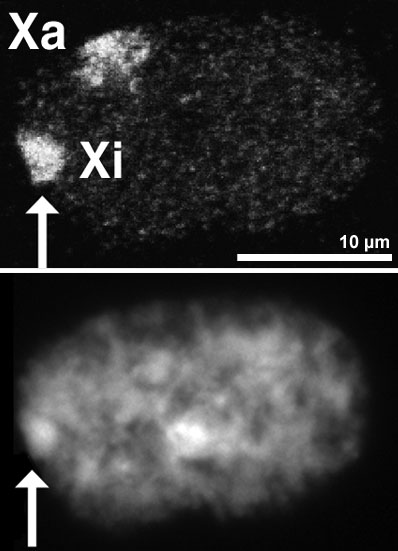
In female mammals, one X chromosome is epigenetically silenced in adult cells by the process of X inactivation (Xi). However, in the pluripotent epiblast cells of the preimplantation mouse embryo, both X chromosomes are active and Xi of the paternal or maternal X occurs at random shortly after implantation (random Xi). By contrast, in very early mouse embryos (and in extra-embryonic lineages), the paternal X chromosome is selectively inactivated (imprinted Xi). So when exactly does the mode of Xi change from imprinted to random during development? On p. 197, Hitoshi Niwa and colleagues examine Xi during the differentiation of inner cell mass (ICM)-derived female mouse embryonic stem (ES) cells. The researchers use forced expression of Cdx2 and Gata6 to induce ES cell differentiation toward trophectoderm (TE) and primitive endoderm (PrE), respectively. They report that random Xi occurs in both TE and PrE cells and in the TE of cloned embryos derived from female ES cells, suggesting that all marks for imprinted Xi must be erased by the time the ICM forms.

Muscle building: a connective tissue workout

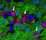
Muscle and its connective tissue are intimately linked during embryogenesis and adult life. Thus, interactions between these tissues might be crucial for their development. To date, the lack of molecular markers for connective tissue fibroblasts has hindered the study of these potentially important interactions, but now, on p. 371, Gabrielle Kardon and colleagues identify the transcription factor Tcf4 as a marker for connective tissue fibroblasts and reveal that connective tissue is an important regulator of myogenesis. By making Tcf4GFPCre mice, which allow genetic manipulation of connective tissue fibroblasts, they show that these fibroblasts regulate both muscle fibre type and maturation. In addition, the researchers unexpectedly discover that low levels of Tcf4 in myogenic cells promote the overall maturation of muscle fibre type. These and other data identify novel extrinsic and intrinsic mechanisms that regulate myogenesis and show for the first time that connective tissue is a vital component of the niche that controls muscle development.

Mitochondrial pathway central to fly apoptosis

Apoptosis (programmed cell death) is essential for development and tissue maintenance in many organisms. In mammals and C. elegans, Bcl-2 family proteins facilitate apoptosis by regulating mitochondrial dynamics but do they play a similar role during apoptosis in Drosophila? According to Kimberly McCall and co-workers, the answer to this question is yes in the Drosophila ovary (see p. 327). During mid-oogenesis in flies, apoptosis is induced in some of the egg chambers in the ovary when nutrients are scarce. The researchers show that, during this event, the mitochondrial networks of ovarian nurse cells undergo extensive remodelling, cluster formation and cluster engulfment by somatic follicle cells. These mitochondrial dynamics, they report, are dependent on caspases, the Bcl-2 family, the mitochondrial fission and fusion machinery and the autophagic machinery. Furthermore, cell death in the ovary is defective in Bcl-2 family mutants. Thus, the researchers conclude, Bcl-2 family proteins do play a major role in controlling both mitochondrial dynamics and cell death in the Drosophila ovary.

Retinal cell fates largely left to chance

Classic experiments in invertebrates suggest that stereotypic patterns of cell division generate specific cell types during development, but the extent to which stereotypic lineages play a part in the developing vertebrate CNS is an open question. Now, Michel Cayouette and co-workers report that stochasticity plays a major role in cell fate decisions in the developing rat retina (see p. 227). In vivo cell-lineage tracing studies show that vertebrate retinal progenitor cells (RPCs) yield retinal clones of varying size and cellular composition. Whether this variability reflects distinct but reproducible lineages among many different RPCs or stochastic fate decisions within a population of more equivalent RPCs is unclear. To find out, the researchers use videomicroscopy to follow the lineages of rat RPCs cultured at clonal density. Their analysis of the reconstructed lineages indicates that fixed probabilities determine the decision of the RPCs to multiply or differentiate. Thus, stochasticity plays a major part in the development of the retina and possibly also of other parts of the vertebrate CNS.

Worming out organiser evolution

Axial organisers, embryonic regions that induce cell fate and establish body axes during development, have been identified in various metazoans but their evolutionary origins and conservation of function remain unclear. The presence of an axial organiser in annelids (ringed worms) has not previously been confirmed, but now Takashi Shimizu and colleagues provide direct evidence that, in Tubifex tubifex annelids, descendants of a single blastomere of 4-cell embryos can function as axial organisers (see p. 283). The first two cleavages of T. tubifex embryos generate four macromeres (A-D) that subsequently divide to generate micromeres. The researchers show that ablation of the D macromere descendants 2d and 4d can inhibit axial development. Co-transplantation of 2d and 4d into ectopic positions, they report, induces secondary axis formation in host embryos; in these axes, neurectoderm and mesoderm derive from the transplanted micromeres, whereas the endoderm derives from the induced host. These studies identify D quadrant micromeres as annelid axial organisers, informing future studies of axial organiser conservation and evolution.

Plus…

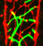
During nervous system development, axon branching allows elaborate synaptic connections to form. Recent advances, reviewed by Daniel Gibson and Le Ma, identify how various axon branching morphologies develop and the common principles that regulate them.
See the Review article on p. 183 of this issue.

Thumbs up (No Ratings Yet)
Loading...

Tags: , , , , , , , , , , , , ,
Categories: Research

Alternative Careers for Developmental Biologists

Posted by , on 21 December 2010

Some of the most popular posts on the Node have been those about career prospects for young scientists. The category pages for job ads and career posts are among the most visited parts of the site, but neither of them has had as many hits as the discussion titled “too many postdocs and PhD students?

In the comments of that post, back in July, Greg Dressler wrote:

“I do think we need to get over the idea that nothing short of an academic career fulfills the ideal goal of our students and post-docs. Most of the folks I went to graduate school with are not in academics anymore, yet they have meaningful and successful careers.”

And James Briscoe added:

“More flexibility is what’s needed and the acknowledgment and encouragement of a diversity of career routes and development paths.”

To follow on these thoughts we’ll profile a range of alternative careers for developmental biologists on the Node. Over the next few months we’ll have posts up from several people who found a career away from the bench. All posts in this series will be tagged altcareers, so you can easily find them all on one page.

We have already approached a few people to ask them to share their story, but if you would like to add your own experience in finding work outside of academic research, feel free to register for the Node and add a post with the altcareers tag, or contact us to get a set of guiding questions if you’d like some help with writing.

Eventually, we’ll summarize all responses in a feature article.

I’ll kick off the series with my own story in a few days. Spoiler: I’ll complain about the phrase “alternative careers”, because for me it was never an alternative to begin with!

Thumbs up (No Ratings Yet)
Loading...

Tags:
Categories: Careers

Feed me!

Posted by , on 16 December 2010

With people in many countries preparing to take a few days off at the end of this month, and other countries starting their summer break, I’m sure many of you have had to deal with the stress of handling your experiments over the holidays. How do you explain to a tank of zebrafish or a flask of cells that you’re going to visit your family for a few days? Do you just not go at all? Is the whole lab leaving feeding instructions with that new postdoc who lives just across the street from the institute and comes in every day anyway?

Let us know via the poll, and leave a comment if you want to explain your answer.

(poll closed and archived)

Thumbs up (No Ratings Yet)
Loading...

Tags: , ,
Categories: Discussion