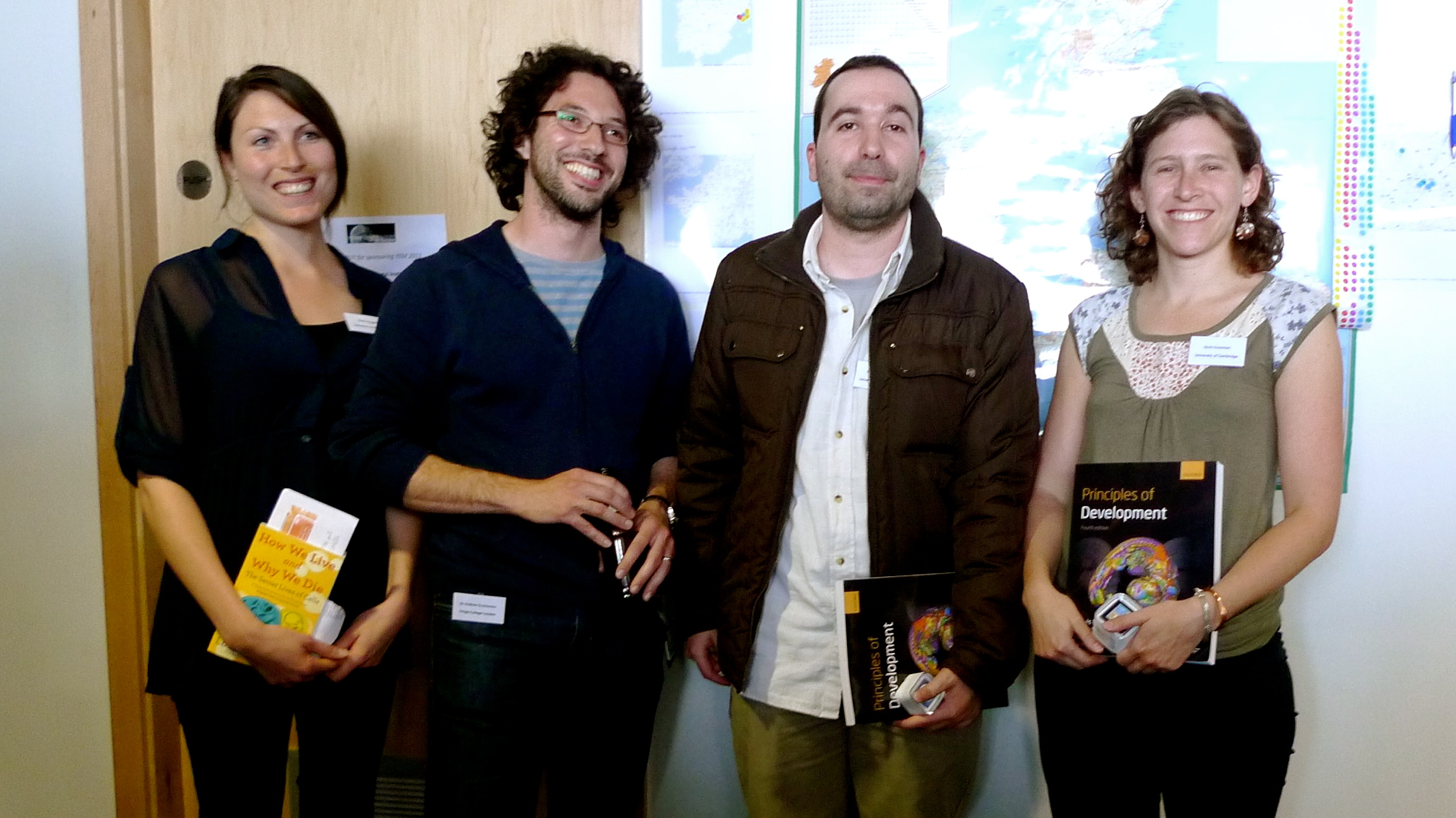
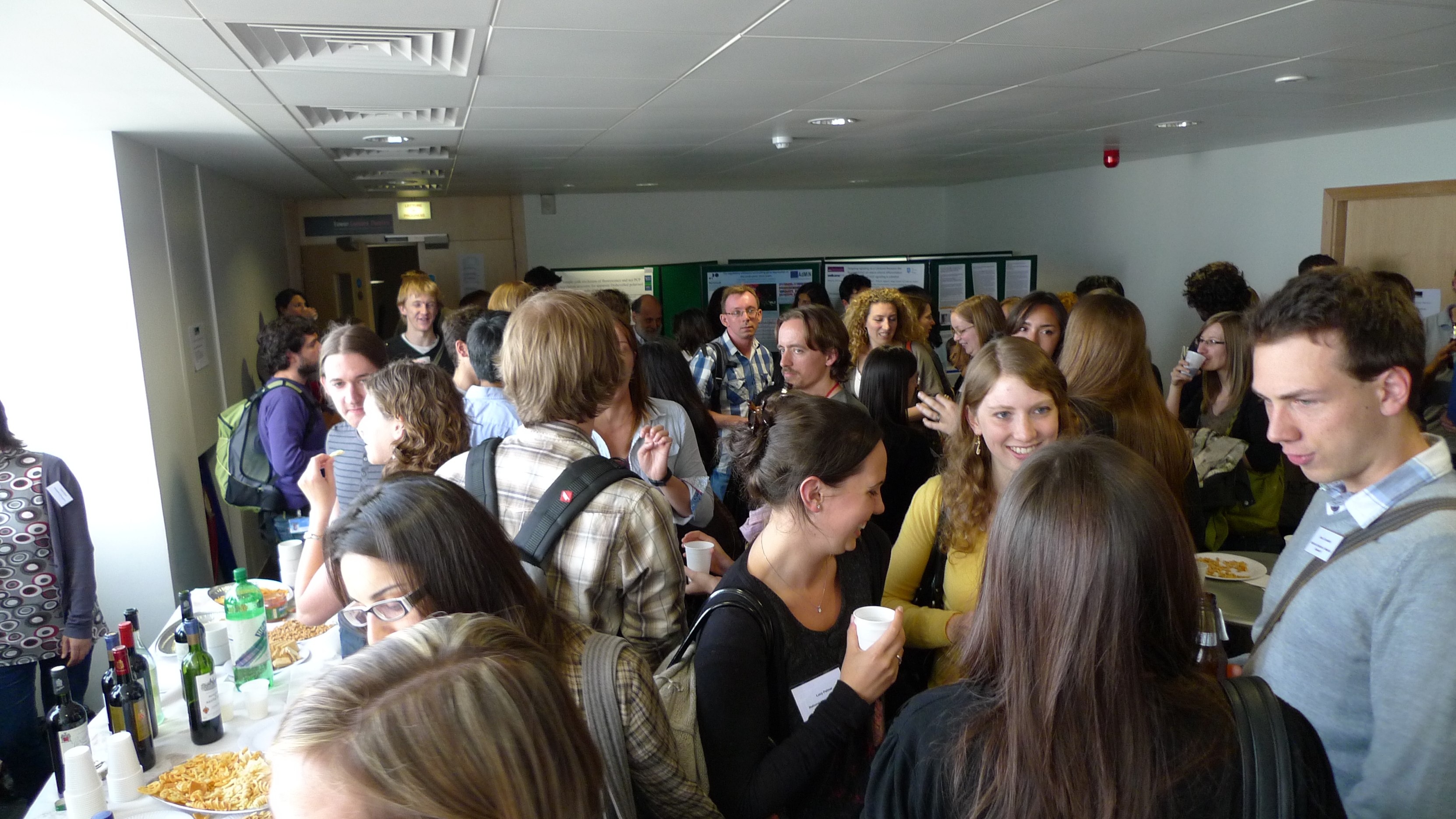
The community site for and by
developmental and stem cell biologists

Stem cells and developmental biology: old friends meet again…or did they ever part ways?

Posted by , on 30 June 2010

Ahhh the Node, my favourite part of the embryo: nice cup shape you can lie back in and get a whirly cilia massage…. OK, on with the post.

So it seems that everyone is working on stem cells now. They’re all the rage. Students come through for a rotation and ask “do you work on stem cells?”, grants with some aim that includes stem cells seem to do better than regular dev bio grants, and of course the papers….well the papers speak for themselves. But is this such a surprise and does this have any bearing on those of us who study embryonic development but haven’t cultured a stem cell other than maybe to make a knockout mouse? The answer is that it depends what you care to study, and whether you care if there is a difference between the two.

Because really stem cell biology is but an integral part of developmental biology. Stem cells, especially the non-cultured variety, are a normal part of embryonic development (or in the case of cancer stem cells, a rather unfortunate return). Isolating and culturing these stem cells is really figuring out how to freeze them in that transient state. In fact one might as well call development embryonic stem cell allocation (or something more clever). Most of you don’t need convincing, as many of the giants in stem cell biology were first giants in developmental biology (Rossant, Melton, Martin, Keller, and so on), and they simply applied the principles of embryology that they had developed to isolate the specific cell types that do their thing in the embryo. Not really simple, but you get the point.

When there were no stem cell journals, papers on stem cells or progenitors were published in development journals. Now of course we still have developmental biology journals, but a flurry of stem cell-specific journals have appeared, some bearing influential imprints, and these have done very well. I do however like the new trend, espoused recently by Development, of enthusiastically marrying back the two fields, like a happy homecoming or two good friends who parted ways years ago. It’s natural, it makes sense, and it certainly fits with what is hot right now in stem cell biology, which is helping stem cells find a path to a particular lineage and coax them become the cell type you wish to have in the dish. Sound familiar? Indeed the reverse trend is also taking place, with the stem cell journals publishing papers with a very developmental angle.

Of course the promise of therapeutics that stem cells bring distinguishes part of that field from developmental biology, but that’s the reason for it, often not the actual research that goes behind it. (Although the irony is that perhaps developmental biology’s fruits will severely diminish the therapeutic potential of stem cells: who needs the chance of a teratoma when you can just reprogram that skin cell straight into a nice neuron? )

So good friends meet again, hang out for a while, get to know each other again. As stem cell biology advances along lineage paths, and developmental biology takes a dip in the stem cell pool, we will do this with the comforting thought that we’re all doing the same thing: discovering where we come from and how we got here.

Thumbs up (7 votes)
Loading...

Tags: ,
Categories: Discussion

Navigate the archive

Use our Advanced Search tool to search and filter posts by date, category, tags and authors.

What do students need to know about developmental biology ?

Posted by , on 29 June 2010

This is a question that I keep asking myself. I am starting work on the third edition of my textbook “Essential Developmental Biology”. Over the years the quantity of published material in developmental biology keeps rising exponentially. Papers nowadays are extremely detailed and technical compared to the way they were in the 1980s when the shape of current molecular-genetic developmental biology was being established.

People always tell me that the thing they like about my books is their brevity and conciseness. But brevity can’t be maintained without being extremely selective, and this inevitably offends people whose research topics get left out.

Suppose an undergraduate (or beginning graduate student) has taken a lecture course in developmental biology, comprising maybe 40 hours of contact. What do you expect them to know when they appear in your lab as a PhD student ?

Do they need to know about sea urchin as well as mouse fertilization ?

Zebrafish pancreas development as well as mouse pancreas development ?

Enhancer traps in mouse as well as Drosophila ?

Would you expect them to know what the amnion is ? What are the properties of neuronal stem cells ? Which end of a Hydra is the head ? Anything about Dictyostelium ?

Or none of the above…?

Is there areally a core set of principles of Developmental Biology as a science, or is it just a bunch of topics that can be varied without limit depending on the interests of the instructor ?

If anyone has comments on this matter, and particularly about what they would like to see included in, or left out of, my third edition, please let me know.

Thumbs up (7 votes)
Loading...

Tags: ,
Categories: Discussion

Lindau Nobel Laureates Meeting

Posted by , on 29 June 2010

Have you ever met a Nobel Laureate? 650 lucky young researchers are currently spending the week with 59 previous Nobel Prize winners from the fields of physiology or medicine, physics and chemistry. Each year, a group of Nobel Laureates meet in Lindau, a German town (and island) at Lake Constance, close to the Swiss and Austrian borders. They share ideas from their respective fields with each other, and with a selection of young researchers.

The young scientists that are accepted to attend the meeting have undergone a rigorous selection process, and about forty thousand applied to fight for the 650 spots. If that sounds like a difficult way to get invited to attend a meeting, remember that the selection process for the speakers was even harder: they have to already have a Nobel Prize!

The meeting programme is a dizzying collection of big names from all areas of science. Developmental biologist Christiane Nüsslein-Volhard presented yesterday (pictured) in the same session as climate scientist Paul Crutzen. Different fields, and speakers you would not normally find at the same meeting, but the goal of this meeting is to cross disciplinary boundaries.

For those of us not at Lindau, there are plenty of ways to keep up with what’s going on via the web. The most convenient way to follow the meeting while it going on is through the special meeting website run by Nature and Spektrum, who have a team of bloggers present at the meeting. The page also aggregates all Twitter activity and photos related to the meeting, displays video interviews, and links to live streams of the lectures. Another good site to visit – even after the meeting is over – is the official Lindau Nobel Laureate Meeting website itself, which contains information about the format and history of the event.

The reports coming out of Lindau this week make it sound like an amazing event. I wonder how many of the young researchers who got to attend will later go on to win a Nobel Prize – even if just to be invited again!

(Photo of Christiane Nüsslein-Volhard speaking at the meeting, by gedankenstuecke on Flickr.)

Thumbs up (No Ratings Yet)
Loading...

Tags: ,
Categories: Events, News

Changes in Wellcome Trust funding

Posted by , on 28 June 2010

Biomedical researchers in the UK are seeing some changes in the way their research is funded. The Wellcome Trust, the UK’s largest charity funding agency for biomedical research, is phasing out the majority of its existing biomedical science grants and replacing them with “Investigator Awards”.

The last applications for project grants, programme grants, equipment grants, biomedical resources grants, technology development grants, university awards, and flexible travel awards are due on July 30 2010. After that date, researchers who would normally apply for any of these grants now need to look into the application process for Investigator Awards (starting October 2010), which is a bit different.

There will be two types of Investigator Awards: one for New Investigators, who have recently been appointed to their first academic post, and one for Senior Investigators. In a long FAQ page, the Wellcome Trust answers a lot of questions people may have about the scheme. If you’re thinking of applying for one of the new grants, that’s a page you should definitely read.

Is anyone in the UK currently preparing to apply for these grants? Or hurrying to get in an application for the old grants that are being phased out? Let us know what you think of the changes, and good luck with all your applications!

(And if there are any recent changes in research funding in your country, feel free to post an update to the Node.)

Photo credit: Dave McClear on Flickr.

Thumbs up (No Ratings Yet)
Loading...

Tags: , , ,
Categories: Discussion, Funding, News

Editorial about the Node in Development

Posted by , on 25 June 2010

Coinciding with the launch of the Node, Development Editor-in-chief Olivier Pourquié, Executive editor Jane Alfred, and the Node’s Community Manager Eva Amsen (that’s me!) wrote an editorial for the current issue of Development. It explains why we set up this site, and who is writing and reading it (that’s you!).

The picture I added is of our Node banner, which travels around a bit to conferences and other places where we want to promote the Node. It’s now back in Cambridge, where it will visit the Gurdon Institute in less than two weeks – but you will hear more about that later. Here is the editorial:

Development is excited to announce the launch of a new community website for all biologists interested in developmental biology, called the Node: a place where the developmental biology community can discuss, debate and deliberate issues relevant to developmental biology and to other research fields that this community’s work informs and touches upon.

In 2009, to assist with the recruitment of our new Editor in Chief, Olivier Pourquié, Development asked many of you in the developmental biology community for your feedback on the journal and whether there was anything about it you would change.

Your response?
Amongst other things, you told us that we needed to improve our website. And we did. In January 2010, we launched Development’s new-look website at dev.biologists.org, featuring a new design, new navigation tools and functionality, and new online-only features. But there was also a consensus amongst the community that you wanted your community journal, Development, to be just that: an online place where you could interact, gossip, write about what was going on in your world and share news about jobs, research advances and events; somewhere that would be a community-based, one-stop shop for developmental biologists.

Our answer? The Node
The Node is a new not-for-profit community website for developmental biologists, which is being launched by Development. At its helm is Eva Amsen, Development‘s Online Editor and the Node’s Community Manager, who is an active science blogger and writer with a track record in community building, and at its heart is a blog that will be open to the community. All that community members need to do to participate on the Node is to register, be approved by Eva and you are then free to post your news on the site. In addition, anyone can leave comments on any of the posts. We will be posting too: news about top research published in Development and in other community journals; deadlines and updates for conferences and funding programmes; reports from conferences we are attending; interviews with our Editors and authors; as well as other posts of general relevance and interest to the community. Sometimes we will invite specific people to contribute something on a particular topic, but we do want everyone in the community to feel free to sign up and contribute something spontaneously. Nobody knows better what is going on in the world of developmental biology than the people actively doing the research, so we’re happy to give you a chance to address your colleagues directly by signing up for the Node and posting your news and events.

So will you participate?
This is the million-dollar question because the Node will only flourish and grow if you, the community, participate. The Node is entirely funded by Development‘s not-for-profit publisher, The Company of Biologists, as a service to the community, but we’re inviting all of you to contribute content. There are already many interesting posts on the Node, which we encourage you to go and browse. This is because some of the community have been kindly helping us to road test and develop the site over the past few months and have also written some great posts for the site’s launch. For example, you can find tips for blogging from conferences, learn about some useful online science communication tools, read updates of some hot new papers in the field, catch up on presentations at the British Society for Developmental Biology conference and find out about how two of Development‘s travelling fellowship recipients are faring in their host labs in Germany and India.

To keep up with what’s new on the Node, you can bookmark the site, subscribe to the RSS feed, follow us on Twitter or look at the regularly updated highlights from the Node featured on Development‘s homepage.

We hope and believe that you will grow to like the Node. You can think of the Node as a coffee break, and use it to catch up with other scientists, to find out what’s new in the field. When you’re done, you can get right back to work by using the links in the Node’s sidebars to navigate to recent papers, your favourite databases and community journals, or find out more about upcoming conferences. We’ve already received some great feedback on the Node: when we demonstrated it to the community at the recent British Society for Developmental Biology conference in March, the community’s response was overwhelmingly positive, with several of you asking ‘why hasn’t this been done before?!’, which we took to be a good sign.

The Node is your community website, not ours, so please sign up, join in and enjoy. And if you like it, tell your colleagues and collaborators. And most of all, share your thoughts and feedback with us at the Node.

Thumbs up (1 votes)
Loading...

Categories: News

The Young Embryologist Meeting, 2010. London.

Posted by , on 23 June 2010

May 10th 2010 marked the second anniversary of a very young but promising scientific meeting, the Young Embryologist Meeting (YEM). This meeting was organised by a group of PhD students and young post-docs from London interested in developmental biology but open to everyone. Researchers at this level are just beginning to make decisions on what direction they would like their career to take. By bringing together young developmental biologists in an informal setting it was hoped to create research links that will outlast the time spent together in London. Also the idea was to create a network where exchange of ideas, support and even collaborations can occur. The outcome was a complete success with a packed seminar room full of students, post-docs and PI’s mostly from London but also from places like Barcelona, East Anglia and Cambridge. This massive attendance and the fact that already people are volunteering for next year’s meeting show how much an event like this was needed.

The meeting was held in the Wilkins room buried within the cloisters of University College London and its capacity was certainly stretched. It was opened by a keynote lecture by Peter Lawrence, who travelled down from Cambridge to talk to us about ‘Moments of discovery in a scientists life’. Everyone commented on how this talk was exactly what the conference needed. We were reminded about how it is the moment when one sees something new in nature that is the ultimate goal of research, and that these moments are a highly personal thing that once given to us can not be taken away again. It is this what drives us to keep experimenting and deal with all the frustrations that that can bring.

The meeting itself, an excellent series of short talks from young embryologists, was organised into four separate sessions. Each of session was named after the title of a classic embryological work that set the basis of the work presented in the corresponding session. This was to remind us of the importance in a continual re-appraisal of the core questions in embryology and to ask to what extent they have been answered, and how are we to address them in light of our recent knowledge and new techniques. The talks for each session were then selected with the aim of presenting a wide variety of different systems in which those core questions are being addressed. Through this and the subsequent discussion, it is hoped that ideas on the optimal experimental system in which to propose a particular research objective can be revealed. The talks covered areas ranging from de-novo centriole formation in mouse embryos, through to neurulation in zebrafish, mouse limb development, primary neurogenesis in Xenopus and the isolation of a novel organiser in the chick. Finally we had a series of talks focussing on neural crest development following by substantial amounts of wine and discussion.

The speakers and audience came form all places in London including the Department of Craniofacial Development and the MRC Developmental Neurobiology Department from KCL. In addition, we had talks from the institute of Women’s health and the Institute of Child Health, the NIMR at Mill Hill and the Department of Cell and Developmental Biology (CDB) at UCL. The CDB requires special thanks, as this was the sponsor of this event. The number of departments involved already, the people coming from outside London and the clear interest in organising new versions of the meeting will make the YEM a yearly and larger event. Thank you to everyone who helped to make this years meeting a success, we are sure everyone will agree on at least one thing; next year we will need a bigger room.

The 2010 YEM organising committee

Thumbs up (3 votes)
Loading...

Categories: Careers, Discussion, Events, Research

Selaginella in frame

Posted by , on 23 June 2010

If you are a plant developmental biologist studying the Selaginella spikemosses, you might be interested in this beautiful animation that shows the life cycle of the Selaginella, Selaginella apoda

Life Cycle: Selaginella apoda from Ciaran Moloney on Vimeo.

And if you are captured by the simple beauty of this plant, you might also like a Development cover we published three years ago featuring the lycophyte Selaginella kraussiana.

The image shows the small dorsal leaves, large ventral leaves and the shoot branching pattern of this plant and was submitted by Harrison et al. to accompany their paper on how the growth of leaves from the meristem of S. kraussiana differs to that of flowering plants; a study that provided a framework for future comparative studies of meristem and leaf development in land plants.

Thumbs up (No Ratings Yet)
Loading...

Tags: , , , ,
Categories: Images, Research

In Development, Vol 137 (Issue 14)

Posted by , on 23 June 2010

Here are the research highlights from the current issue of Development. You can find these on the Development site but we thought it would be useful to have them posted on the Node, too.

Brainy signals for actin dynamics

During brain development, neurite outgrowth and neuronal migration establish the brain architecture needed for brain function. Now, Eric Olson and colleagues reveal a regulatory feedback loop that links the cytoskeletal changes that provide the mechanical force needed for neurite outgrowth and migration to nuclear gene transcription during mouse brain development (see p. 2365). Myocardin-related transcription factors (MRTFs), the expression of which is forebrain enriched, translocate to the nucleus in response to actin polymerisation and cooperate with serum response factor (Srf) to regulate the expression of cytoskeletal genes. The researchers show that either Mrtfa orMrtfb is sufficient to support brain development but that the brain-specific deletion of both produces brain abnormalities similar to those caused by Srf deletion. These abnormalities, they report, are accompanied by dysregulation of the actin-severing protein gelsolin and of the kinase Pctaire1, which cooperates with Cdk5 to initiate a kinase cascade that governs cytoskeletal rearrangements. The researchers suggest, therefore, that MRTFs couple two signalling pathways that modulate cytoskeletal dynamics during neurite outgrowth and neuronal development.

Proliferation’s not over ’til the Fat-Hippo sings

During development, transitions from proliferating, undifferentiated cells to quiescent, differentiated cells are tightly regulated to ensure that organs reach the correct size. Kenneth Irvine and colleagues now reveal that Fat-Hippo and Notch signalling influence this important transition during optic lobe development in Drosophila (see p. 2397). Like the vertebrate nervous system, the Drosophilaoptic lobe develops from neuroepithelial cells, which function as symmetrically dividing neural progenitors. The Fat-Hippo signalling pathway, which contains the large cadherin Fat and the serine/threonine kinase Hippo, regulates the transcription of cell proliferation and survival genes. The researchers report that neuroepithelial cells in the Drosophila optic lobe undergo a cell-cycle arrest that is regulated by Fat-Hippo signalling before converting to neuroblasts. They also identify a role for Notch signalling in committing neuroepithelial cells to become neuroblasts. These and other results suggest that, by arresting the cell cycle, Fat-Hippo signalling contributes to the accumulation of Delta, which modulates Notch signalling and triggers neuroepithelial differentiation. A similar mechanism might be involved in vertebrate neural development.

Pluripotent stem cell derivation gets a (2i-)LIFt

Pluripotent mouse embryonic stem (ES) cells are obtained directly from the mouse epiblast, while pluripotent embryonic germ (EG) cells can be derived from unipotent mouse primordial germ cells (PGCs) by epigenetic reprogramming. But how similar are EG and ES cells? On p. 2279, Azim Surani, Austin Smith and colleagues report that these cells share a conserved molecular and developmental ‘ground state’. ES cells can be established using the cytokine LIF combined with the inhibition of GSK3 and of mitogen-activated protein kinase signalling (so-called 2i-LIF culture). The researchers show that pluripotent mouse EG cells can also be efficiently established using 2i-LIF culture. Then, using the same conditions, they derive rat EG cells for the first time. These cells express similar markers to rat and mouse ES cells, they report, and can contribute extensively to chimeric rats. Together, these findings raise the possibility that 2i-LIF culture could be used to derive EG cell lines with pluripotent ground state properties from other species, including humans.

(more…)

Thumbs up (No Ratings Yet)
Loading...

Tags: , , , , , , , , , , , , , , ,
Categories: Research

BSDB-BSCB Meeting Report Part II

Posted by , on 21 June 2010

As announced in my last post, here is part two of the BSDB-BSCB Spring Meeting Report. It deals with two presentations on networks of transcription factors (TFs). During development, such dynamic networks of TFs and signaling molecules establish and maintain the spatio-temporal patterns of gene expression characteristic for the developing tissue. Using high throughput approaches in this post-genomics era, we now have the opportunity to comprehensively analyze and model these networks and try to link network features to function.

Part 2: Transcription Factor Networks

Eileen Furlong (EMBL, Heidelberg, Germany) presented her lab’s approach to globally decipher the combinatorial action of TFs in cis-regulatory modules (CRM). Using ChIP-on-chip with antibodies against five TFs required for mesoderm development in Drosophila, they generated a high-resolution, genome-wide dataset, describing TF occupancy during 10 hours of early embryonic development. They then used a subset of this data along with the corresponding in vivo activity data of characterized enhancers to train a machine learning algorithm. Using this approach, they were able to correctly predict the spatio-temporal expression of CRMs not included in the training set, based solely on their combinatorial TF binding profiles, in 80% of the tested cases.

Marion Walhout (University of Massachusetts Medical School, Worcester, USA) presented her team’s global analysis of 34 basic helix-loop-helix (bHLH) TFs in C. elegans. They set out to measure all the parameters describing a bHLH TF’s function: which bHLH partner it dimerizes with; where and when the TF is expressed; which DNA sequences it predominantly binds to; and whether it might preferentially regulate genes involved in certain processes. They then combined all of these parameters for every bHLH TF in an integrated network, from which they predicted and experimentally confirmed the function of a specific bHLH, and systematically compared the parameters among all possible bHLH pairs. These analyses linked certain TF dimers to specific processes and interlinked a subset of dimers with each other, uncovering overlapping and specific functions.

I found these presentations inspiring as they combined both previously and newly generated data to try to move beyond merely looking for patterns towards attempting to predict the behavior of the system. However, since these data sets and models are highly complex, it is not always possible to uncover clear-cut trends or rules of behavior.

In part three, my final post on this meeting, I will cover talks on several topics: Stem cells, limb development and evo-devo.

Thumbs up (1 votes)
Loading...

Tags: , , , , ,
Categories: Events

Teaching embryology to undergraduates

Posted by , on 18 June 2010

I am employed as a graduate teaching assistant for a first year biomedical sciences course at Auckland University in New Zealand. The class is called Cellular and Developmental Biology and my job is to help with tutoring the laboratory classes. For my lab streams I’m the one who stands at the front and gives instructions, works with the demonstrators to assist and instruct the students, then grades the assignments afterwards.

With the word ‘developmental’ in the title, as you can imagine, there are lectures about embryology as part of the course. As the aim of the lab section is to support and extend what is taught within the lectures we naturally include an embryology class too. During the three hour session each student constructs a three dimensional model of a human four week old embryo (“today we’re making babies!”). Details are given here on the Auckland Uni website and the instructions are copyright to Colin Quilter 2003.

Each embryo is made of modelling clay called Du Kit and are baked before being returned to the students, giving everyone something to take home and keep. Teaching the class is a lot easier when you’ve been through it before so all demonstrators and tutors are given a modelling clay kit and the instructions then sent home to have a go. Here is the clay sitting on my dining room table ready for me to start.

Lumps of modelling clay

The clay comes in different colours so that each germ layer or type of blood vessel can be colour coded. Normally we’d use dissecting instruments to help with the modelling but I don’t have a dissection kit at home so I raided the kitchen drawers.

The first section to make, and one of the hardest, was the neural tube. It takes a bit of playing for the clay to warm up and also I was still getting the hang of how to work it at this point. The instructions are to scale at this point so the best method is to lay the clay on the page to see if it matches, but it’s easy to make the embryo flat on one side while doing this whereas an embryo should, of course, be rounded and somewhat smooth.

Neural tube

Then the notocord and gut sections are attached, giving the main internal structure of the model. It’s kind of cool how putting on the auditory and optic vesicles make it look more like a human.

Neural tube with foregut etc added

At this point I discovered that my home has a hazard we don’t usually face in the lab:

Cat climbing on the table

After shutting the cat out of the room I started putting on blood vessels. The colour coding really helps here, purple for mixed blood, red for oxygenated and blue for deoxygenated. The next picture has the aortic arches and other arteries attached.

Embryo model with some blood vessels

Since the embryo is getting oxygen from the mother rather than the lungs the blood flow is different to what I’m used to having studied mainly adult physiology. I found this all a bit confusing at first but being able to trace the different vessels with their colour coding to and from the heart to the various other places they go really helped clarify what was happening. I would never have figured this out so well from just pictures, there’s something about physically laying the tubes down in the right place that sticks it into my brain.

Then I just needed to add somites along the back and the mesonephros and trigeminal ganglion. An actual embryo will have 43 somite pairs but we accepted any number as long as they were correctly paired and positioned and the sizes were appropriate. Mine has 14 pairs.

Completed embryo model

So there’s the end product, a beautiful four week old embryo. There are a few tissue types missing from this model to make it easier to see what’s going on, so it’s not technically a complete embryo. However after looking at the marking schedule and checking everything over I think I’ve done a good job, I’d give myself ten out of ten for this effort.

The students on the whole do seem to enjoy this lab, particularly when they get the finished product back at the end. And I was actually surprised at how much I learnt about how everything fits together and what turns into what after both making my version and teaching it to the students, so I think it’s an effective method for teaching this stage of human development.

Thumbs up (18 votes)
Loading...

Tags: , , ,
Categories: Images, Resources