We are delighted to announce that we are offering an opportunity for a 3-month internship to work on the three community sites – the Node, preLights and FocalPlane. This is being offered as a Professional Internships for PhD Students (PIPS) placement to students on the BBSRC DTP program in the UK, or on similar programs where an internship forms part of the PhD training.
If you have a passion for science communication and writing, as well as a love for all things biology, this could be the perfect internship for you! You’ll get an insight into what it’s like to work in the online scicomm environment, and you’ll have the opportunity to come up with ideas for content, talk to potential authors about writing for us, help community site users with their posts, and run the relevant social media accounts. As this internship is based in a publishing company, you’ll also learn about how science publishing works from the inside.
Unlike many other mammalian tissues, adult skeletal muscle has a remarkable aptitude for regeneration. Even after severe and repeated damage, functional skeletal muscle can be regenerated within a month.Such robust skeletal muscle restoration has been attributed to a population of Pax7+ stem/progenitor cells named satellite cells, which proliferate in response to injury, differentiate and fuse to generate mature muscle fibres – the mature, multinucleated, functional cells of the muscle (Collins and Kardon, 2021). In this second instalment, I discuss the highlights from the following two articles:
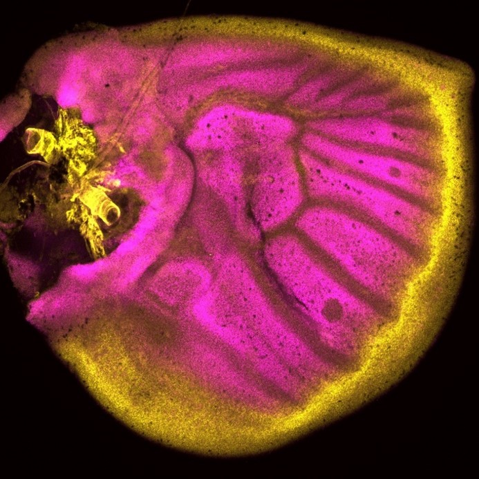
J. C. T. Church spent most – if not all – of his career in East Africa, with some time at Makerere University College Medical School, Uganda, and then at University College in Nairobi, Kenya, where he worked as an orthopaedic surgeon. When not in the hospital, the “enthusiastic teacher” saw it fit to poke holes in the local population of fruit bats (Church and Noronha, 1965). In 1970, JEEM/Development published J.’s latest revelations from the fruit bat Eidolon helvum regarding the ability of skeletal muscle to regenerate following injury. Back then, satellite cells were well known but it remained to be determined whether they were indeed the source of restoring muscle fibre nuclei (myonuclei) during regeneration. Muscle regeneration had also largely been descriptive and, in his publication, Church provided perhaps the first quantitative report of cell populations in the skeletal muscle using electron-microscopy (EM; Fig. 1). Church’s approach was to perform single or repeated muscle ‘crush lesion’ experiments, followed by light and EM imaging, to identify and count the ratio of satellite cells to myonuclei. Although the number of muscle fibres remained stable, Church observed increased satellite cells, fibroblasts and myonuclei during regeneration following both single and double lesions. Overall, this study suggested that satellite cells could provide myonuclei to restore muscle fibres and, importantly, self-renew to sustain the population and regenerative response – a key behaviour of stem/progenitor cells.
Fig. 1. “(A) A light photomicrograph of normal bat web muscle, in transverse section, showing numbers of muscle fibres (mf), nerve fascicles (n), and a satellite cell (S), hardly distinguishable at this magnification, ×450. (B) A light photomicrograph, magnified from Fig. 1A, showing the satellite cell (S), an endomycial fibroblast (f), a myonucleus (m) and a capillary (c). × 1700. (C) An electron photomicrograph of the cells in Fig. 1B, showing the satellite cell (S), fibroblast (f), myonucleus (m) and a pericyte (p) adjacent to the capillary (c). × 4500. (D) An electron photomicrograph, enlarged from Fig. 1C, showing the satellite cell (S) lying under the muscle fibre basement membrane (bm), with its inner plasma membrane lying adjacent to that of the syncytium (pp). × 21500.” Taken from Fig. 1 of Church, 1970.
Let us now leap forward 50 years or so. Fortunately for chiropterophiles, fruit bats are now an uncommon model system for skeletal muscle regeneration research (and regenerative biology in general), with mice and zebrafish a more popular choice instead. It took the invention of inducible genetic labelling and ablation of satellite cells in the 2000s to finally confirm that satellite cells are necessary and sufficient for muscle regeneration (Collins and Kardon, 2021). EM has been complemented by state-of-the-art imaging of genetically manipulated (e.g. transgenic reporter) animals and it’s become increasingly evident that other cell populations, in addition to satellite cells, are needed for regeneration. Whether satellite cells contribute to muscle homeostasis, however, has been far less clear.
Flynn and colleagues’ study (University of Wisconsin School of Medicine and Public Health), published in Development earlier this year (Flynn et al., 2023), comes at a time of some conflicting evidence in the field when the ablation of satellite cells in adult muscle may or may not lead to muscle degeneration – even in the absence of injury. Evidence that muscle homeostasis is preserved when satellite cells are lost suggests that some other stem/progenitor population can contribute myonuclei for tissue maintenance. Using immunofluorescence and genetic labelling, Flynn and colleagues show that a population of Hoxa11-expressing interstitial stromal cells directly contribute to muscle fibres postnatally. Through genetic lineage-tracing experiments, the team confirm that this Hoxa11-expressing population doesn’t go through a satellite cell-like state (i.e. they are not Pax7+) and, therefore, they are an independent progenitor population. In response to injury, Hoxa11-lineage cells minimally contribute to muscle generation (Fig. 2), contributing to the growing body of evidence of satellite cell-independent muscle formation.
Fig. 2. “Hox11 lineage makes minimal contribution to regenerating myofibers after injury.” Taken from Fig. 9. in Corey et al., 2023.
These articles illustrate a joint interest in understanding the cells that give rise to functional muscle. While J. C. T. Church contributed to the knowledge of satellite cells as a stem/progenitor population in regenerating muscle, the search for muscle-generating cell populations still continues many (many) years later, with Flynn and colleagues identifying a whole new population of Hoxa11-expressing cells that maintain muscle homeostasis. Join me again tomorrow for another trip down memory lane, when we look at early studies of wound healing and the important role of the extracellular matrix.
References
J. C. T. Church, R. F. Noronha. The use of the fruit bat in surgical research. East Afr Med J. July 1965; 42 (7): 348-55.
J. C. T. Church; Cell populations in skeletal muscle after regeneration. Development 1 April 1970; 23 (2): 531–537. doi: https://doi.org/10.1242/dev.23.2.531
Brittany C. Collins, Gabrielle Kardon; It takes all kinds: heterogeneity among satellite cells and fibro-adipogenic progenitors during skeletal muscle regeneration. Development 1 November 2021; 148 (21): dev199861. doi: https://doi.org/10.1242/dev.199861
Corey G. K. Flynn, Paul R. Van Ginkel, Katharine A. Hubert, Qingyuan Guo, Steven M. Hrycaj, Aubrey E. McDermott, Angelo Madruga, Anna P. Miller, Deneen M. Wellik; Hox11-expressing interstitial cells contribute to adult skeletal muscle at homeostasis. Development 15 February 2023; 150 (4): dev201026. doi: https://doi.org/10.1242/dev.201026
Do you feel as if a novel imaging approach would give you new insights into your sample or provide a new way to answer your research questions? Imagine you could take your research question to any institute and work with the experts to unlock the power of imaging technologies and get new data and insights. Well, you can – with Euro-BioImaging.
What is Euro-BioImaging?
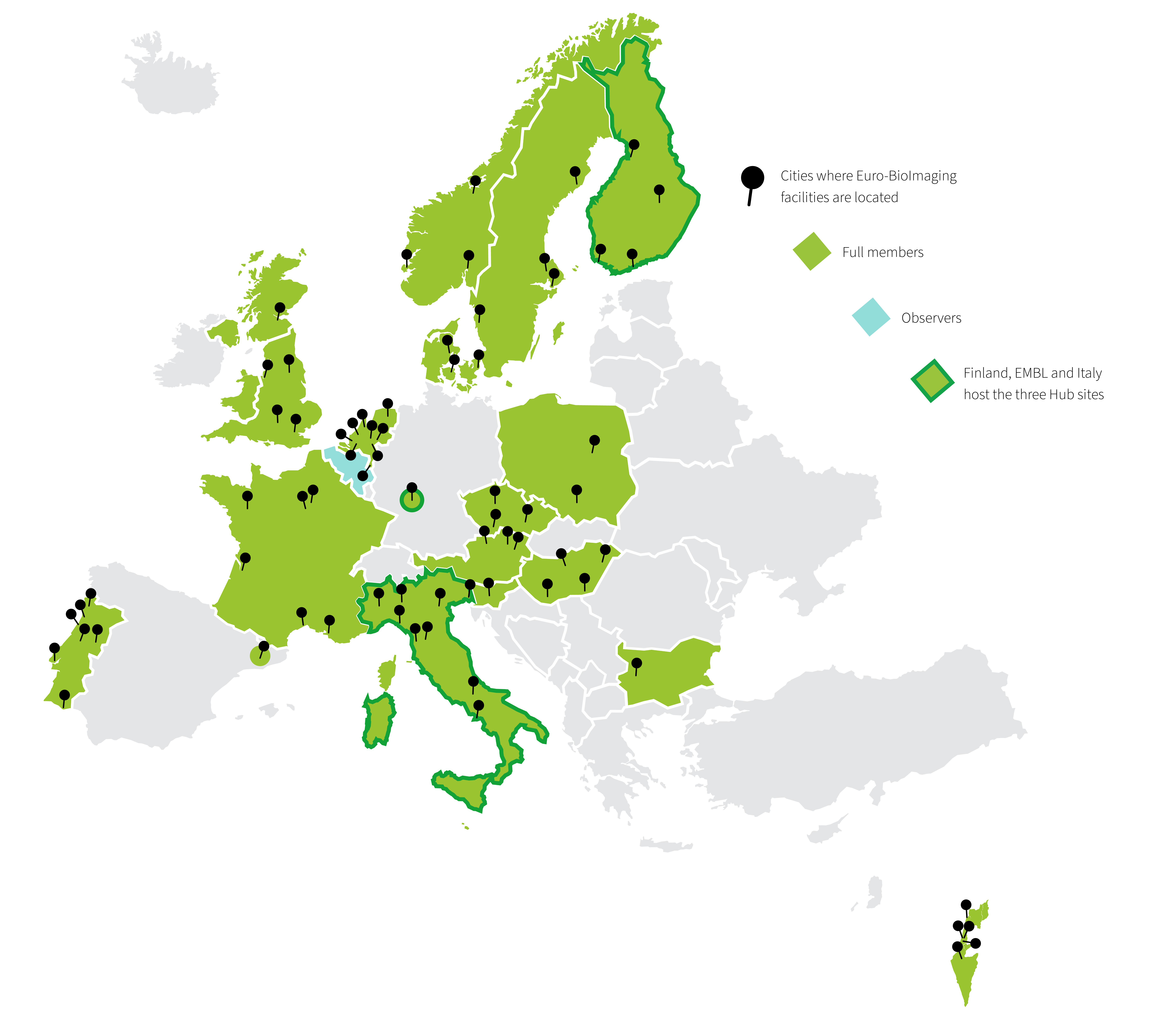
Euro-BioImaging is the European research infrastructure for biological and biomedical imaging that functions as the gateway to over 170 world-class imaging facilities across Europe. Through Euro-BioImaging any researcher, from anywhere around the world, can get access to imaging technologies, image data services and training.
It builds on a set of already existing national and international facilities of excellence in imaging technologies, the Euro-BioImaging Nodes, which provide physical or remote access to imaging technologies, deliver training and support the users at all the stages of their research projects.
Together, over 500 core facility staff work at Euro-BioImaging Nodes and support researchers. We are a fabulous resource for researchers across scale, from atoms to humans, and in diverse disciplines in the life sciences.
Figure 1: Map of Euro-BioImaging member countries, facilities and Hub sites.
How does Euro-BioImaging support researchers?
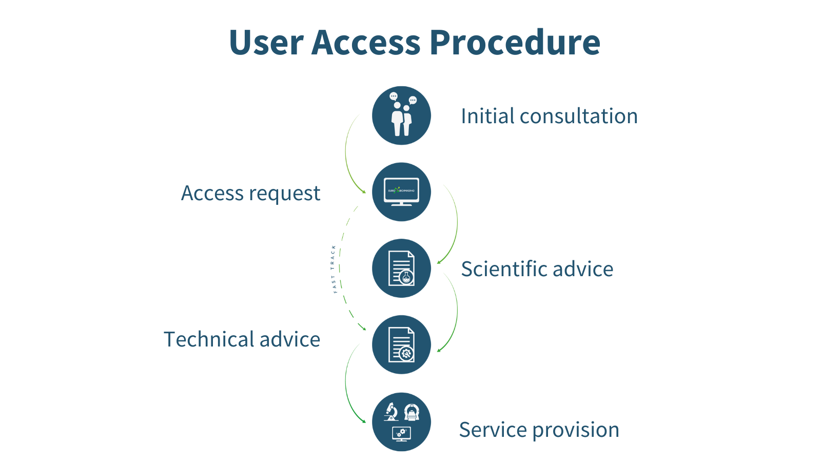
Every researcher, independent of research area, level of expertise, and geographical location, can apply for Euro-BioImaging services whenever they have a project requiring imaging technologies or expertise which they do not have ready access to at their home institute. The expert staff at the Euro-BioImaging Hub can help future users choose the right technology and facility for their research question in the first step of the User Access procedure. Applying for user access is a highly collaborative process in which a researcher has multiple opportunities to hone their experiment and get scientific and technical input from reviewers and technical experts at the imaging facilities before the application is accepted and service provision begins.
Figure 2: The Euro-BioImaging User Access procedure.
An accepted Euro-BioImaging project can be a game-changer. It democratizes access to high-end imaging technologies to push a research question and publication to the next level, and is a starting block towards acquiring new skills, expertise and scientific insight. Euro-BioImaging Users benefit from the expertise of the imaging scientists at the Euro-BioImaging facilities when it comes to sample preparation, experimental set-up and data analysis. Depending on the scope of the project and selected technology, users may also learn skills that they can take back to their home institute.
These skills open doors, especially for early career researchers. Being selected for Euro-BioImaging user access is also a good endorsement of the underlying scientific question or application. Undertaking a project at a Euro-BioImaging facility proves a researcher’s ability to plan and carry out an experiment from start to end.
What imaging technologies are available?
Through the large number of facilities, Euro-BioImaging can offer access to the full range of imaging technologies in the biological and biomedical imaging field. Our technology portfolio covers everything from the nano- to the tissue- and organism scale. We are constantly adding new technologies, making sure that the latest cutting-edge imaging technologies, such as MINFLUX and spatial transcriptomics, are available in open access to all researchers.
Figure 3: The Euro-BioImaging technology portfolio
Harnessing the imaging revolution
The Euro-Bioimaging technology portfolio ranges from light and electron microscopy on the biological imaging side to an expanding range of applications of biomedical imaging, from plant and ex-vivo imaging to animal and human imaging applications.
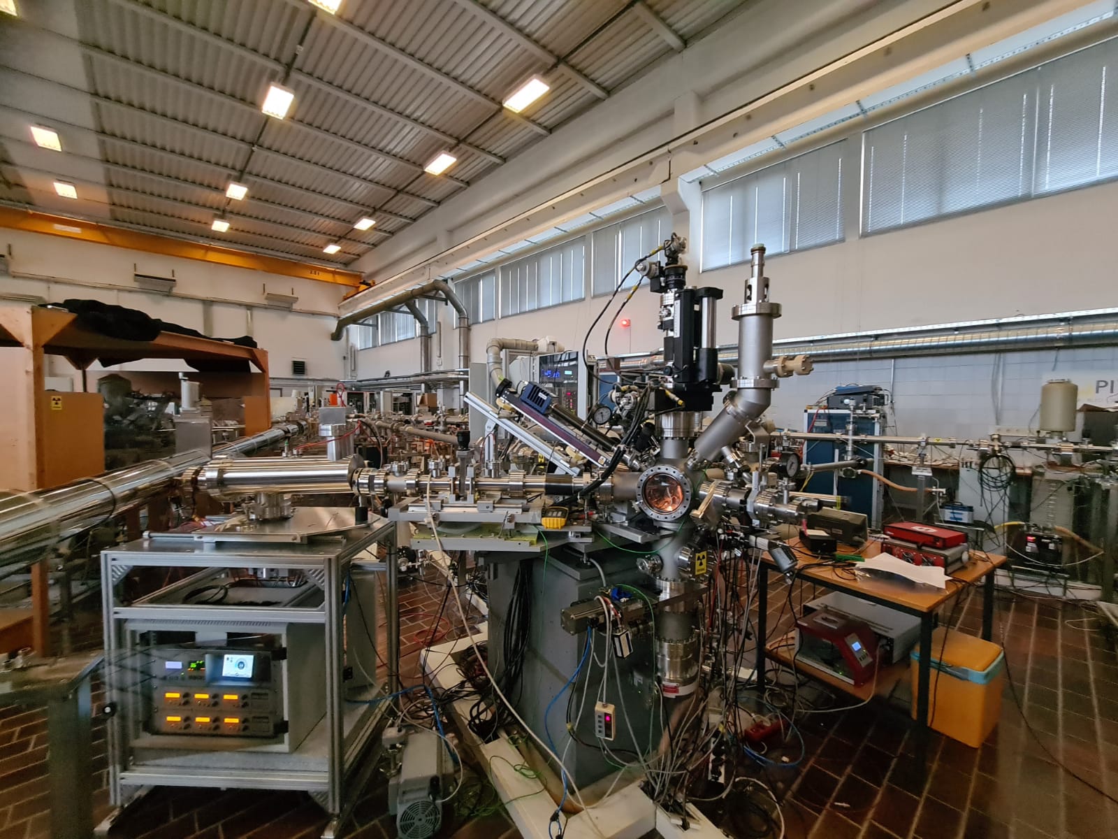
Electron Microscopy
Our Electron Microscopy portfolio covers cryo-EM techniques for ultrastructural exploration, such as cryo-electron tomography (cryo-ET), as well as the full complement of volume EM techniques, such as FIB-SEM, Array Tomography and Serial Blockface SEM. Many of our facilities also specialise in correlative methods, Correlative X-ray Imaging and EM (CXEM) and correlative light and electron microscopy (CLEM).
Light Microscopy
In light microscopy, our Nodes offer everything from basic confocal microscopy up to single molecule location approaches and intravital imaging. Our light microscopy techniques allow for 3D live cell imaging, tracking, high content screening, and include a variety of functional imaging techniques to explore protein dynamics in living cells. Recently we have added a number of new and highly requested methods, such as MINFLUX, Single Particle tracking and Lattice Lightsheet microscopy to our portfolio.
Model Systems
Euro-BioImaging facilities also offer access and support with a wide range of model systems and how to get the best imaging results out of them, from Drosophila and zebrafish to mouse embryos and organoid systems. Here access to instruments is complemented by technical expertise of facility staff, to support specialized sample handling.
Support Technologies
And of course, we also provide access to adaptive and support technologies, such as laser- based microdissection, Feedback Microscopy, high-speed imaging, microscopes at high biosafety levels, and specialized sample preparation methods, such as Tissue Clearing and Expansion Microscopy.
Euro-BioImaging can also support you if you want to explore the physical and chemical properties of your samples, through access to a range of methods such as MassSpec Imaging, Atomic Force Microscopy, and chemical imaging, such as µ-XRF and μ-PIXE.
Figure 4: Micro-PIXE at the Jožef Stefan Institute, part of our SiMBION Node in Slovenia
How will Euro-BioImaging enhance my research?
So, when you read about a cool, new microscopy method in the literature, you can now allow yourself, not just to imagine, but test the impact that method could have on your research question. If it’s a technology that Euro-BioImaging offers, you can always apply. Because the idea behind Euro-BioImaging is to make the best imaging resources available to all researchers, providing new answers to scientific questions and increasing the impact of research.
What about image data analysis?
Image data analysis is an integral part of any experiment and is therefore usually integrated into the experimental concept at Euro-BioImaging at an early stage. Experts at the Nodes help users extract their data and set up image analysis pipelines, typically preparing for image analysis and data extraction, sometimes even before the actual experiment begins. In addition, Euro-BioImaging offers its users Image Data Analysis (IDA) as a stand-alone service through expert Image Analysts at the Nodes, irrespective of where the image data was acquired.
Users can contact our Nodes when they need:
• Biological and biomedical image data analysis support
• Image registration, segmentation, tracking and more
• Data workflows, bespoke analysis tools and machine learning methods
• Access to high performance computing and specialized software
Before you submit a proposal, feel free to browse our technologies and services, view our user stories to get a feel for what Euro-BioImaging can offer, and reach out to us for help in choosing the right imaging method.
What other opportunities are available?
Training
If you’re not ready to undertake a full experiment with Euro-BioImaging, why not start with a training course? Euro-BioImaging Nodes offer a wide range of training courses – covering the full spectrum of biological and biomedical imaging technologies as well as sample preparation and handling, and image data analysis. Some courses are taught remotely and virtually, increasing their accessibility. Taking a course at a Euro-BioImaging facility is a great way to learn a new skill or improve your technique and to build your network. Here’s an overview of training courses available at Euro-Bioimaging Nodes.
Community building
In addition, Euro-BioImaging organizes regular events focusing on imaging for the benefit of the entire community. You are welcome to join our weekly “Virtual Pub” – a free weekly lecture series, open to all imaging enthusiasts. Topics include new biological and biomedical imaging technologies, image analysis, and other topics of interest.
In addition, we organize a “User Forum” twice a year to highlight the importance of imaging to different research areas. These events feature keynote presentations from prominent scientists as well as presentations from users at our Nodes. We usually record them and make the content available on our YouTube channel.
And finally, we are present at many conferences and community events. So we hope to meet with you and talk face-to-face about the wonderful opportunities Euro-BioImaging can provide. Until we meet in person, you can always reach out to us by email info@eurobiomaging.eu or sign up for our Newsletter to stay informed.
In this latest SciArt profile, we talked to Sonhita Chakraborty about her scientific background in plant biology, how science and art blend into each other, and her first solo art exhibition.
Can you tell us about your background and what you work on now? I’ve worn different hats in the field of plant biology for over 10 years now. After completing my PhD in plant molecular biology, I joined the publishing company Elsevier as a scientific editor. I really enjoy reading and thinking about molecular pathways over a piping hot cup of coffee.
Mitodelic – digital illustration of a mitochondriun. This piece was on the ‘Molecular Cell’ journal cover for their special issue on mitochondria
Were you always going to be a scientist? I didn’t think so. After a trip to Disney World, when I was 10, I would tell everyone who would care to listen that I was going to become an animator for Disney. I think I believed it too. In the last year of my high school I was convinced to pick a more “practical ” career choice and pursue art as a hobby. Out of fear of missing out on opportunities I chose to rendezvous with biology instead of art.
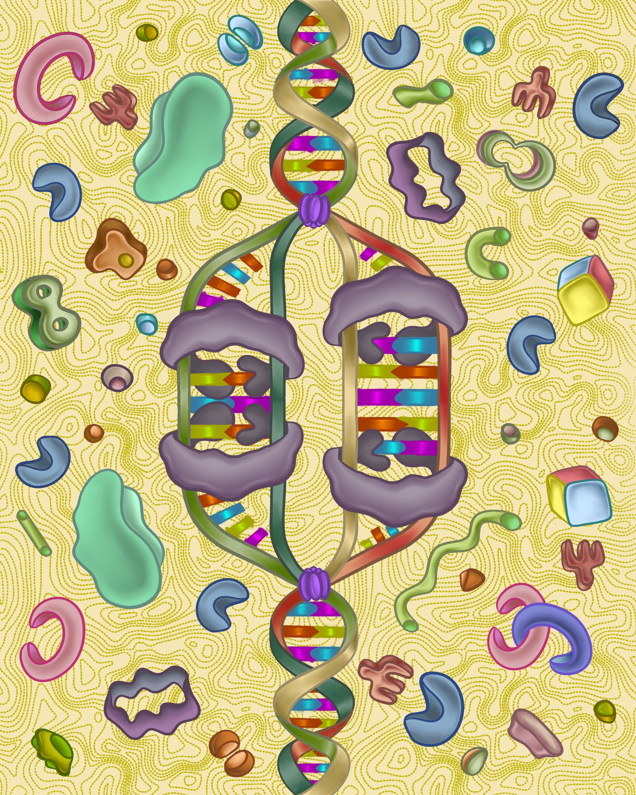
Replication Dream – An artistic and whimsical representation of DNA replication
And what about art – have you always enjoyed it? People often ask me when I first started making art and that’s hard to pinpoint. The happiest memories of my childhood are when I was making art. Some of my early “masterpieces” were “frescos” that I painted as a toddler onto the underside of my parent’s dining table. Stationary stores and art supplies will always delight me.
What or who are your most important artistic influences? I have been spellbound by David Goodsell’s paintings of cells from the get go. On Instagram I’ve come across a cornucopia of very talented and imaginative scientists, artists and science artists who constantly inspire me. I’ve also noticed that feeling grounded and connected with nature can really stoke my creative flame.
Optic Fascination – digital illustration and tribute to the founders of the microscope
How do you make your art? Right now I mostly make art on my iPad but up until 2-3 years ago I used to water colour. I miss the feeling and urgency of pushing wet paint around on a page but digital art let’s me do so much more and explore other artistic aspects of myself.
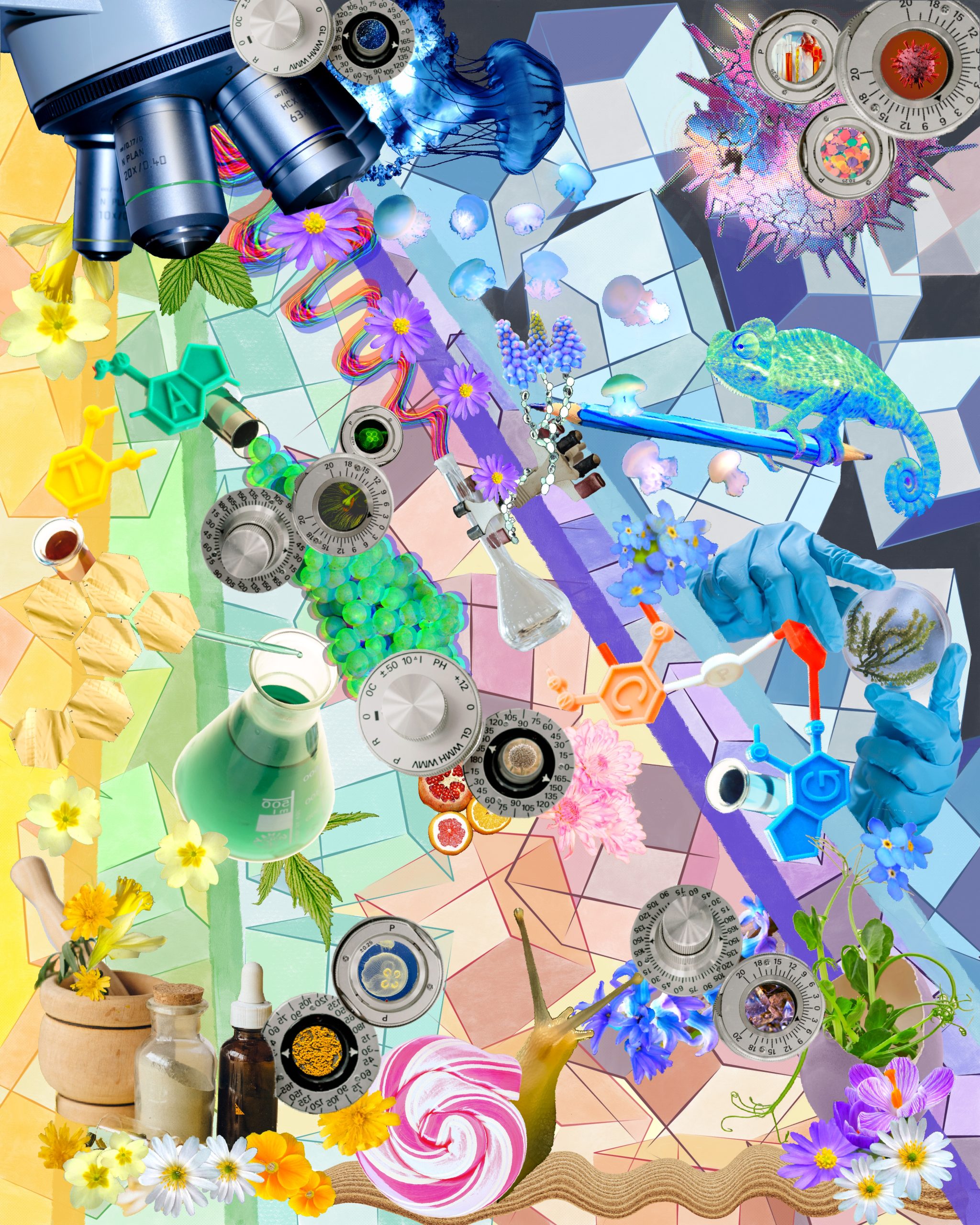
Big Science – digital collage from publicly available images
Does yourart influence your science at all, or are they separate worlds? That’s a great question! Science and art are definitely not separate in my books and they blend into each other. The amazing science I read about at work gives me a lot of interesting ideas, some of which have become journal cover art. My art doesn’t seem to influence my science (yet). I’ll be starting a postdoc in the fall so I’m not sure what sort of artistic inspirations I will draw from my science (and vice versa) – I can’t wait to find out!
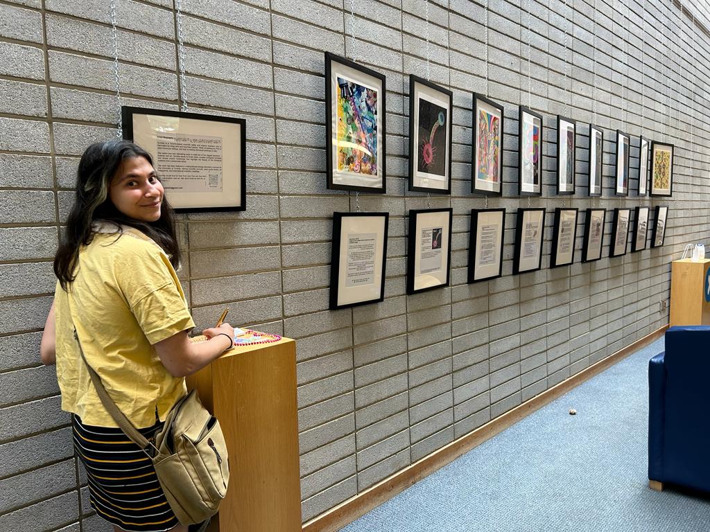
What are you thinking of working on next? I just had my first solo art exhibition at the local library. At work I’m designing a few art covers for different journals. I’m also illustrating for a biology student group at the North Carolina State University. I have an endless list of personal artistic projects I’d like to make time for so I won’t be running out of projects to work on anytime soon.
First exhibition hosted by the Toronto Public Library
In some ways, it’s difficult to imagine a better model of regeneration than the planarian flatworm. These comical, googly-eyed creatures have captivated scientists for centuries with their remarkable regenerative potential (Dalyell., 1814; Abeloos, 1930). Famously, planarians can be cut into small fragments, each of which is capable of regenerating the full body, (including the brain, eye-spots and intestine), an ability owed to the distribution of potent adult stem cells, known as neoblasts, which are distributed throughout the body and ‘hyperproliferate’ following amputation (Ivankovic et al., 2019; Lee et a., 2022). In this first instalment, I discuss the highlights from the following two articles:
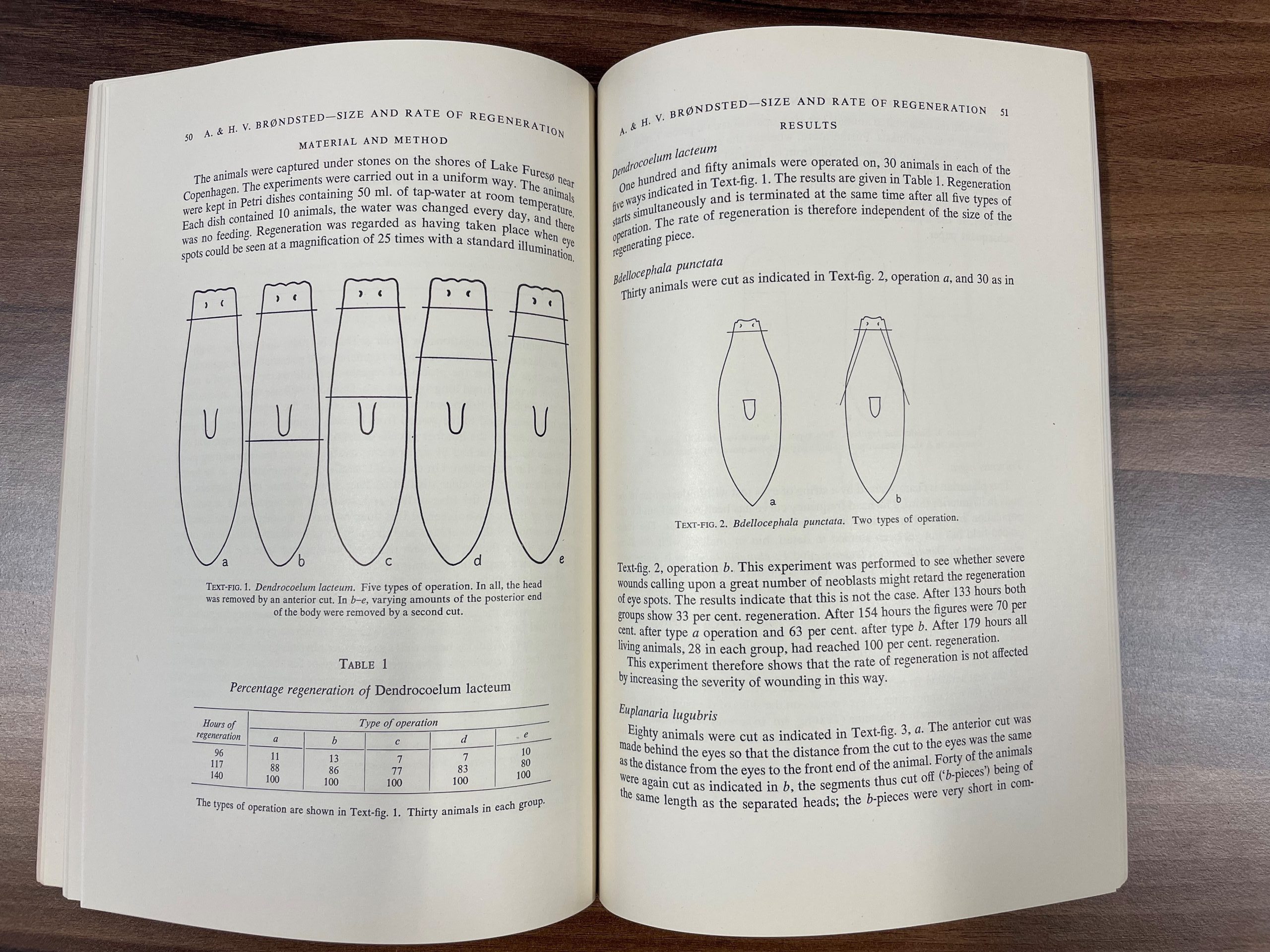
Danish couple, Agnes and Holger Brøndsted, married in 1919 and had the culmination of their work studying planarian fragment size regeneration published in March 1954, in the first issue of the second volume of JEEM/Development (Brøndsted and Brøndsted, 1954). At this time, the regenerative ability of planarian fragments was well established and the presence of neoblasts was accepted. However, Agnes and Holger’s article came at a time of confusion in the field, when conflicting evidence made it unclear whether the number of cells in each fragment contributed to the speed at which the fragment regenerated. They set out to address this question with the minimalistic approach of cutting animals into fragments of different sizes and measuring the length of time eye-spots took to regenerate. Their work began by circling the shores of Lake Fures near Copenhagen, Denmark, lifting rocks to collect planarians from the wild. They ended up with a collection of a few hundred animals from four different species: Dendrocoelum lacteum, Bdellocephala punctata, Euplanaria lugubris and Polycelis nigra. The authors highlight that these four species do not reproduce asexually by fission, removing a confounding variable from their studies. For reasons, I admit, aren’t so clear to me, they dissect each of the species in different ways to produce fragments of different sizes (Fig. 1). However, regardless of the size or shape of the fragment, the rate of regeneration was the same, showing conclusively that the number of cells (and thus the number of neoblasts) in the fragment does not contribute to regeneration speed, suggesting that neoblasts are unlike to migrate within the fragment.
Fig. 1. Agnes Brøndsted, H. V. Brøndsted; Size of Fragment and Rate of Regeneration in Planarians. Development 1 March 1954; 2 (1): 49–54. doi: https://doi.org/10.1242/dev.2.1.49
Around 70 years later, planarians are still a potent and popular organism for studying regeneration; however, the variety of species used as models has largely converged on two species: Schmidtea mediterranea and Dugesia japonica. Significant technical advances, not least of which the culture of planaria in the lab, have allowed researchers to understand the cellular and molecular mechanisms of stem cell behaviour during regeneration and mean that hunting for critters under rocks is now a pastime, rather than a scientific necessity.
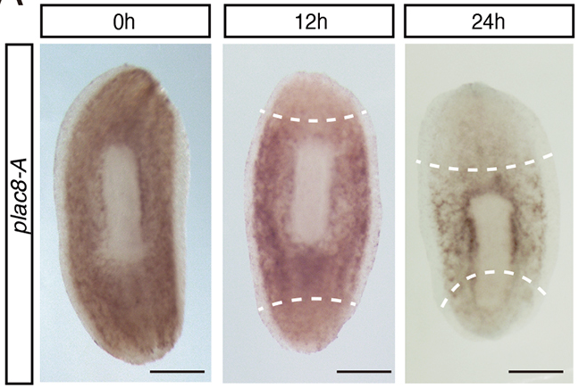
Last year in Development, Hayoung Lee (Kyoto University) and colleagues capitalised on modern innovations, such as markers for neoblasts and genetic knockdown experiments, to understand the role of a specific gene, plac8-A, during regeneration in D. japonica (Lee et al., 2022). Unlike the species used by the Brøndsteds, D. japonica can reproduce asexually via fission. The authors of the present study employ this fission phenomenon as a measure of cell proliferation and show that knockdown of plac8-A increases the number of fission events. Using imaging and expression analyses, Lee and colleagues demonstrate that plac8-A is expressed by neoblasts and is downregulated before the neoblasts proliferate (Fig. 2). Finally, through chemical treatments, Lee and colleagues show that ERK signalling acts upstream of plac8-A and inhibits plac8-A expression during regeneration via the JNK signalling pathway. Together, these findings demonstrate that plac8-A acts as a molecular switch that regulates neoblasts’ regenerative behaviour.
Fig. 2. Expression pattern of plac8-A mRNA during early regeneration determined by whole-mount in situ hybridization. Taken from Lee et al., 2022.
Together, these two articles address a similar question: What influences neoblast proliferation during planarian regeneration? The Brøndsteds show that, following amputation, not all cells hyperproliferate and that the quantity of cells doesn’t regulate this process, while Lee and colleagues determine that plac8-A must be downregulated in order for neoblast hyperproliferation to occur and loss of plac8-A can induce neoblast proliferation across the whole organism. I hope you enjoyed the post and join me again tomorrow when we will be revisiting the archives to look back on skeletal muscle regeneration.
References
M. Abeloos. Recherches expérimentales sur la croissance et la régénération chez les Planaires. Bull. biol. 1930; 64, 1–140.
Agnes Brøndsted, H. V. Brøndsted; Size of Fragment and Rate of Regeneration in Planarians. Development 1 March 1954; 2 (1): 49–54. doi: https://doi.org/10.1242/dev.2.1.49
J. G. Dalyell. Observations on Some Interesting Phenomena in Animal Physiology, Exhibited by Several Species of Planariae: Illustrated by Coloured Figures Of Living Animals. 1814. Edinburgh: Archibald Constable & Co.
Mario Ivankovic, Radmila Haneckova, Albert Thommen, Markus A. Grohme, Miquel Vila-Farré, Steffen Werner, Jochen C. Rink; Model systems for regeneration: planarians. Development 1 September 2019; 146 (17): dev167684. doi: https://doi.org/10.1242/dev.167684
Hayoung Lee, Kanon Hikasa, Yoshihiko Umesono, Tetsutaro Hayashi, Kiyokazu Agata, Norito Shibata; Loss of plac8 expression rapidly leads pluripotent stem cells to enter active state during planarian regeneration. Development 1 February 2022; 149 (3): dev199449. doi: https://doi.org/10.1242/dev.199449
The field of regenerative biology has grown considerably since the millennium and, with the creation of the International Society of Regeneration Biology a couple of years ago (Poss and Tanaka, 2021), you’d be forgiven for assuming that it’s a relatively modern field. However, a quick peak through the archives of Development, or the Journal of Embryology and Experimental Morphology (JEEM) as it used to be known, demonstrates that regeneration was – and is – a key focus in the journal since its conception in 1953.
In honour of the inaugural ISRB meeting starting today, this one-week series will take a retrospective look back through some of the earliest regeneration articles published in Development, comparing the research questions, approaches and technologies to more recent publications.
Here are the posts in the series:
Planarians aplenty Learn about a Danish couple that enjoyed long walks on the beach collecting flatworms and the work of Hayoung Lee, Kiyokazu Agata, Norito Shibata and colleagues.
Muscle memory lane We meet East Africa-based chiropteran-crusher, J.C.T. Church, and take a whistle-stop tour through the work of Corey Flynn, Deneen Wellik and colleagues.
Time heals all wounds In this collagen-centric third instalment, we discuss the work of amateur guinea pig tattoo artists, together with Filipa Simões, Paul Riley and colleagues’ study of cardiac regeneration.
A budding tale Introducing zoologist, engineer, Lieutenant and author David Newth, and his work on epimorphic tail regeneration, complemented by recent studies by Momoko Deguchi, Taro Fukazawa and Takeo Kubo.
Hands-on hard graft We revisit Dr. D.R. Newth’s newts and their mysterious limb regeneration abilities, compared with Takashi Takeuchi, Haruka Matsubara and colleagues’ modern perspective.
Go fish The last post says “goodbye and thanks for all the fish”, featuring work from a Nobel Prize winner and Lili Zhou, Ken Poss, Massya Mollaked and colleagues.
The archive at The Company of Biologists offices. (No Ratings Yet) Loading...
With the recent advances in human stem cell-derived embryo models, a team of researchers have suggested that perhaps it is time to redefine what a human embryo is.
Good summary of our piece! In response to some comments, I would also note that we're not suggesting 'changing' the definition of the embryo per se, but rather, unifying the plethora of existing definitions, while additionally stressing that embryos can be made several ways… https://t.co/NqWtYbAyaF
A featured preLight is on the preprints ‘Cell polarity linked to gravity sensing is generated by protein translocation from statoliths to the plasma membrane’ by Takeshi Nishimura et al. and ‘Amyloplast sedimentation repolarizes LAZYs to achieve gravity sensing in plants’ by Jiayue Chen et al.
News from the community
Learning from our friends over at ZebrafishRock, the Node would like to be more intentional in celebrating the various achievements of people in the developmental and stem cell biology community. We have trawled through social media (which is a bit all over the place nowadays) to look for any relevant news in the past month, but fill in this form if you know someone who deserves a mention, and we’ll consider sharing the piece of news in the next installment of ‘Developing news’.
Fantastic #PhD defense by @FriedaLeesch@PauliGroup on the 'characterization & function of maternal ribosomes during development'! As a mentor, these are the days I cherish most – to see a student grow & develop into a mature, independent scientist! Super well done, Dr. Leesch! pic.twitter.com/T96Cnzr9Bn
since i sort of buried the lede in the thread linked here (seriously, go read it. it's way more important than this tweet), i'm proud to say i've been promoted to associate professor. but don't worry, i'm still the same old ass prof. :)https://t.co/0rA74vObsupic.twitter.com/Rs90wUkwYm
— Tim Mosca (he/him) (@drosophilosophy) July 23, 2023
Awards:
We are so happy to add our appreciation and congratulations to Sally Moody for the Lifetime Achievement Award! Thank you for everything you’ve done and all the frogs you’ve wrangled! ❤️🐸 pic.twitter.com/IQYoRmIcU5
Learning from our friends over at ZebrafishRock, the Node would like to be more intentional in celebrating the various achievements of the wonderful people in the developmental and stem cell biology community.
In the latest ‘Developing news‘, we have trawled through social media (which is a bit all over the place nowadays) to look for any relevant news in the past month — newly minted PhDs, promotions, awards — but we know we’ve definitely missed some. That’s why we want to hear directly from you!
If you know someone who deserves a mention, fill in this form, and we’ll consider sharing the piece of news in the next installment of ‘Developing news’.
A recent paper in Science Advances titled ‘Spatial and temporal regulation of Wnt signaling pathway members in the development of butterfly wing patterns’ explores the expression and function of Wnt signaling pathway members in setting up butterfly wing patterns. We caught up with first author Tirtha Das Banerjee and corresponding author Antόnia Monteiro from the National University of Singapore to learn about the behind the paper story.
Antónia Monteiro and Tirtha Das Banerjee
What was known about Wnt signalling and the butterfly wing patterning before your work?
Tirtha: Wnt signalling is a fundamental signalling pathway that regulates cell communication, cell growth, and cell proliferation in metazoans. A lot is known about this pathway in classical model systems such as Drosophila, but little is known in other systems, such as in butterfly wings. In butterfly systems a few Wnt ligands, primarily WntA and Wnt1, had been associated with the development of bands and eyespots, but most of the other ligands in the pathway, and their receptors, had not been examined in any species.
Antónia: Back in 2006, in my lab at Buffalo, we had visualized Wg/Wnt1 (using an antibody against the Wnt1 protein in humans) at the center of butterfly eyespots, but it wasn’t until we were able to produce a transgenic line, expressing two copies of wg back to back, that folded upon each other when transcribed, that we were able to knock-down this gene to observe its effects on eyespots. A graduate student in my lab in Singapore, Nesibe Özsu, worked on this, and she was able to see smaller eyespots developing when the double stranded RNA was transcribed inside cells via a heat-shock. Tirtha, however, used CRISPR to try and get stronger phenotypes on the wing.
How did this project get started? And Tirtha, what brought you to Antónia’s lab?
Tirtha: This work was partially inspired by my previous work on venation patterning published in Development in 2020 where I observed a very dynamic pattern of Armadillo (Arm) in the larval wings of butterflies. Since Arm is an important component of Wnt signaling, I hypothesized that the ligands (the Wnts) and their receptors (the Frizzleds) might also show dynamic patterns of expression as the wing develops. I started examining their expression, one by one, and uncovered that these other components of Wnt signaling are also extremely dynamic across both larval and pupal wings.
I visited Antónia’s lab back in 2014 during a summer internship program from my graduate studies at NIT-Durgapur. Back then, I visualized the expression of two transcription factors, Engrailed and Spalt, using antibodies that produced some interesting patterns. Even though these early stains were extremely blurry and saturated with colors, they were super cool to me. I proposed an hypothesis for butterfly venation patterning based on the data but due to the limited time of my internship (of 2 months), I was unable to continue the work. Later after graduation I continued this work, which later got published. I was hooked into the evo-devo of wing vein (and color) patterning ever since.
Why did you choose the butterfly wing as a model to undercover the complexities of Wnt signalling?
Tirtha: Developing butterfly wings are simple 2D sheets of cells where numerous ligands, receptors, signal-transducers, and transcription factors orchestrate the specification of extremely complex patterns (of colour) that will be visible in the adult wing. Cells send and receive cues, and interact with each other during development, to specify these colour patterns. Since larval and pupal wings are miniature versions of adult wings, it is easy to map the molecules involved in these signalling processes to the final colour patterns they are likely affecting.
Antónia: I started working on butterfly wing patterns as an honours student in 1990. I thought butterflies would make great genetic and developmental models because they can produce a ton of eggs and the wings are large and flat, like Tirtha said. In addition, they were much prettier than Drosophila, and their intricate colour patterns were the real hook.
Can you summarise your key findings?
Tirtha: In our recent work we found that the expression and function of different Wnt signaling members varies quite a bit during wing development. For example, Frizzled4, one of the Wnt receptors, is expressed quite uniformly during larval development, but is missing from the future eyespot centers, where canonical Wnt signaling, mediated by Armadillo, is taking place. During the pupal stage, however, the expression of Frizzled4 completely changes and now it is co-expressed in the eyespot centers together with Armadillo. During these two stages Frizzled4 is also likely playing different roles: it is involved in the localization of the eyespot centers during the larval stage because its removal leads to eyespot center duplications, and it is involved in wing scale orientation during the pupal stage. We observed similar dynamics for many other genes we tested in the study and proposed mechanisms of how these genes are likely interacting to specify the multiple cell fates on the wings of butterflies.
Antónia: A key realization for me was observing that a patchwork of different Frizzled ligands and receptors is expressed across the whole wing, which means that every cell of the wing is either producing or receiving Wnt signals, but processing them in different ways. How this patchwork of different Wnt ligands and receptors work together will be interesting to investigate in future.
Expression of frizzled4 (magenta) and frizzled9 (yellow) in the larval wing of Bicyclus anynana butterflies.
Did you have any particular result or eureka moment that has stuck with you?
Tirtha: Well one of the exciting moments was when I observed the armadillo CRISPR phenotype showing a double eyespot on the wing. I was also very excited the day I observed the different frizzled patterns on the pupal wings. It made me wonder how nature, over the course of evolution, has been intricately patterning wings by activating certain genes at some location while repressing them in other locations. It’s really incredible, and makes you sit down and think how things which we consider simple are so complex and elegantly tuned at the molecular level.
And the flipside: were there any moments of frustration or despair?
Tirtha: Ohh there were many. As a scientist I believe we all have accepted that there will be more moments of frustration than excitement. For example, for one of the Wnt ligands called Wnt1, I tried to knock it out with over ten different CRISPR guides, and injected over 5000 embryos. I got no results. Doing stainings with the traditional enzymatic in-situ hybridization was also a painstaking job. I am glad at least those days are over with the new HCR technique we have adapted in the lab.
A recent Development paper also used CRISPR to look at WntA and Frizzled receptors in the butterfly wing patterning. How does the two papers complement each other?
Tirtha: The study from Arnaud Martin’s lab is extremely impressive. Their lab has consistently produced papers that have advanced the field of biological colour pattern evolution. The authors have generated a massive amount of information in different species of butterflies on how WntA is likely being transduced via the Frizzled2 receptors. They have also gathered functional data on other receptors such as frizzled, frizzled3, and frizzled4 patterning different aspects of the adult wing scales and venation which are not present in our work. The use of RNAi as a gene knockdown methodology they used would also be extremely useful for other labs working on similar lepidopteran tissues. The gene expression data from their lab and our lab confirms the presence of the different receptors during larval and pupal stages in similar conserved domains strengthening our hypothesis that these receptors are involved in patterning adult wings across butterflies.
Where will this story take the lab?
Antónia: Well, we have only touched the tip of iceberg with the present study. This work has opened up many new avenues for new students to work on. For example, a student in the lab is now testing a hypothesis we proposed on the interaction of Frizzled4 and Arm in larval wings and the function of frizzled2 and frizzled9 in the development of scales and colour patterns. Another student is testing where the rest of the Wnt signalling members are expressed and what function they play. A postdoc in the lab is trying to visualize whether there is a Wnt1 morphogen gradient around the eyespot centers, as hypothesized nearly 45 years ago.
Tirtha, what’s next for you?
Tirtha: Well, I am currently involved in the development of more advanced technologies for spatial transcriptomics that will allow us to multiplex the number of genes we tested in these studies. Basically, we would be able to understand how perturbations to individual members of Wnt signaling affect the expression of other Wnt pathway members or downstream targets. I hope the upcoming work will have broad applicability across different model systems.
Our Congenital Anomalies Cluster invites you to join them for an exciting hybrid event at the Advance Training Centre at MRC Harwell on the 23rd and 24th of November.
The meeting title ‘From Cardiac Gene Variant to Mouse Model’ is a good summary of the aims of this informal meeting which are to:
Create a framework for accurately modelling and phenotyping human congenital heart defects in mouse.
Link human and mouse phenotyping.
The target audience is varied and expected to comprise clinicians, clinical geneticists and developmental biologists interested in finding new genes for congenital heart defects and validating them in mouse models.
For more information and to register for the meeting please download our programme
 (No Ratings Yet)
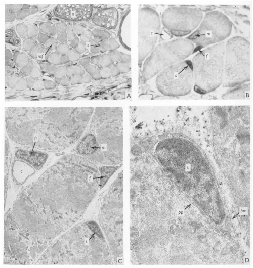

 (2 votes)