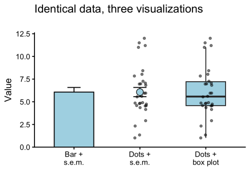
Thanks to community campaigns (#barbarplots) and opinionated papers (Drummond & Vowler, 2011; Weisgerber et al 2015) the dynamite plunger plot (a bar plot together with an error bar) has been abandoned as the default graph. The main reason to reject bar plots is that they display only an abstraction of the actual data and therefore oversimplify it. For full transparency and interpretability, the all data should be displayed. This can be achieved effectively by displaying the data as dots (or other symbols).
Figure 1: Identical data, visualized in three different ways. The bar plot conceals the data and is an oversimplification. Showing all observations as dots improves transparency. The data-points can be accompanied by statistics, e.g. mean and standard error of the mean (s.e.m.) or a box plot.
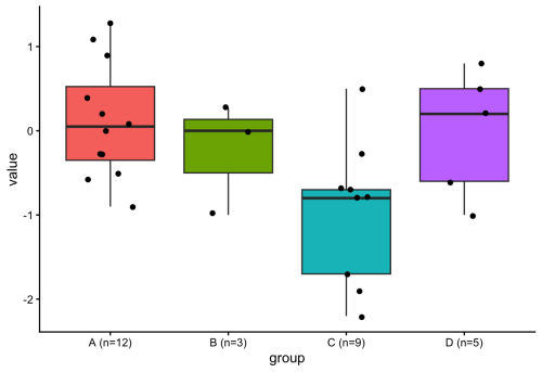
The dot plots are often accompanied by a graphical statistical summary. Common statistics are the mean or median. A more comprehensive statistical summary is provided by the box plot. The box plot was first proposed by Mary Eleanor Spear in her book “Charting Statistics” and publicised by the work of John W. Tukey. Open source tools, such as the user-friendly web tool BoxPlotR, have contributed to a wider adoption of box plots in publications. The box plot is characterised by 5 values, the median, the two borders of the box that indicate the IQR, and two whiskers. The whiskers can reflect multiple things, but most commonly indicate the most extreme data-point that is maximally at 1.5 x IQR from the border of the box (Krzywinski & Altman, 2014).
Figure 2: Four conditions with varying numbers of observations. When n is 5 or smaller (conditions B & D), the box plot does not add any information.
Since a box plot summarises the data distribution with 5 values, it does not add any information when the data consists of only 5 or less points. This can also be seen in figure 2 for conditions B & D. Adding a box plot to a condition that has only 5 datapoints would be similar to adding the mean for only 1 datapoint. Since some datasets have variable numbers of observations per condition, it would be ideal to only display the box plot when sufficient observations (n>5) are present. To do this in R with {ggplot2}, I considered defining a new geom (if you are interested in that, I recommend this tutorial), but then I realized that it can be done by filtering the data within the box plot function (inspired by the work of June Choe on {ggtrace}, see also this video: https://youtu.be/dUBnitXf5mk).
The trick is to use a filter() function within the geom_boxplot() definition to keep only the conditions for which n>5 (aggregated for each condition by group_by(group)). Here’s the R code:
#Filtered box plot, only drawing a box for conditions that have n > 5
ggplot(demo_data, aes(x = group, y = value, fill = group)) +
  geom_boxplot(
    data = ~ .x %>% group_by(group) %>% filter(n() > 5)
  ) +
  geom_jitter(width = 0.2, size = 1.5) +
  theme_classic() +
  theme(legend.position = "none")
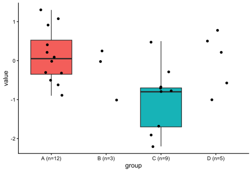
The resulting plot only shows a box plot when n>5:
Figure 3: Similar to figure 2, but the box plot is only shown for conditions where n>5.
The use of a filter() function within the definition of a geom is an elegant method for getting rid of the box plot when the number of observations is too low. In general, this approach is very powerful and gives more control over plotting with ggplot2. There’s probably a ton of other applications, and one that comes to mind is to filter data based on some criterion and changing the color, e.g. for outliers. And, fun fact, Figure 1 was also created using the filter() function. Check out the R code (for all plots) here: https://github.com/JoachimGoedhart/Unboxing-data
by Irene Amblard, Thamarailingam Athilingam, Alejandra Guzman-Herrera,Dimitra Mouzourou, and Andrew Plygawko
As members of the British Society for Developmental Biology, its annual meetings are always a highlight. This year’s ‘from Molecules to Morphogenesis’ meeting was held at University of Warwick in March, bringing together an inspiring range of talks spanning classical and modern developmental biology as well as a wide range of model systems.
Day 1: Seven Decades of Developmental Biology
On the first day we were all welcomed by a heartwarming introduction from Marysia Placzek (BSDB chair) talking about the history of the BSDB and the pioneer work of researchers that have shaped this society since the beginning, such as Philip Ingham who was present in the audience. The first session thus commemorated Seven Decades of Developmental Biology with a great line up of invited speakers highlighting the power of revisiting classical developmental questions with new technologies.
Starting with Denis Duboule remarkably showing in vitro embryo models retaining axis elongation and A-P polarity despite removing Hox gene function but showing defects in endodermal and mesodermal fate. Ruth Lehmann talked about the role of Nanos in early specification and maintenance of the Primordial Germ Cell (PGC) program in Drosophila embryos. Cliff Tabin added an evolutionary perspective on duck syrinx formation and morphological asymmetry through different types of Shh gradients. Yun Xia gave a fascinating talk on using ‘assembloids’ to study the interplay of cell repertoires in kidney development and disease progression. After a short coffee break, Alicia Hidalgo spoke about Toll signalling in brain plasticity and degeneration in flies. A standout talk for me was Norbert Perrimon’s on a decade long effort (also in flies!) to lay a framework of metabolic crosstalk between growing tumours and the host tissues that result in cachexia, a complex wasting syndrome. Andy McMahon described the underlying principles to direct iPSCs and generate human kidney organoids.
The last talk of the day was Valentina Lorenzi from the Wellcome Sanger Institute, who received the Beddington Medal for her impressive PhD work characterising spatiotemporal developmental trajectories of the human reproductive tract using cutting-edge technologies, but also for her work advancing and communicating women’s health as the former president of the Cambridge Femtech Society and through a collaborative zine called ‘Pelvic Matters’.
The Annual General Meeting followed, where all BSDB members get to hear from the committee what the society has been doing for the past year. All members also vote on changes to the constitution and on new committee members. The day ended with welcome drinks and dinner to encourage networking across all attendees.
Day 2: Cell Identity & Gene Regulation, Patterning & Morphogenesis
The first session of day two on Cell Identity and Gene Regulation, chaired by Vicki Metzis, began with Edith Heard on the dynamics of X-chromosome inactivation (XCI), followed by two short talks from Amruta Vasudevan, examining a Wnt/Nodal/Notch temporal module underlying symmetry breaking in mouse gastruloids, and Oliver Davis, highlighting the role of FoxG1 in regulating chromatin accessibility and neural fate in cerebral organoids. Joshua Brickman talked about using in vitro models to study chromatin remodelling and Sox2 activity. Following a short break and snacks, the session continued with Hilary Ashe, discussing ribosomal pausing and protein synthesis control in Drosophila embryos, and two more short talks. Christos Kalaitzis on the Hox code mediating regional diversity of vagal neural crest cells, and Daniel Goszczynski on dual developmental origins of mammalian PGCs.
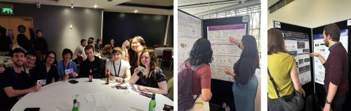
There were also two rounds of flash talks from poster presenters, spanning varied topics including nuclear mechanosensing, transgenic avian lines, cell size scaling, cell morphology and more. During lunch time, the poster session provided a fantastic opportunity for delegates to dive deeper into the science, spark new conversations, and discover exciting work being done across the community. With packed rooms, lively discussions, and an exceptionally high standard of posters, the poster sessions were a highlight of the meeting.
The afternoon session covered Patterning & Morphogenesis under the lens of model organism diversity, chaired by Shankar Srinivas. Ranging from Drosophila to Arabidopsis, this was a unique opportunity to learn about how very distinct systems develop and shape their body plan or organs (e.g. mouse heart, fly blastoderm and epithelium, chick and shark telencephalon, mayfly eye, marsupial trunk, plant roots!). Several talks also highlighted the question of developmental timing. Kenzo Ivanovitch showed how early versus late primitive streak progenitors make distinct parts of the heart. Sergio Menchero from the Crick Institute gave the Dennis Summerbell Award lecture, showcasing his study on temporal diversity in marsupials’ developmental programs and how they prioritise differentiation of necessary structures for their survival. Dana Fakhreddine presented her work identifying heterochronic differences in molecular events shaping the distinct telencephalon identities. Erik Clark refined the classical morphogen-gradient models by adding a temporal lock mechanism to explain how striped expression is appearing in the fly embryo. Lastly, Bert De Rybel presented work from his lab on Arabidopsis root meristem, introducing the idea of developmental timers during development and aging.
After dinner two medal winners were announced. First, the Wolpert Medal was awarded to Prof. Neil Vargesson from U. of Aberdeen to recognise his outstanding contributions to our understanding of chemically induced birth differences for the past 20 years. Neil’s work has led to changes in health policies and help for affected families in the UK and abroad. His commitment to public engagement through numerous outreach activities has raised awareness of developmental biology and medicine safety. Next, the Waddington Medal lecture by this year’s awardee, James Briscoe from the Crick Institute. James’ fundamental discoveries have shaped our understanding of how morphogens work. He has also promoted developmental biology through numerous roles, including as Director and then Editor-in-Chief of the journal ‘Development’ from the Company of Biologists.
To end the evening, we had a Student/Postdoc ECR social event that lived up to expectations in every way. A science-themed pub quiz drew 120 attendees into teams that brought their competitive best. The questions ranged from easy to tricky and delightfully niche, a favourite was ‘when dealing with chick development, what does YSL stand for?’ and no, the answer is not Yves Saint Laurent but Yolk Syncytial Layer. Spirits were high and competition was tough, but one team came on top: ‘Shut up nurds!’ (a nod to Joshua Brickman’s talk). Perhaps the most creative team name came second, ‘Bad Scientists Doing Biology’ or, of course, B.S.D.B., and in third place we had the ‘Mighty-Chondria’ team. A great way to end day two!
Day 3: Cell Communication & Fate, Organogenesis & Regeneration, Conference Dinner
Wednesday morning started with the Cell Communication & Fate session, chaired by David Turner. This series of talks opened with Sally Lowell summarising elegant work from her lab generating new tools to track the neighbourhood of a cell and discussed how these tools can be applied to diverse model systems. Following this, a series of fascinating talks covered distinct systems where cell fate is controlled by intrinsic mechanisms, such as post-transcriptional modifications shaping neural crest fate as presented by Lara Busby, and extracellular cues provided by the environment, such as mechanical cues generated by neighbouring cells. On this topic, Stefan Harmansa presented his work on how mechanical forces shape epithelium architecture in the drosophila wing disk by combining experimental and modelling approaches. Vikas Trivedi shared recent work from his lab investigating the role of temperature on the shape of the zebrafish epithelial layer during gastrulation. Nine flash-talks concluded this session by covering a diversity of questions, ranging from the impact of metabolic alterations to the role of cellular geometry.
After lunch and the second poster session of this meeting, we returned for the afternoon session on Organogenesis & Regeneration, chaired by Timothy Saunders. This session underscored how the study of diverse systems furthers our general understanding of morphological and regenerative processes. Beautiful work from Emily Noël focused on the importance of ECM asymmetry in shaping the developing zebrafish heart. Cristina Newnes on muscle development in Drosophila embryos, showed the power of established model systems. Meanwhile, the comparative analysis of muscle development in zebrafish versus sharks from Peter Currie’s lab, the study of head scale formation in diverse reptiles by Rory Cooper, and the in vitro generation of hindlimb progenitors from hPSCs by Sude Uyulgan, all made clear how much there is to learn from studying novel or less conventional systems.
The Marie Johansson Prize was introduced for the first time by Corinne Houart, in memory of an outstanding Postdoc in her lab to recognise leading ECR researchers contributing to developmental biology. The first recipient of this prize is Giulia Boezio from the Crick Institute for her exceptional work on spinal cord patterning and establishing new techniques in complex embryonic tissues in vivo, as well as her contributions to public engagement, mentoring, and network. The Tickle Medal was then awarded to Cynthia Andoniadou from KCL for her incredible work as a developmental endocrinologist on the pituitary gland, building the human pituitary atlas, and championing female scientists in the UK and internationally.
Ending the last full day, we had the long-awaited Conference Dinner, during which Marysia Placzek acknowledged and thanked the meeting organisers with a special gift for making this meeting happen. She then announced the poster prize runner ups and winners: Gareth Moore (£150) and Luke Simpson (£300) in the postdoc category, Achira Karunaratna (£150) and Noura Maziak in the student category. Noura won the big prize of an all-expenses-paid trip to attend the Society for Developmental Biology meeting in the US! The new members of the BSDB committee were also announced, congratulations and welcome to Paula Alexandre, Gi Fay (Geoffrey) Mok, and Teresa Rayon. Finally, it wouldn’t be a BSDB meeting without lots of scientists cheering and dancing the night away!
Day 4: Human Development & Disease
Our final session of the conference mercifully started at 9.30, giving us extra time to recover after the night before. This session was all about Human Development & Disease, chaired by Anahi Binagui-Casas. Sarah Teichmann showed the incredible efforts of the Human Developmental Cell Atlas, which has mapped >450 cell types across pre- and postnatal development, an invaluable resource for the community. This work also highlighted an underlying theme across the conference that many progenitor populations and lineage commitments emerge earlier in development than previously anticipated. Presentations from Lila Allou, Teresa P. Silva, and Lorenz Studer also made clear, in a sometimes-poignant manner, how garnering a deeper understanding of developmental processes such as organogenesis and axis formation can help us to better understand how such processes change in the context of congenital diseases. Kathy Niakan shared the molecular mechanisms regulating the first cell fate decisions in human embryos but also made a noteworthy remark about supporting model organisms, highlighting that “we wouldn’t be able to know what to look for [in humans] if it wasn’t for work in other organisms”.
Overall, this was an enjoyable and memorable meeting thanks to all the attendees, speakers, organisers, and everyone in the community coming together to share their passion for developmental biology. Looking forward to next year’s BSDB meeting in Edinburgh!
P.S. Check the programme to learn more about the speakers, the BSDB website for more information on the awards and to become a member, and the BSDB social media to relive the highlights of the 2026 meeting! (@bsdb.socials on Instagram and TikTok)
A press release from the University of Colorado Anschutz on a study published in Development
Written by David Kelly
Researchers at the University of Colorado Anschutz may have identified why many cancer patients say food suddenly tastes unpleasant during treatment.
The study, published today in Development, found that a class of targeted cancer drugs known as tyrosine kinase inhibitors (TKIs) can change how taste buds are maintained—reducing the ability to taste sweet foods and altering flavor perception overall. While the study was conducted in animal models, researchers believe similar changes likely occur in humans.
The findings offer the clearest explanation to date for a common but often overlooked side effect of cancer treatment.
What researchers found
Using mouse models and lab-grown taste tissue, scientists studied the cancer drug cabozantinib and discovered:
Taste buds as a whole were not damaged or reduced in number by drug treatment
The composition of cells inside taste buds shifted
Drug treatment reduced the number of cells that detect sweet tastes
Drug treatment increased the number of cells that detect bitter and savory (umami) tastes
Mice lost their preference for sweet-tasting solutions
Researchers identified an unexpected cause: a protein called KIT.
While TKIs are used to block cancer growth pathways, they also unintentionally block KIT—an important regulator of taste cell development.
When KIT is blocked:
Sweet-sensing cells fail to develop properly
Bitter/umami-sensing taste cells take their place
The proportion of sweet and bitter taste bud cells is very tightly controlled. When this proportion is altered, taste perception may drastically change.
“If you lose the sweet component of everything you eat, your entire sense of taste becomes distorted,” said senior author Linda Barlow, PhD, professor of cell and developmental biology at CU Anschutz.
Why it matters
TKIs are important anti-cancer drugs that significantly extend survival in several types of advanced cancers. However, an estimated 10% to 50% of patients taking these drugs experience taste changes, known as dysgeusia.
Though often considered minor, the impact of dysgeusia can be significant:
Loss of appetite
Weight loss
Poor nutrition
Social withdrawal and reduced quality of life
“It’s difficult for them to enjoy a meal with their family and friends,” Barlow said. “Nothing tastes good to them so they withdraw and become isolated. Isolation leads to depression.”
Study co-author Elaine Lam, MD, professor of medicine and medical oncology at the CU Anschutz Cancer Center, said the drugs are meant to block blood vessels developing in tumors, effectively starving them. Unfortunately, they also cause unintended consequences.
“People don’t eat and they lose weight. This sometimes leads us to reduce or interrupt the dose of their drugs,” said Lam, a kidney cancer specialist. “This research is important because it identifies the underlying mechanisms that affect taste. Now we have to figure out the best way to treat this.”
Lam said possible solutions include designing cancer drugs that avoid blocking KIT or developing treatments to protect taste function.
What’s next
Future research will focus on confirming these findings in patients and identifying ways to prevent or reduce taste changes.
Bottom line
Targeted cancer drugs called tyrosine kinase inhibitors may not destroy taste buds—but they can change their cellular makeup, shifting the balance away from sweet-sensing cells and potentially changing how food tastes.
The study in Development is titled “Tyrosine kinase inhibitors affect sweet taste and dysregulate fate selection of specific taste bud cell subtypes via KIT inhibition”. The lead author is Christina M. Piarowski, PhD. Additional authors are Jennifer K. Scott, Courtney E. Wilson, PhD, Heber I. Lara, PhD, Ernesto Salcedo, PhD, Andrew S. Han, Peter J. Dempsey, PhD and Jakob von Moltke, PhD. The study is available here.
This press release was originally published on the University of Colorado Anschutz news page.
We are pleased to announce that the Physics of Living Matter conference is back in Cambridge for its 19th edition!
This will be on the 24th and 25th of September 2026, at the Centre for Mathematical Sciences (Wilberforce Rd, Cambridge CB3 0WA, UK).
As per tradition, the conference will showcase a diverse set of biological problems that are tackled through the lens of the physical sciences and in addition to an exciting programme presented by renowned international speakers, oral presentations will be selected from the submitted abstracts.
The PLM series started 19 years ago from an interest in promoting the interface between the Life and Physical Sciences in Cambridge. Over the years, PLM has grown from a local meeting to a popular international event that attracts interdisciplinary scientists from all around the world.
Confirmed speakers for this edition are:
Nancy Kleckner (Harvard University, USA) – Bragg Lecture
Rosana Collepardo (University of Cambridge, UK)
William Durham (University of Sheffield, UK)
Zena Hadjivasiliou (Francis Crick Institute, UK)
Pulin Li (Whitehead Institute – MIT, USA)
Jean-Leon Maitre (Institute Curie, FR)
Jeremie Pallaci (Institute of Science and Technology Austria, AT)
Marco Polin (IMEDEA UIB-CSIC, ES)
Victror Sourjik (Max Planck Institute for Terrestrial Microbiology, DE)
Peter Swain (University of Edinburgh, UK)
Berta Verd (University of Oxford, UK)
Andrea Weisse (University of Edinburgh, UK)
The call for abstracts is open now! You can submit an abstract for a talk or a poster presentation using this form. The deadline for abstracts is 12 of June.
We will be opening the egistrations in the next few days; if you are interested, please fill in this form for updates.
For further information, you may check our website, or contact Maria Bargués-Ribera at admin@physbiol.cam.ac.uk.
We are looking forward to welcoming you to PLM19!
Best regards,
The organising committee
James Locke (Sainsbury Lab, University of Cambridge, UK) Teuta Pilizota (Department of Physics, University of Cambridge, UK) Ben Steventon (Department of Genetics, University of Cambridge, UK)
We are delighted to launch our new webinar series, Macro to micro: quantitative plant imaging across scales, with Alex Johnson and Joe McKenna. In this series, we’ll be highlighting the latest research using imaging to investigate questions in plant biology. We’ll also hear talks from imaging experts from outside the plant biology field.
You can find more information on the webinar series, including how to register for event notifications and to sign up to give a talk, on our dedicated webpage.
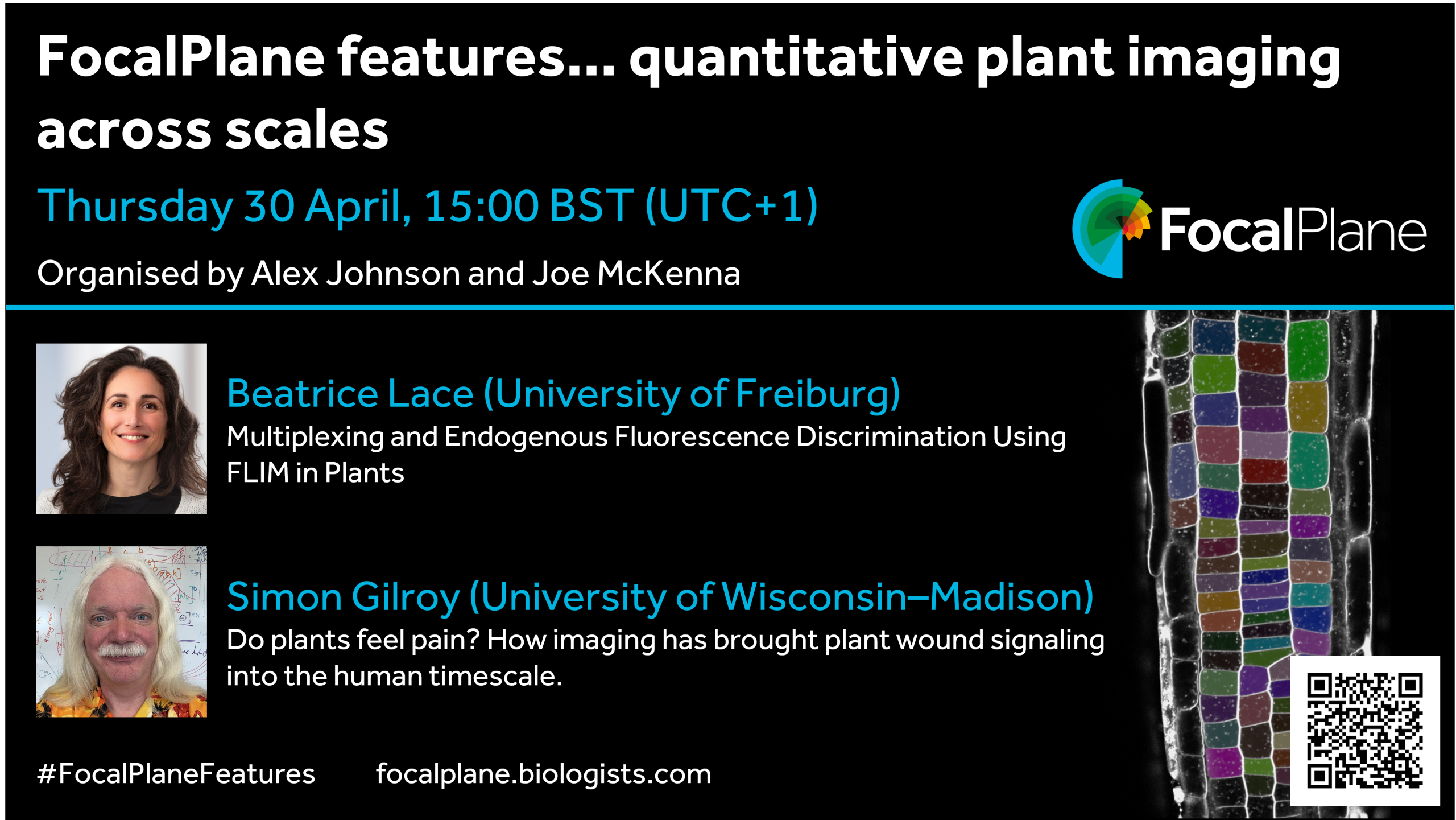
Our first webinar is on Thursday 30 April at 15:00 BST and will feature talks from Beatrice Lace and Simon Gilroy. Beatrice will present, ‘Multiplexing and Endogenous Fluorescence Discrimination Using FLIM in Plants’ and Simon will ask, ‘Do plants feel pain? How imaging has brought plant wound signaling into the human timescale’.
We are happy to announce the forthcoming workshop entitled “Roadmap for EvoDevoMec”, Nov. 2nd – 5th, 2026, Université Paris Cité.
The link with evolutionary history of an organism is key to understand embryonic development. Much of the focus has been so far on genetic circuitry. However, it has become increasingly clear that physical constraints are an essential aspect at the basis of morphogenetic processes. We have now reached an exciting stage where it becomes possible to integrate mechanical considerations in our view of how development has been shaped during evolution: EvoDevMec.
This new approach has recently raised great interest, but biological questions still need to be reformulated with precision. In addition, due to its integrative nature, it poses a scientific and technical challenge: What type of experimental systems could be used? How could we define experimental proof of concepts? How can theoretical biophysical models help integrate various questions and approaches?
We aim at discussing these questions in an informal context, i.e. with chalk talks. The workshop is limited to 30 persons, from PhD students to senior scientists from various countries. We will welcome biologists using a breadth of models (from unicellular organisms to animal and plant models), physicists (theoreticians, numericians, experimentalists), and mathematicians.
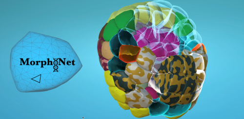
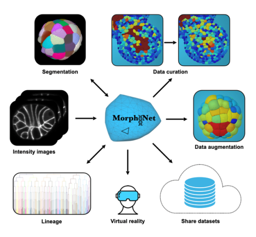
Our ‘Featured resource’ series aims to shine a light on the resources that support our research – the unsung heroes of the science world. In this post, we learn about the data and functionalities available at MorphoNet 2.0, and hear about new initiatives they are developing.
MorphoNet 2.0: breaking the glass ceiling of 3D+time bioimage curation
Modern microscopy now allows us to image living systems in three dimensions over time. From early embryonic divisions to complex tissue morphogenesis, we can follow every cell with remarkable spatial and temporal resolution.
Yet a critical bottleneck remains. Even with powerful AI-based segmentation tools, such as Cellpose or StarDist, errors are inevitable. In large 3D+time (3D+t) datasets, a seemingly small error rate quickly translates into thousands of mis-segmented cells, broken lineages, and ultimately misleading biological conclusions.
Correcting these errors — a process known as curation — is often so time-consuming that it becomes the real “glass ceiling” of quantitative developmental biology.
MorphoNet 2.0 was designed to break this glass ceiling.
From segmentation to curation
MorphoNet 2.0 is a major evolution of the original platform. Rather than proposing yet another segmentation algorithm, it focuses on a problem that is just as critical, but far less addressed: how to efficiently assess, correct, and validate 3D and 4D segmentations at scale.
The key idea is simple: automated segmentation is only useful if biologists can trust and refine its output. MorphoNet 2.0 makes this process fast, efficient, interactive, and accessible to non-programmers.
A hybrid architecture built for 3D+t data
Unlike the original web-based MorphoNet, version 2.0 is a standalone application designed to run on standard research workstations and laptops. Its architecture deliberately bridges two complementary worlds:
a high-performance 3D interface, powered by the Unity game engine, for smooth exploration of thousands of objects in real time
a Python backend integrated with powerful scientific image-processing libraries, enabling advanced image analysis, editing, and AI-based segmentation directly on raw data.
Letting the data highlight its own problems
A major challenge in large 3D datasets is simply knowing where to look. Manually inspecting every cell is unrealistic.
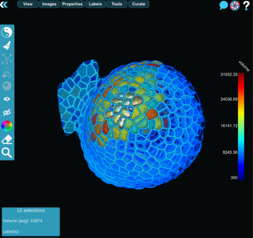
MorphoNet 2.0 addresses this by computing unsupervised quality metrics for each segmented object, such as volume, shape regularity, boundary intensity, or temporal stability. These properties are projected, as color maps, directly onto the 3D meshes or cell lineages, turning potential errors into visible outliers.
While these metrics are not absolute measures of correctness, they act as intelligent guides, directing experts toward regions that deserve closer inspection.
Curation as a local, interactive process
Once a potential error is identified, MorphoNet 2.0 enables rapid local, targeted corrections rather than global reprocessing.
This is achieved through a modular plug-in system that allows users to perform a wide range of operations, including (but not limited to):
re-segment specific regions using AI or classical methods
split fused cells or merge over-segmented fragments
remove specifically small objects
propagate corrections across time in 3D+t datasets
edit and repair cell lineages.
Because plug-ins operate only on user-defined regions of interest, most corrections take seconds rather than hours. This transforms curation from a batch process into a true human-in-the-loop workflow, with immediate visual feedback.
MorphoNet 2.0 provides an open plug-in architecture that enables contributors to develop and share custom plug-ins tailored to their own segmentation, tracking, or lineage-repair challenges.
Tested on real biological datasets
This paper demonstrates MorphoNet 2.0’s successful use on five previously published datasets spanning insects, plants, nematodes, echinoderms, and ascidians. These case studies show how the platform can:
reveal hidden segmentation errors in datasets long considered “finished”
significantly improve segmentation quality through iterative correction
polish cell lineages to a level suitable for studying subtle biological variability.
In one example, rare lineage errors scattered across thousands of cells were identified and corrected in a few hours — even in datasets comprising several thousand segmented objects — a task that would have been practically infeasible with traditional tools.
Why this matters for AI and data reuse
High-quality 3D ground truth data are essential for training and benchmarking modern AI models. Yet producing such datasets is extremely costly when curation tools do not scale.
By making 3D+t curation feasible, traceable, and accessible to biologists, MorphoNet 2.0 directly addresses this gap. It helps turn raw automated outputs (“silver truth”) into reliable, reusable datasets (“gold truth”) that can support reproducible analysis, fair algorithm comparison, and community challenges.
Who is MorphoNet 2.0 for?
MorphoNet 2.0 is designed for:
developmental and cell biologists working with complex 3D or 4D imaging data
imaging facilities producing reference datasets
researchers developing or benchmarking segmentation and tracking algorithms.
No programming skills are required to use the platform, but its open, Python-based plug-in architecture allows advanced users to extend it and share new tools with the community.
Looking ahead
MorphoNet 2.0 positions complex 3D curation as a fast and manageable activity rather than a never-ending technical burden. By combining intuitive 3D interaction, unsupervised quality assessment, and local correction, it offers a practical solution to one of the most persistent bottlenecks in modern bioimage analysis.
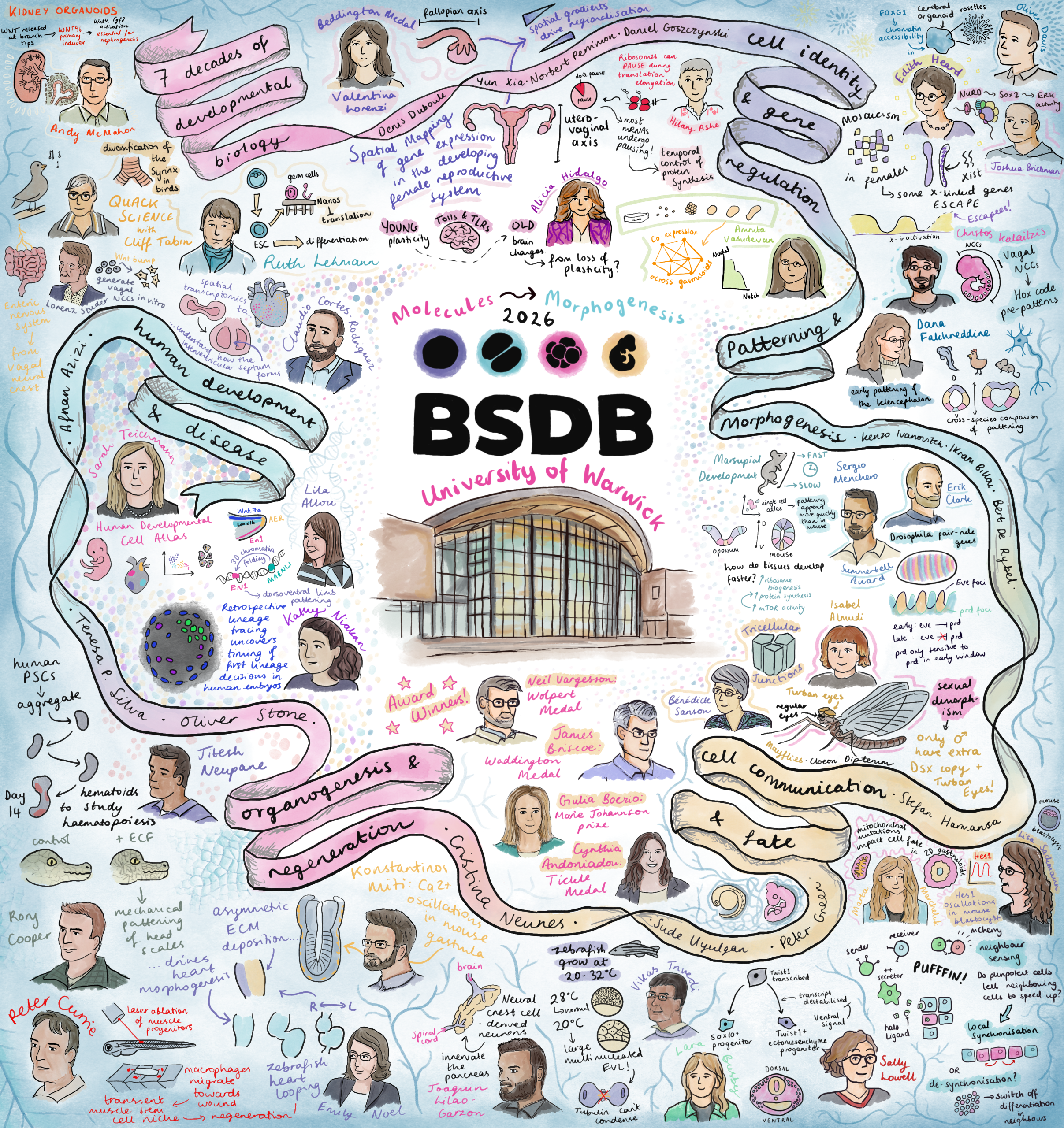
I recently attended the BSDB Spring Meeting, and decided to spend most of my time at the conference sketching. The result is this illustrated summary – featuring portraits of almost all the speakers alongside drawings of elements from their talks, whether that’s a model organism, a signalling pathway, or a particularly striking image from their work.
I initially felt quite anxious and self-conscious to be drawing in public. After all, most of the time when I’m creating art at home, it tends to look rough right up until the last moment when it all comes together. I also normally paint from static references, which conference speakers definitely are not! But, I finally decided to take the leap after numerous conversations with veteran conference illustrator Alex Cagan, who urged me to go for it. Once I started, I could feel that each drawing was turning out a little better than the last. I have always been a visual learner and definitely felt more engaged while sketching, rather than frantically scribbling notes as I would normally do!
What started as 30+ separate illustrations on my iPad slowly turned into this after I got home from the conference. My original intention was to share each as a separate piece. However, none of the illustrations felt complete enough for this (I will definitely have to work on my drawing speed in the future), so my solution was to combine them all into one large piece.
This isn’t intended as a scientific summary, more as a memento or a snapshot of what it felt like to sit in those sessions, surrounded by so much brilliant developmental biology. If you spot yourself in there, I hope I have done you justice! And I apologise if I missed out on drawing you – at times, I couldn’t quite keep up.
I’m interested in using illustration to share the joy of developmental biology, as I’ve tried to do here, but I also think it can be a wonderful tool for communicating complex scientific ideas to those who might not otherwise engage with them. This conference reminded me how much exciting work there is to communicate. Thanks to all the speakers and organisers for making it such an enjoyable meeting!
This year the popular Placental Biology Course returns online from 14 to 18 September.
This online course is designed for a diverse audience, including students, postdoctoral researchers, established academics, medical and veterinary healthcare professionals, and industry specialists with an interest in the latest developments in placental biology.
The programme includes pre-recorded lectures and practical sessions delivered by leading experts in the field, allowing participants to engage with cutting-edge research at their own pace. Each day also includes live Q&A sessions, offering a valuable opportunity to interact directly with speakers and deepen understanding.
In addition, attendees can take part in Fellowship Workshops and informal Meet and Greet sessions to build connections within the community. Participants are also invited to submit abstracts for consideration in flash talks or virtual poster presentations, providing a platform to showcase their own work.
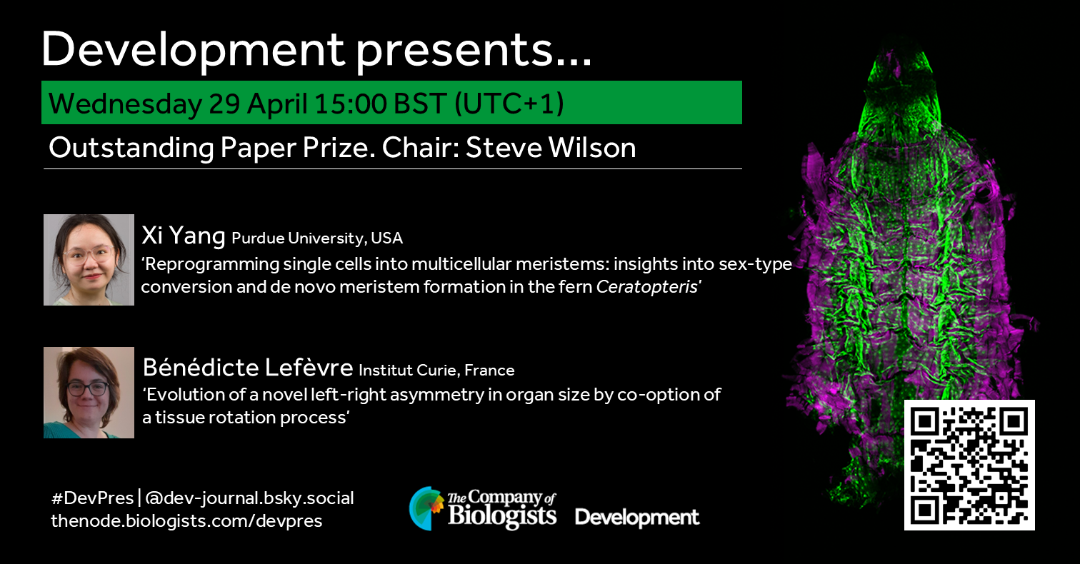
Join us in April to learn about the three papers named as finalists of Development’s 2025 Outstanding Paper Prize. In this webinar two of the papers will be presented by its first author and chaired by Deputy Editor, Steve Wilson. Learn more about the finalists in our Editorial and read the full shortlist of nominated papers in Development’s subject collection.
Wednesday 29 April – 15:00 BST (UTC+1)
Xi Yang (Purdue University, USA) ‘Reprogramming single cells into multicellular meristems: insights into sex‑type conversion and de novo meristem formation in the fern Ceratopteris’
Bénédicte Lefèvre (Institut Curie, France) ‘Evolution of a novel left-right asymmetry in organ size by co-option of a tissue rotation process’
The third finalist, Christopher De Bono (INSERM, France),will present ‘Multi-modal refinement of the human heart atlas during the first gestational trimester’ later in the year.
At the speakers’ discretion, the webinar will be recorded to view on demand. To see the other webinars scheduled in our series, and to catch up on previous talks, please visit: thenode.biologists.com/devpres
 (2 votes)
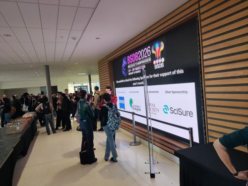
 (No Ratings Yet)