PhD position opened in Switzerland for a developmental / cell biologist
Posted by Michel Milinkovitch, on 11 March 2016
Closing Date: 15 March 2021
Evolutionary developmental genetics of reptile skin colour
In the context of evolutionary developmental biology analyses of reptilian skin colours and colour patterns, we offer one PhD position for an outstanding, highly motivated, and creative experimental wet-lab biologist with strong skills in developmental biology and cell biology.
Michel Milinkovitch’s group at the University of Geneva (UNIGE) integrates the expertise of developmental biologists, evolutionary biologists, computer scientists and physicists for an improved understanding of the mechanisms generating a diversity of skin colours and colour patterns in reptiles.
We have recently shown (Saenko et al. 2013) that the extensive variation of skin colours and patterns in Sauropsida reptiles is generated by precise co-localisation of interacting pigmentary and nano-structural elements. In this framework, we have also shown (Teyssier et al. 2015) that chameleons shift colour through active tuning of a lattice of guanine nanocrystals, which photonic effect is filtered by a layer of pigments. In addition, we have built extensive transcriptomic and genomic resources (Ullate-Agote et al. 2014; Tzika et al. 2015) for mapping colour mutations in our new model species of snakes and lizards. For example, we recently mapped and identified the mutation responsible for the amelanistic mutation in corn snakes (Saenko et al. 2015).
The successful candidate will (i) participate to linkage mapping of multiple colour and colour pattern mutations in snakes and lizards and (ii) use molecular/cell/developmental biology methods (microscopy, immuno-histochemistry, in-situ hybridisation, transcriptomics, in-vivo assays, ex-vivo cultures, etc.) to characterise the effects of these mutations on neural-crest cell migration, as well as on the physiology of pigmentary and structural-colour cells. The new PhD student will also interact with a physicist PhD student who is mathematically modelling the reaction-diffusion processes that generate colour patterns in snakes and lizards.
Candidates must have a Master in biology or biochemistry. Skills and experience with developmental biology and/or cell biology are mandatory. Skills in biophysics are useful. The successful candidate will have a genuine interest for organismal biology and will appreciate interactions with physicists and computer scientists.
The University of Geneva (UNIGE) is world-renowned for its research in Biology and Physics. UNIGE is among the top 1% best universities in the world and the Faculty of Sciences is ranked 32th world best (Shangai Academic Ranking of World Universities).
PhD students are remunerated according to the standards of UNIGE, which are very generous when compared to other international programs.
Geneva is an international city occupying a privileged geographical situation.
Candidates must send their application – in the form of a single PDF file including a brief letter of interest, a CV, as well as contact information (not support letters) of two persons of reference – to:
Prof. Michel Milinkovitch (Michel.Milinkovitch@unige.ch), Laboratory of Artificial & Natural Evolution (www.lanevol.org), University of Geneva, Switzerland.
Refs: Saenko, Teyssier, van der Marel & Milinkovitch. Precise colocalization of interacting structural and pigmentary elements generates extensive color pattern variation in Phelsuma lizards. BMC Biology 2013, 11: 105; Teyssier Saenko van der Marel & Milinkovitch. Photonic Crystals Cause Active Colour Change in Chameleons. Nature Communications 6: 6368 (2015); Tzika, Ullate-Agote, Grbic & Milinkovitch. Reptilian Transcriptomes v2.0: An Extensive Resource for Sauropsida Genomics and Transcriptomics. Genome Biol. Evol. 7: 1827-1841 (2015); Ullate-Agote, Milinkovitch & Tzika. The genome sequence of the corn snake (Pantherophis guttatus), a valuable resource for EvoDevo studies in squamates. Int. J. Dev. Biol. 58: 881-888 (2014); Saenko, Lamichhaney, Martinez Barrio, Rafati, Andersson & Milinkovitch. Amelanism in the corn snake is associated with the insertion of an LTR-retrotransposon in the OCA2 gene. Scientific Reports 5, 17118 (2015) .
 (No Ratings Yet)
 (1 votes)
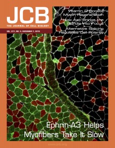 Each adult mammalian skeletal muscle has a unique complement of fast and slow myofibers, a consequence of cell-autonomous and non-cell-autonomous patterning decisions during development. Intriguingly, following either acute muscle regeneration or deinnervation/reinnervation, the proportionality and patterning of muscle fibers is largely preserved, suggesting a mechanism for ongoing maintenance of fiber type specification during homeostasis and regeneration/repair. We have recently reported that interactions between the repulsive guidance ligand ephrin-A3 [which is expressed on all and only slow (Type I) myofibers] and EphA8 [an ephrin receptor expressed by terminal Schwann cells at all and only neuromuscular junctions of fast (Type II) myofibers] promote and preserve slow myofiber identity by preventing stable innervation of slow muscle fibers by fast motor neurons [

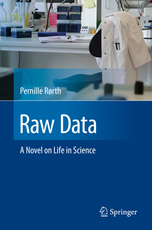 I’ve just finished reading ‘