The final webinar of 2024 features two early-career researchers working on gene regulation and will be chaired by Development’s Senior Editor, Alex Eve.
Wednesday 4 December – 15:00 GMT
Madalena M. Reimão Pinto (Biozentrum) ‘The regulatory landscape of 5′ UTRs in translational control during zebrafish embryogenesis’
Gabriel Aughey (University College London) ‘Characterisation of an RBL2-associated neurodevelopmental disorder sheds light on neuronal cell-cycle exit and re-entry’
At the speakers’ discretion, the webinar will be recorded for viewing on demand. To see the other webinars scheduled in our series, and to catch up on previous talks, please visit: thenode.biologists.com/devpres
Queer people, trans individuals in particular, remain significantly underrepresented in STEM and academia. In August 2023, Nick wrote an Honest Conversations post on the Node entitled ‘Coming Out of my Cage and I’ve Been Doing Really, Really Good’, discussing his experience of being a transgender scientist, the importance of the support he received from his lab, and the freedom he found in living authentically.
In this Voices piece, we hear from two PhD students who identify as trans. They discuss their experiences navigating academia, issues prevalent in academia that trans people still face, and the support systems they have found that have empowered them on their journeys.
Of being a trans immigrant – an uphill journey through science
by Aflah Hanafiah
As an 8th year PhD student, my journey to climb this career ladder consisted of numerous hurdles, setbacks, and emotional and mental rollercoasters. I was lucky enough to navigate myself out of one of the most queerphobic countries in the world. The UCLA Williams Institute ranks Malaysia 115th of 175 countries based on social acceptance. The Human Rights Watch (HRW) reported on certain Malaysian laws criminalizing same-sex sexual acts and transgender people’s gender expression and sentencing those who found guilty with jail time, fines, and whipping. These laws and the generally conservative culture that is hegemonic in the country easily subject queer people to discriminations and violence, especially towards trans femme people. I grew up learning very early on that expressing myself outside of the heteronormative traits would make my life difficult, so I learned to conform the best I could.
I spent my high school years throwing myself into schoolwork and getting my grades up so I could qualify for a government’s scholarship that would later take me to the USA where I eventually completed my bachelor’s degree in molecular biology. This opportunity allowed me to not only pursue my career goals, but it had also provided me with the space to start addressing my queer identity that I had long suppressed. During my time at Rochester Institute of Technology (RIT), I met many people, student body and faculty included, who were accepting and affirming of queer identities in general. They essentially created a safe space for people like me to feel comfortable in my own skin. However, there was still lingering stigma behind being openly queer and pursuing post-graduate education. I was reminded of this as I applied to several graduate programs. During my interviews, I pivoted strongly to presenting as the gender that appeared on my legal documents so I could avoid the possible awkwardness and questions that could arise from my queer identity.
As I went through the process and started my PhD, I felt like I was given another opportunity to further explore my queer identity now that I was in a totally new environment. This was where I started to feel comfortable to use my preferred pronouns, dressed the way that affirmed my gender identity, and explored gender affirming care. By this time, I was already 27 years old and well past my junior PhD years. My decision to undergo hormone replacement therapy (HRT) that depends on my school’s health insurance while at the same time working on my PhD was a difficult one to make. The PhD already came with its own sets of challenges, and I was unsure if I should add to this chaos by physiologically jumbling my hormonal level. However, the persistent gender dysphoria that is always occupying my mind would later drive me to embark on this next phase of my transition. I am fortunate because my student’s health insurance covers gender-affirming care, so I knew that this was an opportunity that I could not pass on.
Though I have not faced much explicit transphobia in my graduate school, which I largely attribute to me being passable enough, my other trans friends on the other hand, have a much different experience. Despite universities improving the workplace environment for queer people and other minorities; prejudice, micro- and macro-aggressions are still prevalent. I have no doubt that if I did not pass as what society deems as a woman, I would have had a very different experience going through life and specifically, working in academia. I am not “out” per se, in graduate school because it never became a question. I do make it a point that I am transgender around other queer colleagues and in queer spaces because I think that it’s important to let people know that they are not alone and that they are safe with me. As I am at the tail end of my PhD, I can’t help but be worried for my future. A slew of anti-trans laws is being passed in almost every state in the country and more companies are reportedly moving away from investing in LGBTQ+ oriented diversity, equity, and inclusion (DEI) efforts. Coupled with my F1 visa status, the career choice that is viable to me are ever narrowing. However, it is times like these that I rely on my chosen family, loved ones, and community in navigating these challenges. I believe that with a supporting community behind you, you can overcome any obstacles.
Searching for others like me: navigating academia as a trans person
By James Lythall
As a queer and trans person in STEM, I’m acutely aware of how few LGBTQIA+ people there are in STEM. There are plenty of heterosexual and/ or cisgender people who I enjoyed working and socializing with, the vast majority of whom have been very supportive. At the same time, I can count the number of queer academics I know in the field of life sciences without running out of fingers, and the number of trans academics in any scientific field on one hand. There are probably many more out there, but the number of visible, openly trans and/or queer academics is vanishingly small. Of course, representation will not solve all the problems facing queer and trans researchers, but it can help us fight the feelings of isolation that are often common amongst under-represented groups. To know that someone like you has managed to succeed, despite the odds, can both be comforting and motivating. On a more practical level, it also means there are people you can ask for advice who are familiar with the problems you may be facing and may have already found solutions to them.
I can’t help but feel that I am having to forge my own path all the time, and whilst that is sometimes exciting, it is often exhausting. There are many people who I feel I can ask for scientific guidance, but almost none who I feel comfortable asking for support on issues I face from being trans. This is not because I believe those around me to be trans- or homophobic, but simply because they are often unaware of specific problems that trans and queer researchers face.
As a researcher, I am acutely aware that my personal experience is not necessarily representative of others- an n of 1 is not much! Frustratingly, there is precious little data available on the experiences of the queer – and particularly trans – people in academia. Much of the current data focuses on the experiences of undergraduate students, often in the US, and often not stratified beyond science and humanities. Both this data and surveys conducted by scientific organizations rarely collect data on the trans status of participants, and group everyone who doesn’t identify as a woman or a man as ‘other’. Nonetheless, the little data that there is suggests that queer and trans people are often underrepresented in STEM compared to in the general population, with one US survey finding LGBTQIA+ people were ~20% less prevalent in STEM fields compared to the general population (1). The same study also reported 70% of academics felt uncomfortable being out at work. Focusing on undergraduates, another study found that LGBTQIA+ undergraduates were 9.4% less likely to remain in a STEM major (2), with this rising to 10% for trans undergraduates (3). Another study reported 45.67% of natural sciences students compared to 14.96% of social sciences students reported being misgendered (4). This is particularly alarming in the light of a new study that has found an association of higher levels of microaggressions (including misgendering) and worse mental health outcomes in trans adults in the UK (5).
Despite these gloomy statistics, I am optimistic. I do believe science is slowly becoming a more welcoming place for queer and trans people. I have had some very positive experiences and moments of connection as a trans person in STEM, and in academia. I have had lecturers who have gone above and beyond to create a welcoming teaching and learning environment for trans and queer students, as well as offering considerable personal support to me and other queer students. I have also been encouraged by the acceptance and support for trans and queer people that other students have offered, such as using and advocating for more inclusive language to challenging the heteronormative and cisnormative narratives that often pervade medical sciences. More recently when I was applying for PhDs, one potential supervisor went out of their way to ensure that only my preferred name would be used throughout the application process, and another offered to correct a colleague when they noticed they had got my pronouns wrong.
I would like to finish by suggesting a few things that I think allies can do to help improve LGBTQIA+ experiences in the academic world. Firstly, if you have teaching responsibilities alongside your research, include queer, trans and intersex people whenever possible. On a more individual level, ask and listen to what your LGBTQIA+ students and colleagues need and try to avoid making assumptions. Another important thing you can do is look into how your institution collects data on students and staff and whether this includes appropriate gender and sexuality choices, including options to not disclose. This makes institutional data much more helpful for researchers trying to understand the experiences of queer and trans people in STEM.
These are just a few suggestions, but there are numerous excellent articles out there on how to support queer and trans students and colleagues that I would encourage you to read. Equally, I would also encourage you to think about what you can do outside of the academic sphere to support LGBTQIA+ people. Visible and meaningful support for LGBTQIA+ people has never been more important, particularly in the UK where hate crime rates continue to rise and transphobic rhetoric has become increasingly commonplace in media and politics. Building a better academia also means building a better society in large.
References:
1: Freeman, J. B. (2020). Measuring and Resolving LGBTQ Disparities in STEM. Policy Insights from the Behavioral and Brain Sciences, 7(2), 141-148. doi:10.1177/2372732220943232
2: Hughes, B. E. (2018). Coming out in STEM: Factors affecting retention of sexual minority STEM students. Science Advances, 4(3), eaao6373. doi:doi:10.1126/sciadv.aao6373
3: Maloy, J., Kwapisz, M. B., & Hughes, B. E. (2022). Factors Influencing Retention of Transgender and Gender Nonconforming Students in Undergraduate STEM Majors. CBE—Life Sciences Education, 21(1), ar13. doi:10.1187/cbe.21-05-0136
4: Whitley, C.T., Nordmarken, S., Kolysh, S. and Goldstein-Kral, J. (2022), I’ve Been Misgendered So Many Times: Comparing the Experiences of Chronic Misgendering among Transgender Graduate Students in the Social and Natural Sciences. Sociol Inq, 92: 1001-1028. https://doi.org/10.1111/soin.12482
5: Wright, T., Lewis, G., Greene, T. et al. The association between microaggressions and mental health among UK trans people: a cross-sectional study. Soc Psychiatry Psychiatr Epidemiol (2024). https://doi.org/10.1007/s00127-024-02775-2
from Irene Karapidaki, Béryl Laplace-Builhé and Michalis Averof
What is this?
These are crustacean embryos injected with mRNA encoding a red fluorescent protein bound to membranes. On the left, the fluorescent protein is localised on the plasma membrane, on the right it is trapped in the endoplasmic reticulum and/or Golgi.
How was this image made?
These images were made while testing different membrane localisation tags in the marine crustacean Parhyale hawaiensis. One-cell stage embryos were injected with mRNAs encoding mScarlet3 fused to a Lyn tag, directing protein myristoylation and palmitoylation (on the left), or a signal peptide and the CD8 transmembrane domain (on the right). The embryos were allowed to grow for a day and then imaged live on a confocal microscope.
Why should people care about this?
Targeting fluorescent proteins to cell membranes allows us to visualise the shape and behaviours of cells in living embryos, as they build the body. See for example this amazing movie, showing the choreography of cells as they build sensory organs in the fish embryo: https://thenode.biologists.com/ready-steady-cooooooonga/research/
Can I do this in my favourite research organism?
The problem is that existing membrane-localising tags do not work equally well in all species. The SP-CD8 tag for example (on the right) gives good plasma membrane localisation in Drosophila, but gets stuck in the secretory pathway in our crustacean (Parhyale) embryos. In non-conventional model organisms, one needs to test several tags to find a good one.
To facilitate this process we generated a toolkit of 11 membrane-localising tags, which can be screened rapidly by microinjecting mRNA in your species of interest. Comparing results obtained in different species will help to identify tags that work well in a wide range of eukaryotes. If you are interested in trying the toolkit and joining our comparative screen, please get in touch.
My name is Katie Pickup and I wanted to introduce myself as I’m a new Reviews Editor at The Company of Biologists. I am going to be working with Development and the Node, as well as with three of the Company’s other journals (Journal of Cell Science, Disease Models & Mechanisms and Journal of Experimental Biology). I’m really looking forward to being exposed to a broad range of bioscience topics in this role.
My research background is mainly in stem cell biology. I did my PhD at the MRC Human Genetics Unit at the University of Edinburgh investigating the role of DNA methylation in pluripotency and differentiation of mouse embryonic stem cells in Richard Meehan’s lab. I used 3D models of differentiation including gastruloids and embryoid bodies to understand the impact of DNA hypomethylation on cell lineage trajectories and exit from pluripotency. I’m particularly interested to see where stem cell-based embryo model research goes over the next few years, both scientifically and from the regulatory side. I’m also excited for the opportunity to branch out in my new role as an editor and learn more about other areas of developmental biology across the spectrum of model organisms.
I’m really looking forward to getting to know the wider community better through working with authors on Review articles and other front-section journal content like interviews and poster articles. I also can’t wait to travel to lots of different conferences and workshops across the world, so hopefully I’ll get to meet some of you there! In the meantime, feel free to get in touch via email, LinkedIn or X.
When I joined the Zon lab in June 2021, my mentor, Leonard Zon, shared an insightful piece of advice: “A good project always has two questions, one you can answer and one you dream of answering.” In this post, I’ll focus on that dream.
In brief, the question we managed to answer – at least a little bit – is how to instruct the expression of the “eat-me” signal driven by Calreticulin (CALR) and its complementary “don’t-eat-me” signal, driven by beta-2-microglobulin (B2M). In our study (https://www.science.org/doi/10.1126/science.adn1629) , we showed that high levels of reactive oxygen species (ROS) leads to high surface presentation of Calr, which, in turn, leads to high levels of interaction with a macrophage, and clearance of the stressed hematopoietic stem and progenitor cell (HSPC). On the flip side, TLR3 can modulate the expression of CALR together with B2M. Here, the balance between these molecules leads to a scenario where a macrophage interacts with the HSPCs, but does not “eat” them. This intricate signaling impacts clonal diversity, revealing a potential avenue for future immunotherapies targeting mutant or cancerous stem-cell populations while sparing healthy ones.
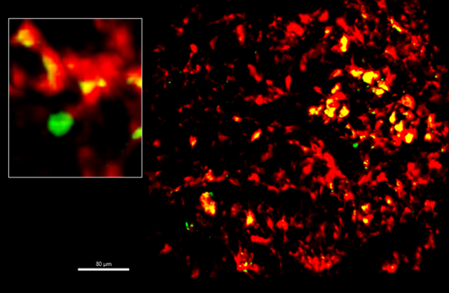
Macrophage and long-term hematopoietic stem cell (LT-HSC) interaction in mammals. Life imaging of calvarium bone marrow from Mds1GFP/+ Flt3Cre (MFG) mouse showing macrophages (F480+) in red and LT-HSCs in green.
Our uncharted territory lies in harnessing macrophages to selectively target malignant clones. We found higher B2M expression in HSPCs from acute myeloid leukemia (AML) patients with malignant stratification, suggesting that malignant clones may exploit B2M to evade macrophage clearance. This paves the way for drug development aimed at eliminating pre-leukemic and leukemic cells via macrophage-mediated clearance. This idea may also be further explored in aging studies, in which one could teach the macrophage to eliminate aged progenitor cells.
Another aspect of our study that could be further explored relies on a key finding that repetitive elements (RE), including Ltr, are the endogenous ligand of the Tlr3 and triggers b2m expression via the tlr3/irf3 pathway. We observed that these endogenous REs promoted high levels of ISG15, a gene linked to the type I interferon response.
Given the evolutionary conservation of RE and B2M, we explored the significance of this mechanism in both fish and humans, focusing on pathogen infections that are a common threat to both species. Specifically, we examined the role of TLR3 signaling in inducing “emergency granulopoiesis,” a protective process that accelerates neutrophil production during severe infection. Upon poly I:C stimulation, neutrophil populations increased.
Although further studies are needed to strengthen the relevance of this phenomenon, our results suggest that viral stimulation may confer a better fit against opportunistic pathogens by promoting granulocyte differentiation. This observation gets more fascinating if one considers that this increase of type I response could not only alter the emergency granulopoiesis, but also contribute to innate immune training. Seminal studies have shown that type I IFN signaling mediates neutrophil trained innate immunity, mainly in the context of solid cancer. This therefore suggests that RE-triggered type I interferon may play a role in trained immunity—a concept previously explored in cancer but now potentially relevant in other systems.
Over the summer, I had the opportunity to conduct research in Vivian Li’s Lab, focusing on the role of WNT signalling in the response of intestinal stem cells to injury, under the expert guidance of Dr. James Wilmouth Jr. As a medical student, I am deeply appreciative of the opportunity to gain hands-on experience in fundamental research through undertaking a project over the course of two months. I am truly inspired by the continuous innovation at the Francis Crick Institute, driven by a diverse community of committed scientists who embrace collaboration and interdisciplinary approaches.
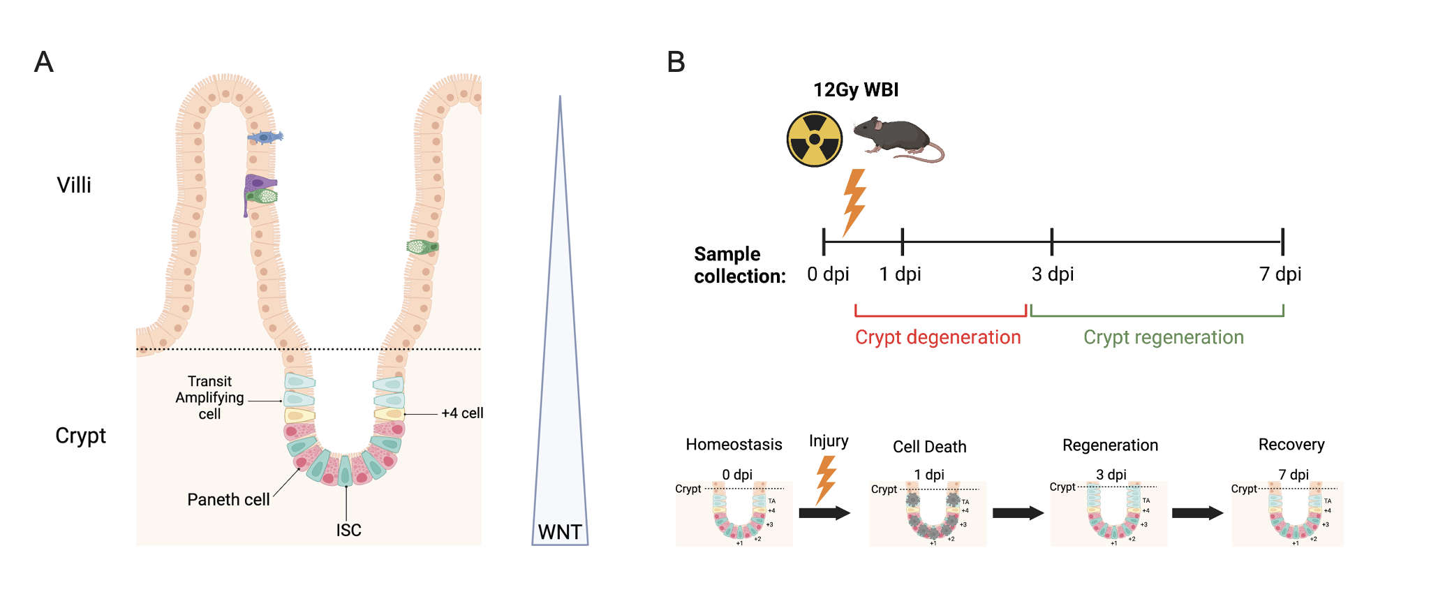
One research axis within the Li Lab focuses on how canonical WNT signalling maintains intestinal stem cells (ISCs) and influences cell fate decision. The gut epithelium can be organised into an architecture of crypts and villi (Fig 1A). Crypts are found at the base, extending into the villi towards the gut lumen. ISCs are located within the crypt and responsible for driving renewal of the gut epithelium every 3-5 days during homeostasis. High WNT signalling in the crypts maintains ISC homeostasis before decreasing along a gradient towards the villi. Publications have demonstrated that whilst inhibiting WNT signals causes degeneration, WNT overactivation induces adenoma formation. Therefore, fine-tuned WNT signalling is crucial for maintaining ISC homeostasis.
Figure 1. A) Intestinal epithelium during homeostasis. B) Model of intestinal injury. TCF/Lef:H2B:mCherry reporter mice were exposed to 12Gy of whole body CSM radiation. Intestine or isolated crypts were collected at 0 dpi (pre-irradiation control), 1 dpi, 3 dpi, and 7 dpi.
For this project, I examined the role of WNT signalling in ISCs during injury-induced regeneration. We utilized whole-body irradiation to simulate ISC injury-induced regeneration in mice. This model results in crypt degeneration at 1-day post-irradiation (dpi), followed by regeneration at 3 dpi, and recovery by 7 dpi (Fig 1B). Intestinal tissues were harvested for crypt isolation at four time points: 0 dpi (pre-irradiation control), 1 dpi, 3 dpi, and 7 dpi. These samples were then used to investigate three transcriptional signatures throughout the regenerative process: Classical Intestinal Stem Cell (ISC), Revival Stem Cell (RevSC), and WNT target genes.
I performed quantitative PCR (qPCR) to analyse the temporal changes in gene expression during the regenerative process (Fig 2). Results indicated that classic ISC markers were downregulated at 1 dpi following injury and recovered to near homeostatic levels by 7 dpi. RevSC markers showed an induction at 1 dpi, followed by a decline toward baseline levels by 7 dpi. Interestingly, WNT target gene expression remained relatively stable throughout the 7-day period.
Figure 2. qPCR analysis of isolated crypts from 0, 1, 3 & 7 dpi. A) Classis ISC markers (Lgr5, Olfm4) decrease after injury at 1dpi, but recover close to homeostatic levels by 7 dpi. B) RevSC markers (Trop2, Anxa1, Clu, Sca1) increase after injury at 1-3dpi and 3/4 targets recover back to homeostatic levels by 7dpi. C) Most WNT target genes (3/4) remain constant over all timepoints. One target, Cd44, showed increased levels from 1-3dpi and recovered to homeostatic levels by 7 dpi. Graphs represent means ± SEM.
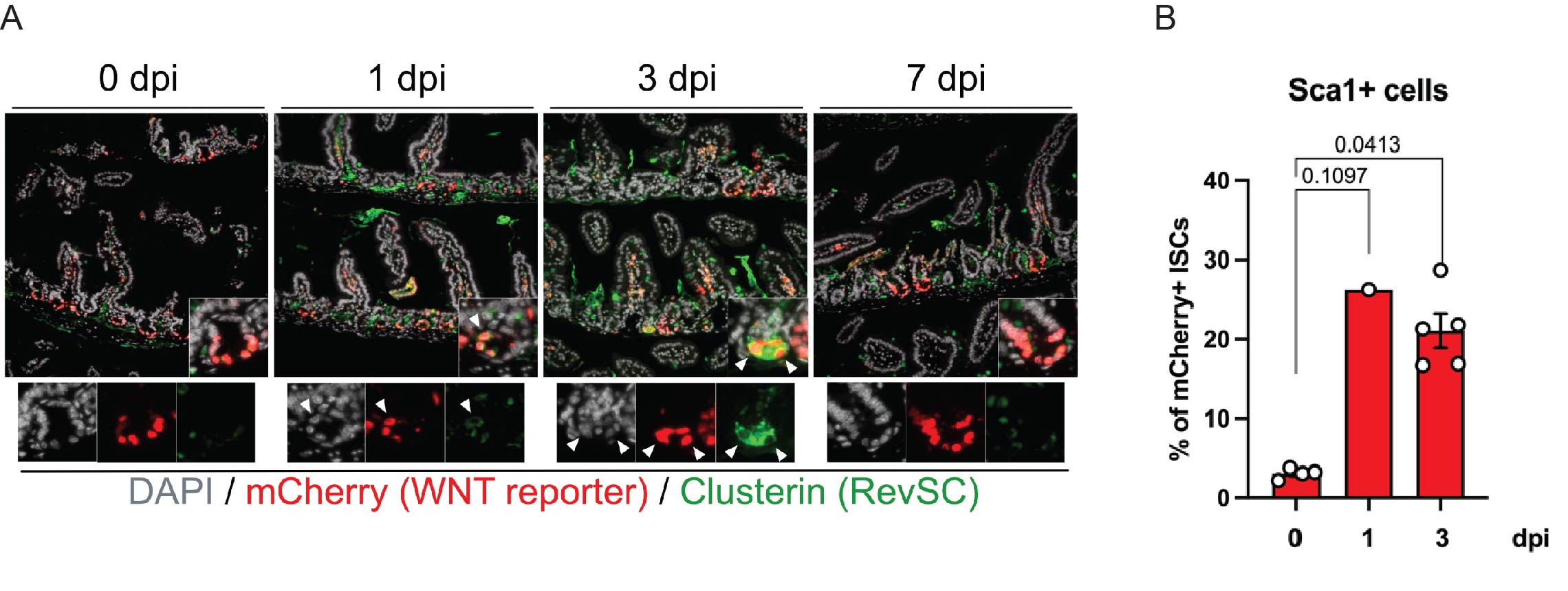
These initial results suggest that both Yap-driven RevSC signature and certain WNT targets are expressed during injury-induced regeneration. This contradicts the current understanding, which proposed an antagonistic relationship between YAP and WNT during ISC regeneration up on injury. To further investigate the co-expression of RevSC markers and WNT in ISCs, we utilised immunohistochemistry (IHC) and flow cytometry of WNT reporter mice in the irradiation induced regeneration model (Fig 3).
Figure 3.A) Co-expression of WNT and Clusterin upon injury. mCherry (WNT reporter) in red, Clusterin (RevSC marker) in green, DAPI in grey. mCherry and Clusterin colocalised at 1dpi, with stronger signals overlapping at 3dpi. B) Quantifying the percentage of WNT-high ISCs that are positive for Sca1 (RevSC marker) by flow cytometry. Graphs represent means ± SEM. Statistical analysis were conducted by two-way ANOVA in (B).
To spatially characterise WNT-high cells in the crypts co-expressing the Revival Stem Cell (RevSC) signature, I conducted IHC using a TCF/Lef:H2B;mCherry reporter system. The reporter has Tcf/Lef binding sites which drive the expression of the H2B:mCherry fluorescent protein, enabling visualisation of active WNT signalling. I observed colocalisation of mCherry and Clu (RevSC marker) at 1dpi, which was more pronounced at 3dpi (Fig 3A). These results suggest that WNT-high ISCs express RevSC markers during regeneration. In order to quantify how often this happens during regeneration, I utilised flow cytometry. By gating for mCherry+ ISCs (WNT-high ISCs), I found there was a significant increase in the percentage of mCherry+ ISCs expressing the RevSC marker Sca1 at 3dpi (~20%) compared to 0 dpi (~3%) (Fig3B).
In conclusion, my project demonstrated that a proportion of WNT-high ISCs co-express RevSC markers during injury-induced regeneration. Although preliminary, these results highlight that it is necessary to further investigate the role of YAP in the interplay between WNT signalling and the RevSC signature in ISCs during regeneration. This work would provide clarity into which signals dictate ISC survival during regeneration.
This project has provided me with invaluable insight into the scientific process. I have learned advanced techniques at the Li lab, including organoid culture and maintenance. I would like to thank my supervisor, James, for his exceptional mentorship in building my critical thinking, guiding my experiments, and supporting my data analysis and interpretation. I would also like to extend my thanks to the Francis Crick Institute and the Medical Research Foundation Rosa Beddington Fund for their generous support, which has enabled me to contribute to this exciting field of research.
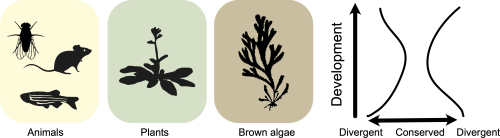
Brown algae are a group of complex multicellular eukaryotes, unrelated to animals, plants and fungi. It follows that brown algae evolved the process of multicellular development independently, offering a unique opportunity to investigate shared principles underlying developmental evolution across the tree of life. One such principle is the hourglass model of embryo evolution. The hourglass model describes a pattern of evolutionary conservation and divergence during embryogenesis, where the divergent earlier and later stages are bridged by a conserved mid-embryonic period (Duboule 1994). Such hourglass patterns have been observed in animal, plant and fungal development (Drost et al. 2017). But what about brown algae?
In our recent paper, “A transcriptomic hourglass in brown algae” (Lotharukpong et al. 2024), we asked a simple question: does brown algal development follow the hourglass model? By profiling the developmental transcriptome in two Fucus species, we observe an hourglass pattern in brown algal embryogenesis (Fig 1). The conserved mid-embryonic period is underpinned by the reduced expression of evolutionarily younger genes and the presence of more broadly expressed, potentially pleiotropic genes. We also explored the transcriptome across life cycle stages in brown algae with differing levels of morphological complexity. Crucially, in morphologically simple Ectocarpus species without canonical embryos, multicellular development itself appeared to constrain transcriptome evolution, suggesting how such embryo hourglass patterns may have emerged in the first place (at least in brown algae). Overall, this work gives evidence for the hourglass model as a general principle underlying developmental evolution across the tree of life.
But as we all know; a lot went on behind the paper.
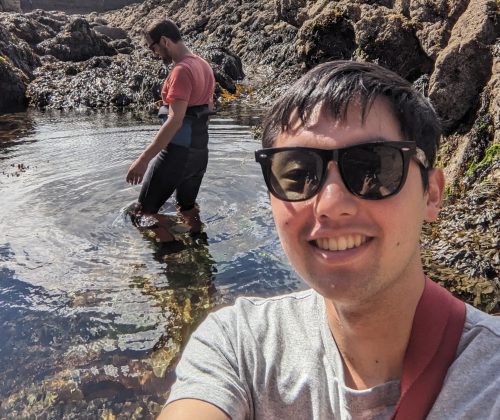
To test the hourglass model in brown algae, we first had to literally find morphologically complex brown algae that undergo embryogenesis (Fig 2). Between France and Germany, Rémy Luthringer (co-author) had to go collect reproductively mature adults to produce embryos and cultivate them in the lab. It was a challenge to culture Fucus embryos beyond the earliest embryonic stages, since they require a lot of care to ensure healthy growth. This was all the more difficult for species such as F. distichus, which required up to half a year to reach the latest embryonic stage used in the study.
Figure 2. Rémy (left; phycoculturalist) and Sodai (right; bioinformatician) sampling brown algae in the wild.
Bioinformatics in the spotlight:
We then faced several hurdles on the bioinformatics side when piecing this study together, which required a sustained push on software development. In particular, we wanted to infer the evolutionary age of each gene, which is needed for computing the transcriptome age index (a key metric for evolutionary transcriptomics). Existing approaches were not suitable nor computationally scalable to current large databases such as the National Center for Biotechnology Information (NCBI) non-redundant (nr) database and did not account for potential database contaminations among other confounding factors in gene age inference. We therefore teamed up with Josué Barrera-Redondo to create GenEra (Barrera-Redondo et al. 2023), a wrapper around the fast and sensitive pairwise sequence aligner DIAMOND v2 (Buchfink et al. 2021), which brought down the search time from months to days. This finally allowed us to infer the gene age in a timely manner for any eukaryotic genome, including the species we used in the hourglass study (Fucus serratus, Fucus distichus, Ectocarpus sp., Laminaria digitata and Saccorhiza polyschides).
On top of this, we extended the functionalities of R packages for evolutionary transcriptomics, such as myTAI (Drost et al. 2018), to accommodate our analyses. New and existing functions in myTAI enabled us to distinguish evolutionary signals from random noise. We hope these efforts will be useful for the wider community interested in asking evo-devo questions using transcriptomic data.
What’s next for the story?
It is an exciting time for brown algal research. A recent study has provided several dozen genome assemblies across major groups of brown algae (Denoeud et al. 2024). Furthermore, functional genetics is now a possibility for multiple brown algal species, fuelling recent findings such the independent evolution of HMG domain genes as a male sex-determining factor (Luthringer et al. 2024). There is also a drive to generate transcriptomic data to understand cell-type and tissue evolution in brown algae, which also evolved cell-types and tissues independently from other complex multicellular eukaryotes. There is still much more to come!
References
Barrera-Redondo J, Lotharukpong JS, Drost H-G, Coelho SM. 2023. Uncovering gene-family founder events during major evolutionary transitions in animals, plants and fungi using GenEra. Genome Biol. 24:54.
Buchfink B, Reuter K, Drost H-G. 2021. Sensitive protein alignments at tree-of-life scale using DIAMOND. Nat. Methods 18:366–368.
Denoeud F, Godfroy O, Cruaud C, Heesch S, Nehr Z, Tadrent N, Couloux A, Brillet-Guéguen L, Delage L, Mckeown D, et al. 2024. Evolutionary genomics of the emergence of brown algae as key components of coastal ecosystems. :2024.02.19.579948. Available from: https://www.biorxiv.org/content/10.1101/2024.02.19.579948v2
Drost H-G, Gabel A, Liu J, Quint M, Grosse I. 2018. myTAI: evolutionary transcriptomics with R. Bioinformatics 34:1589–1590.
Drost H-G, Janitza P, Grosse I, Quint M. 2017. Cross-kingdom comparison of the developmental hourglass. Curr. Opin. Genet. Dev. 45:69–75.
Duboule D. 1994. Temporal colinearity and the phylotypic progression: a basis for the stability of a vertebrate Bauplan and the evolution of morphologies through heterochrony. Development 1994:135–142.
Lotharukpong JS, Zheng M, Luthringer R, Liesner D, Drost H-G, Coelho SM. 2024. A transcriptomic hourglass in brown algae. Nature 635:129–135.
Luthringer R, Raphalen M, Guerra C, Colin S, Martinho C, Zheng M, Hoshino M, Badis Y, Lipinska AP, Haas FB, et al. 2024. Repeated co-option of HMG-box genes for sex determination in brown algae and animals. Science 383:eadk5466.
It’s now been 13 months since I started my lab, marking the end of my ‘New PI Diaries’ series here on the Node. The journey has so far been both overwhelming and rewarding, and I’ve enjoyed the chance to reflect and share my experiences in this blog. Overall, it’s been a lot—but mostly a good lot. My time gets split between a lot more responsibilities and I have less time to be in the lab. Both mentoring and teaching now take up much more time than during my postdoc, but luckily, I already know I enjoy both.
However, one aspect that was almost completely new to me was managing lab finances. In this blog, I want to share some examples of situations I encountered and the lessons I learned, along with a few key takeaways. To provide some context, my Independent Junior Group Leader position comes with support that includes a PhD student position, a technician, a yearly consumables budget, and some startup funds. Comparing lab resources across universities and countries is difficult. For instance, my startup budget is considerably smaller than those typically offered at US universities but potentially bigger than that available at other institutes or universities. However, I also benefit from funded positions and an equipped lab. In addition, many larger pieces of equipment and facilities are shared within the institute and can be used free of charge or at a low rate, reducing my need to purchase new equipment.
But let’s talk people first. In addition to support from the ZMBP, I was able to secure an Emmy Noether grant from the DFG. This grant covers my salary for six years, along with funding four years of two PhD students and a postdoc, allowing for steady growth and stability in the coming years. However, I quickly ran into an unexpected issue: the PhD student position from the University is paid a lower salary compared to the positions provided by the DFG. Different enough to feel unfair but unfortunately it is common. Similarly, PhD positions in different disciplines get compensated at different rates. Now, I cannot change decisions of the University or the state of Baden-Württemberg, but I figured there must be a workaround. Once I started asking around, I learned that people have come up with a variety of solutions. Talking with fellow group leaders and our Head of Central Services I learned that we could reassign the unused % of the technician position together with some flexible lab budget to equalize the PhD salaries. So, it’s important not to hesitate in discussing challenges; I certainly wasn’t the first to face this problem, and people had already identified potential solutions.
Apart from people, we’re spending money on consumables and equipment. The ZMBP account, which includes a startup and a yearly budget, allows me flexibility in spending. Additionally, last year I received one time support from a collaborative project. This account had its own constraints, while flexible, the funds must be used within two months, adding some pressure. Finally, the Emmy Noether provides consumables funding, albeit with more limited flexibility since it is there to support the group and project and any equipment should be provided by the University. As a result, I got a crash course into which account to use for what purchase. In short: for me it’s best to save the ZMBP account for computer and equipment purchases as those cannot be made from all other accounts.
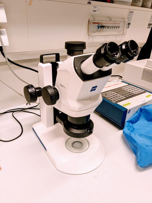
Most of the necessary equipment was actually already there, but I found several items that could significantly make our lab’s work easier and more efficient. Before purchasing these I first spent some time looking around, checking what is available already, what our need really is, and what options we have for financing the things we need. And although I still don’t have a precise overview of our total finances, we’re in a fortunate position where I don’t have to stress excessively about money. I do feel it’s important for everyone in the lab to know the price of some of our reagents and equipment. Investing in reagents, such as 2x mixes for genotyping and colony PCR, is worth the costs right now for the time they save as long as people are aware of their cost. We’re in a good position, but I want to avoid wasting funds that could be better spent elsewhere to make our days easier. Eventually using lab funds we purchased two new stereomicroscopes to dissect Arabidopsis embryos, additional pipettes, and computers. Each piece of equipment or expensive kit is an investment that I think will pay off and that we can afford in our current situation. However, I remain careful as, well, I lack experience, and you never know what’s up ahead.
Here are my early-stage financial takeaways:
People are expensive. Value the positions you have and hire carefully. In addition, sometimes spending more on consumables and equipment is worth the time saved for your team.
Keep track of your accounts. Understand what’s in each account, what you can spend it on, and where your money is going. Is it what you expected?
Use your funds. Once you have a clear overview, consider what tools could make lab operations smoother. I initially waited to determine which investments would be beneficial for everyone in the lab but then decided to make some investments.
And my personal goals:
Have a more complete overview of the lab finances. I have a rough idea of what we’re spending and what’s available but I still have a long way to go. Luckily I have plenty of people to ask for advice.
Get experience. While enthusiasm is important, it doesn’t replace experience. I’m learning a lot and I already feel a lot more confident in making these decisions.
While the lab’s immediate future is secure, I also need to plan for what comes next. To be honest, at this point, anytime I feel like I’m in control of things I realize I just have been forgetting or ignoring something important. I guess that’s life. In general, I think I should spend more time thinking of grants. Next year will be my final opportunity to apply for the ERC Starting Grant, so my focus will be on that. Additionally, in a few years, I’ll need to start searching for a professor position, as my ZMBP Junior Group Leader position and Emmy Noether funding will end in about five years, and securing a professorship in Germany can take some time.
Wish me luck!
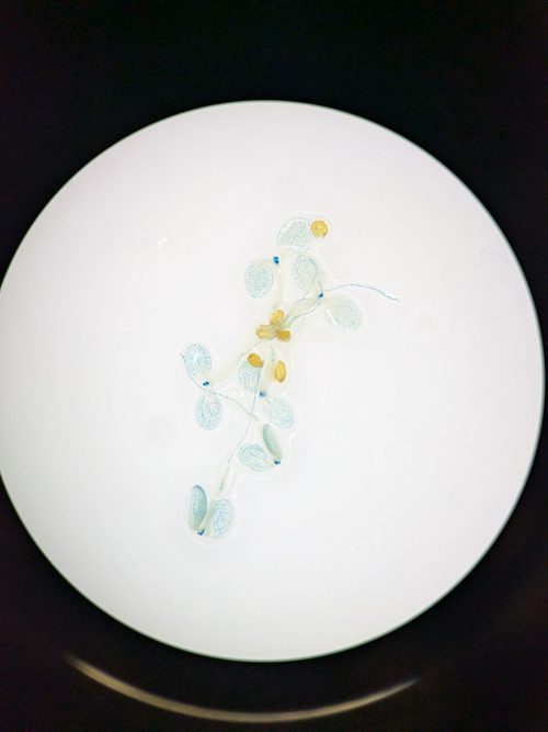
First investment: new stereomicroscope for doing embryo dissection and the beautiful images it takes of GUS-stained seedlings.
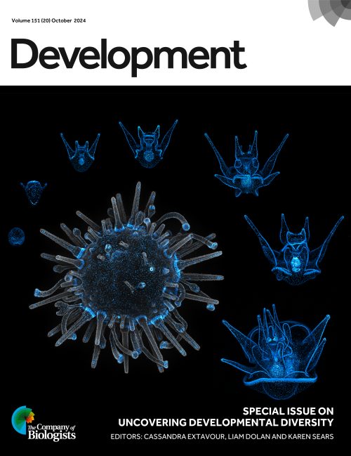
[Editorial from Development’s latest Special Issue ‘Uncovering Developmental Diversity’, edited by Cassandra Extavour, Liam Dolan and Karen Sears.]
Scanning through early issues of the Journal of Embryology and Experimental Biology (the previous name for this journal) reveals the diverse range of organisms that were investigated by developmental biologists in the 1950s and 1960s. However, the rise of molecular genetics in subsequent decades led to a narrowing in species choice to a small repertoire of well-characterised model organisms for which there were genetic tools for functional experimentation. In recent years, however, technological advances, including genome and transcriptome sequencing, flexible genome-editing approaches and high-resolution four-dimensional imaging, provide an opportunity to once again study developmental questions in organisms across all kingdoms of life. Given the current global challenges of climate change and biodiversity loss, it is particularly important that we turn our attention to understanding development in an unstable world.
This important topic was the basis of Development’s Journal Meeting, ‘Unconventional and Emerging Experimental Organisms in Cell and Developmental Biology’ in 2023, which you can learn more about in the Meeting Review published here (Lemke et al., 2024). Fuelled by the success of the meeting, we chose to focus this Special Issue, led by Academic Editor Cassandra Extavour, together with Liam Dolan and Karen Sears as Guest Editors, on a related topic: Uncovering Developmental Diversity. We are particularly delighted that multiple attendees from our meeting have contributed both research and review-type articles to this issue.
The 28 research papers in this Special Issue highlight 32 different organisms from across the multicellular tree of life, featuring cnidarians (Garschall et al., 2024), insects (Matsuda et al., 2024; Bai et al., 2024; Beaven et al., 2024; Pallarès-Albanell et al., 2024) and annelids (Bideau et al., 2024), as well as echinoderms (Barone et al., 2024; McDonald et al., 2024; Jackson et al., 2024; Clarke et al., 2024) and chordates (Gigante et al., 2024; Johnson et al., 2024), including vertebrates (Rees et al., 2024; Pérez-Gómez et al., 2024), many of which are various fishes (Leclercq et al., 2024; Li et al., 2024; Woronowicz et al., 2024; Peloggia et al., 2024; Jin et al., 2024). These articles demonstrate the importance of finding the best model to address a developmental question, such as making use of the curved epithelium in the sea star embryo to investigate cell organisation and packing (Barone et al., 2024) or using the regenerative capacity of annelids to learn more about cell plasticity (Bideau et al., 2024). In addition to annelids, a Perspective in this issue highlights five more ‘extraordinary’ model systems for regeneration across scales from single cells to whole organisms (Accorsi et al., 2024).
Not limited to animals, the Special Issue also embraces a wide array of studies uncovering fundamental developmental processes, such as axis formation and organogenesis in photosynthetic organisms, including brown algae (Vigneau et al., 2024; Boscq et al., 2024), liverworts (Attrill and Dolan, 2024; Attrill et al., 2024; Sakai et al., 2024), and vascular plants such as ferns (Woudenberg et al., 2024) and angiosperms (Mody et al., 2024; Spiegelhalder et al., 2024). Photosynthetic organisms feature heavily in the issue’s review-type content, too, with articles describing how brown algae can inform us about the transition to multicellularity (Batista et al., 2024), how the environment and climate change influence development through the lens of stomata (Chua and Lau, 2024) and what we can learn about the evolution of plant development through the fossil record (Hetherington, 2024).
The evo-devo field, in particular, has benefitted from the appreciation of biodiversity and increased taxonomic sampling. Reflecting this, two Reviews discuss fundamental evolutionary concepts, including how phenotypes can be maintained by different underlying genetic architecture through developmental systems drift (McColgan and DiFrisco, 2024), as well as a cautionary tale of how reports on the low-hanging fruit of simple genetic explanations of evolution should not change our perception that evolution is inherently complex (Cooper, 2024).
A broad selection of available organisms also allows the study of rare evolutionary innovations, such as the ability of Nematostella to degrow in response to food availability (Garschall et al., 2024) or of teleost fish to adapt ionocyte differentiation to regulate osmotic levels within aquatic environments (Peloggia et al., 2024). Adaptive plasticity is also the focus of a Review article describing how organisms assess environmental cues across scales and respond via phenotypic changes (Hill et al., 2024). Furthermore, capturing developmental biodiversity furthers our understanding of complex life cycles (McDonald et al., 2024; Peloggia et al., 2024) – a topic motivating a Hypothesis for unravelling cellular rejuvenation (Berger, 2024). Indeed, studying organisms with metamorphic life cycles allows us to learn about the intrinsic developmental process, such as how the rhinoceros beetle remodels its horn (Matsuda et al., 2024) or neuronal cell survival in Ciona (Gigante et al., 2024).
Importantly, research using emerging model systems relies on new tools that facilitate functional experiments. Our Techniques and Resources section features methods for the delivery of proteins and nucleic acids into oocytes in a variety of species (Clarke et al., 2024), as well as approaches for generating stable genetic lines (Jackson et al., 2024) and tools for quantifying diversity (Mody et al., 2024). However, not all species are amenable to being cultured in the lab, and a Perspective describes the importance of fieldwork for developmental biology in unconventional model systems (Brown et al., 2024). In addition, a Spotlight article describes how modern innovations in stem cell technology might be employed for species conservation (Hutchinson et al., 2024), highlighting how understanding biodiversity is the first step to its preservation, an increasingly prevalent topic in the context of climate change.
Overall, we hope that this issue demonstrates both how technological advances have made it possible to understand development and regeneration in previously intractable organisms, as well as the importance of this pursuit. We continue to ensure Development is an appropriate home for your studies in developmental biology, stem cells and regeneration using any organism. We welcome your submissions.
Maria Victoria Serrano, Stephanie Cottier, Lianzijun Wang, Sergio Moreira-Antepara, Anthony Nzessi, Zhiyu Liu, Byron Williams, Myeongwoo Lee, Roger Schneiter, Jun Liu
Aaron M. Savage, Alexandra C. Wagner, Ryan T. Kim, Paul Gilbert, Hani D. Singer, Erica Chen, Elane M. Kim, Noah Lopez, Kelly E. Dooling, Julia C. Paoli, S.Y. Celeste Wu, Sebastian Bohm, Rachna Chilambi, Tim Froitzheim, Steven J. Blair, Connor Powell, Adnan Abouelela, Anna G. Luong, Kara N. Thornton, Benjamin Tajer, Duygu Payzin-Dogru, Jessica L. Whited
Peggy P. Hsu, Ansley S. Conchola, Tristan Frum, Xiangning Dong, Lila Tudrick, Varun Ponnusamy, Michael S. Downey, Manqi Wu, Mengkun Yang, Yusoo Lee, Emma Niestroy, Yu-Hwai Tsai, Angeline Wu, Sha Huang, Ian A. Glass, Sofia D. Merajver, Jason R. Spence
Ling S. Loh, Joseph J. Hanly, Alexander Carter, Martik Chatterjee, Martina Tsimba, Donya N. Shodja, Luca Livraghi, Christopher R. Day, Robert D. Reed, W. Owen McMillan, Gregory A. Wray, Arnaud Martin
Tim Ott, Amelie Brugger, Emmanuelle Szenker-Ravi, Yvonne Kurrle, Olivia Aberle, Matthias Tisler, Martin Blum, Sandra Whalen, Patrice Bouvagnet, Bruno Reversade, Axel Schweickert
Yuchen Liu, Tianli Qin, Xin Weng, Bernice Leung, Karl Kam Hei So, Boshi Wang, Wanying Feng, Alexander Marsolais, Sheena Josselyn, Pingbo Huang, Bernd Fritzsch, Chi-Chung Hui, Mai Har Sham
Qiao Wu, Jian Zhang, Bing Long, Xiao Hu, Bruna Mafra de Faria, Stephen Maxwell Scalf, Kutay Karatepe, Wenxiang Cao, Nikolaos Tsopoulidis, Andres Binkercosen, Masaki Yagi, Aaron Weiner, Mary Kaileh, Enrique M. De La Cruz, Ananda L Roy, Konrad Hochedlinger, Shangqin Guo
Stephen Spurgin, Ange Michelle Nguimtsop, Fatima N. Chaudhry, Sylvia N. Michki, Jocelynda Salvador, M. Luisa Iruela-Arispe, Jarod A. Zepp, Saikat Mukhopadhyay, Ondine Cleaver
Sera Lotte Weevers, Alistair D. Falconer, Moritz Mercker, Hajar Sadeghi, Jaroslav Ferenc, Albrecht Ott, Dietmar B. Oelz, Anna Marciniak-Czochra, Charisios D. Tsiairis
Theopi Rados, Olivia S. Leland, Pedro Escudeiro, John Mallon, Katherine Andre, Ido Caspy, Andriko von Kügelgen, Elad Stolovicki, Sinead Nguyen, Inés Lucía Patop, Thiberio Rangel, Sebastian Kadener, Lars D. Renner, Vera Thiel, Yoav Soen, Tanmay A.M. Bharat, Vikram Alva, Alex Bisson
Nora Ditzer, Ezgi Senoglu, Theresa M. Schütze, Aikaterina Nikolaidi, Annika Kolodziejczyk, Katrin Sameith, Sevina Dietz, Razvan P. Derihaci, Cahit Birdir, Anne Eugster, Mike O. Karl, Andreas Dahl, Pauline Wimberger, Franziska Baenke, Claudia Peitzsch, Mareike Albert
Audrey J. Marsh, Sergei Pirogov, Abby J. Ruffridge, Suresh Sajwan, Tyler J. Gibson, George Hunt, Yadwinder Kaur, Melissa M. Harrison, Mattias Mannervik
Dimitris Botskaris, Ioannis K. Deligiannis, Ioanna Peraki, Haroula Kontaki, Marianna Stagaki, Matthieu D. Lavigne, Celia P. Martinez-Jimenez, Iannis Talianidis
Surbhi Sood, Aktan Alpsoy, Guanming Jiao, Alisha Dhiman, Charles Samuel King, Gabriella Grace Conjelko, Judy E. Hallett, Sagar M Utturkar, Jill E Hutchcroft, Emily C Dykhuizen
Olga M. Sigalova, Mattia Forneris, Frosina Stojanovska, Bingqing Zhao, Rebecca R. Viales, Adam Rabinowitz, Fayrouz Hamal, Benoît Ballester, Judith B Zaugg, Eileen E.M. Furlong
Yuliia Haluza, Joseph A. Zoller, Ake T. Lu, Hannah E. Walters, Martina Lachnit, Robert Lowe, Amin Haghani, Robert T. Brooke, Naomi Park, Maximina H. Yun, Steve Horvath
Rita Manco, Camilla Moliterni, Gauthier Neirynck, Maxime De Rudder, Corinne Picalausa, Leana Ducor, Montserrat Fraga, Frédéric Lemaigre, Christine Sempoux, Alexandra Dili, Isabelle A. Leclercq
Yingnan Lei, Mai Chi Duong, Nuša Krivec, Charlotte Janssens, Marius Regin, Anfien Huyghebaert, Edouard Couvreu de Deckersberg, Karen Sermon, Diana Al Delbany, Claudia Spits
Julian Weihs, Fatima Baldo, Alessandra Cardinali, Gehad Youssef, Katarzyna Ludwik, Harald Stachelscheid, Nils Haep, Peter Tang, Igor Sauer, Pavitra Kumar, Cornelius Engelmann, Susanna Quach, Philip Bufler, Namshik Han, Milad Rezvani
Milad Rezvani, Kyle Lewis, Susanna Quach, Kentaro Iwasawa, Julian Weihs, Hasan Al Reza, Yuqi Cai, Masaki Kimura, RanRan Zhang, Yuka Milton, Praneet Chaturvedi, Konrad Thorner, Ramesh C. Nayak, Jorge Munera, Phillip Kramer, Brian R. Davis, Appakalai N. Balamurugan, Yeni Ait Ahmed, Marcel Finke, Rose Yinghan Behncke, Adrien Guillot, René Hägerling, Julia K. Polansky, Philip Bufler, Jose A Cancelas, James M. Wells, Momoko Yoshimoto, Takanori Takebe
B. Pardo-Rodríguez, A.M. Baraibar, I. Manero-Roig, J. Luzuriaga, J. Salvador-Moya, Y. Polo, R. Basanta-Torres, F. Unda, S. Mato, G. Ibarretxe, J.R. Pineda
Sara Cannavò, Chiara Paleni, Alma Costarelli, Maria Cristina Valeri, Martina Cerri, Antonietta Saccomanno, Veronica Gregis, Graziella Chini Zittelli, Petre I. Dobrev, Lara Reale, Martin M. Kater, Francesco Paolocci
Alicia Tovar, Scott Monahan, Trevor Mugoya, Adrian Kristan, Walker Welch, Ryan Dettmers, Camila Arce, Theresa Buck, Michele Ruben, Alexander Rothenberg, Roxane Saisho, Ryan Cartmill, Timothy Skaggs, Robert Reyes, MJ Lee, John Obrycki, William Kristan, Arun Sethuraman
Haidong Yan, John P. Mendieta, Xuan Zhang, Alexandre P. Marand, Yan Liang, Ziliang Luo, Mark A.A. Minow, Hosung Jang, Xiang Li, Thomas Roulé, Doris Wagner, Xiaoyu Tu, Yonghong Wang, Daiquan Jiang, Silin Zhong, Linkai Huang, Susan R. Wessler, Robert J. Schmitz
Magdalena Schindler, Christian Feregrino, Silvia Aldrovandi, Bai-Wei Lo, Anna A. Monaco, Alessa R. Ringel, Ariadna Morales, Tobias Zehnder, Rose Yinghan Behncke, Juliane Glaser, Alexander Barclay, Guillaume Andrey, Bjørt K. Kragesteen, René Hägerling, Stefan Haas, Martin Vingron, Igor Ulitsky, Marc Marti-Renom, Julio Hechavarria, Nicolas Fasel, Michael Hiller, Darío Lupiáñez, Stefan Mundlos, Francisca M. Real
Cristofer Calvo, Casey O. Swoboda, Fabian Montecino Morales, Siddhant Nagar, Michael J. Petrany, Chengyi Sun, Hima Bindu Durumutla, Mattia Quattrocelli, Douglas P. Millay
Ismael Moreno-Sanchez, Luis Hernandez-Huertas, Daniel Nahon-Cano, Carlos Gomez-Marin, Pedro Manuel Martinez-García, Anthony J. Treichel, Laura Tomas-Gallardo, Gabriel da Silva Pescador, Gopal Kushawah, Alejandro Díaz-Moscoso, Alejandra Cano-Ruiz, John A. Walker II, Manuel J. Muñoz, Kevin Holden, Joan Galcerán, María Ángela Nieto, Ariel Bazzini, Miguel A. Moreno-Mateos
Sara Di Carlo, Adrian Salas-Bastos, Mariela Castelblanco Castelblanco, Muriel Auberson, Marie Rumpler, Malaury Tournier, Lukas Sommer, Olaia Naveiras, Edith Hummler
Ália dos Santos, Oliver Knowles, Tom Dendooven, Thomas Hale, Alister Burt, Piotr Kolata, Giuseppe Cannone, Dom Bellini, David Barford, Matteo Allegretti
Daniel Medina-Cano, Mohammed T. Islam, Veronika Petrova, Sanjana Dixit, Zerina Balic, Marty G. Yang, Matthias Stadtfeld, Emily S. Wong, Thomas Vierbuchen
Brian Ho Ching Chan, Holly Hardy, Teresa Requena, Amy Findlay, Jason Ioannidis, Dominique Meunier, Maria Toms, Mariya Moosajee, Anna Raper, Mike McGrew, Joe Rainger
Guilherme E. Kundlatsch, Alina S. L. Rodrigues, Vitória F. B. Zocca, Laura A. S. Amorim, Gabriela B. de Paiva, Almiro P. S. Neto, Juliana A. D. B. Campos, Danielle B. Pedrolli
 (3 votes)
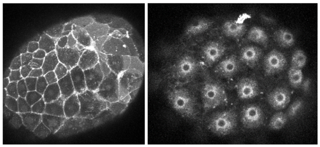
 (No Ratings Yet)