The community site for and by
developmental and stem cell biologists

Fibroblast Growth Factors in Development and Repair, Cancun, Mexico (4th Feb 2015- 7th Feb 2015)

Posted by , on 5 November 2014

4th – 7th February 2015

The major goal of this Fusion Conference is to bring together in a single forum the diverse group of researchers who study fibroblast growth factor (FGF) signaling. This will provide a unique opportunity to discuss new results and to target future research areas. Among the topics to be covered in this FGF signaling in adult tissue homeostasis, repair, regeneration and angiogenesis; 3)  aberrant FGF signaling in developmental/hereditary diseases meeting are: 1) the mechanisms by which FGF signaling governs organogenesis and tissue patterning during embryonic development; 2) the role of including skeletal syndromes, hearing loss and hypogonadism.


Target Audience

Reflecting the pleiotropic functions of FGF signaling in human biology, FGF researchers encompass a wide range of scientific disciplines, including structural biologists, biochemists, cell biologists, endocrinologists, developmental biologists, geneticists, pharmacologists and clinicians.

Important Deadlines

Early bird and Talk Submission deadline– 30 Nov 14

Poster Submission – 21 Nov 14

Last Chance – 28 Nov 14


fusion-conferences

Register at: https://www.fusion-conferences.com/registration28.php

Thumbs up (1 votes)
Loading...

Tags:
Categories: Events

Navigate the archive

Use our Advanced Search tool to search and filter posts by date, category, tags and authors.

A new stem cell film plus more from EuroStemCell

Posted by , on 4 November 2014

Hello! We’ve got lots of new things to share, including a film and fact sheet combo that looks at cell fate, cell identity and reprogramming, a closer look at an unusual model organism, and an interview with stem cell scientist and Parkinson’s disease specialist Malin Parmar.

Also featured in this news update: schools outreach in Scotland and Spain, and six new Spanish translations.

As ever, we’re keen to hear from readers of The Node – on TwitterFacebook, or via our websiteYou can get involved as a writer or translator, tell us about the stem cell events you’re involved in, make comments or suggestions, or just say hello! And for regular stem cell news, why not subscribe to our monthly newsletter?


Cell Fate: Journeys to Specialisation


EuroStemCell’s latest film looks at how specialized cells develop from stem cells.

Cell Fate: Journeys to Specialisation premiered in Heidelberg earlier this month, followed by a lively Q&A session with Andreas Trumpp, featured scientist Thomas Graf, and great questions from the audience.

Read more about the launch and the making of the film.


New fact sheet: Cell identity and reprogramming

Macrophage train track drawing by Vanessa De Mello

Our body contains several hundred different types of specialised cells. Each cell has very specific features that enable it to do its job. Yet every cell in your body contains the same genes – the same biological ‘instruction book’. So what makes each type of cell different? And can we control or change cell identities? How might this help us develop new approaches to medicine? 

Read more


Snail fur: an alternative model organism for stem cell research

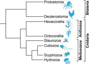
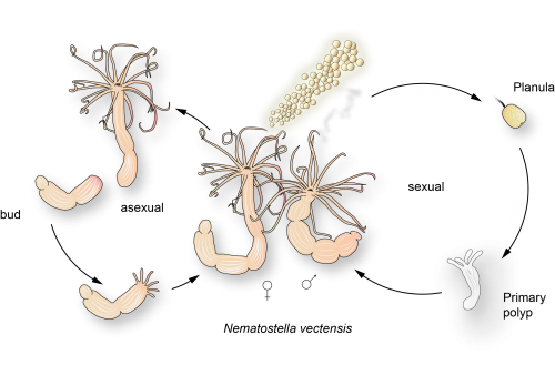
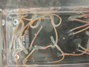
snail fur - hydractinia echinataIn this guest blog post Hakima Flici, a postdoctoral researcher at NUIG’s Regenerative Medicine Institute (REMEDI), tells us a bit more about her particular area of stem cell research…the model organism hydractinia echinata.

Read more

The work of this REMEDI lab also featured recently in a BBC Future news story, The animal that regrows its head (and in Spanish: El animal que regenera su propia cabeza)

Interview with Malin Parmar: cell therapy for Parkinson’s disease

malin ParmarMalin Parmar heads a research group focused on developmental and regenerative neurobiology at Lund University in Sweden. The ultimate goal of her research is to develop cell therapy for Parkinson’s disease.

At this year’s Hydra summer school we spoke to Malin about how she got started in stem cell research, what she’s working on at the moment, and her view of the prospects for treating Parkinson’s disease with stem cells.

Read more


Amazing stem cell questions at Inverkeithing High School

Inverkeithing high school studentsThis Stem Cell Awareness Day PhD student Jamie Gillies joined Richard Axton and Cathy Southworth at Inverkeithing High School in Scotland to share with students the exciting world of stem cell biology and the work of being scientists. Here’s his account of the day, and the intriguing questions the students asked.

Read more


Transdifferentiation workshops for secondary students at CRG

students at CRG transdifferentiation labThe Centre for Genomic Regulation (CRG) in Barcelona has started the school year with a new workshop for high school students. The workshop is taking place every Thursday in the CRG Teaching and Training Lab facilities, a space specifically designed for the training of new researchers and for outreach activities.

Read more


Six new Spanish fact sheet translations

Muchas gracias to our translators!

Subscribe and stay informed

EuroStemCell’s newsletter is sent out monthly – subscribe now for regular updates.

Thumbs up (2 votes)
Loading...

Tags: ,
Categories: News, Research, Video

In Development this week (Vol. 141, Issue 22)

Posted by , on 4 November 2014

Here are the highlights from the current issue of Development:

Meristem maintenance is KNOX so simple

FigureClass I KNOX transcription factors, such as SHOOT MERISTEMLESS (STM) and KNAT1, are known to play a role in the plant shoot apical meristem (SAM), where they are thought to prevent differentiation and hence promote stem cell maintenance. Now, on p. 4311, Urs Fischer and colleagues uncover a role for STM and KNAT1 in another Arabidopsis meristem – the vascular cambium, which is a lateral meristem that gives rise to xylem and phloem cells. They first show that STM and KNAT1 are expressed in undifferentiated cambial cells but also in differentiated phloem and xylem cells. The researchers further demonstrate that xylem fibre formation is reduced in stm and knat1 mutants, suggesting that STM and KNAT1 promote the differentiation of cambial derivatives. In addition, they report that STM and KNAT1 regulate xylem differentiation via transcriptional repression of BLADE-ON-PETIOLE 1 (BOP1) and BOP2, which are not expressed in the SAM. Together, these findings demonstrate that, in contrast to their function in the SAM, STM and KNAT1 promote cell differentiation in the cambium, suggesting that the exact role of these transcription factors in other meristems needs re-examination.

TBX1: at the heart of epithelial properties

FigureDuring its development, the heart tube undergoes rapid elongation, fuelled by the addition of cardiac progenitors from the second heart field (SHF). The gene regulatory networks governing SHF formation have been studied extensively, but little is known about the basic cellular features of SHF cells. Now, Robert Kelly and co-workers show that the transcription factor TBX1, which is implicated in both normal SHF development and congenital heart defects, regulates the epithelial properties of mouse SHF cells (p. 4320). Using immunofluorescence microscopy, they first show that SHF cells in the dorsal pericardial wall constitute an apicobasally polarised epithelium. Transmission and scanning electron microscopy reveal the presence of monocilia on the apical surface of SHF cells and of actin-rich filopodia on their basal surface. Using live-imaging of thick-slice cultures, the researchers demonstrate that these filopodia are dynamic, extending towards and making contact with surrounding tissues. Importantly, they report that TBX1 plays a crucial role in regulating these epithelial cell features; cell shape, cell polarity and filopodia dynamics are perturbed in Tbx1-/- mutants. These exciting findings suggest that TBX1-mediated control of epithelial state is crucial for heart development.

Mesogenin 1 masters the presomitic mesoderm

FigureDuring development, neuromesodermal (NM) stem cells give rise to both neural cells and paraxial presomitic mesoderm (PSM) cells, but what dictates PSM fate? Here, Terry Yamaguchi and colleagues show that a single transcription factor – mesogenin 1 (Msgn1) – acts as a master regulator of PSM development (p. 4285). They show that the overexpression of Msgn1 in mouse ESCs cultured as embryoid bodies (EBs) is sufficient to drive PSM differentiation. Microarray and ChIP-seq analyses of Msgn1-overexpressing EBs confirm that Msgn1 controls the expression of key regulators of PSM development, including those involved in epithelial-mesenchymal transition and segmentation. Importantly, the researchers demonstrate that Msgn1 overexpression in NM stem cells in vivo biases fate towards the PSM; the contribution of these cells to the neural tube is reduced while the number of PSM cells is dramatically increased. Finally, the authors show that Msgn1 overexpression can partly rescue the PSM differentiation defects observed in Wnt3a−/− embryos, suggesting that Msgn1 functions downstream of Wnt3a as master regulator of PSM fate. Given the role of the PSM as a precursor for a multitude of cell types, this finding has important implications for the fields of cellular reprogramming and regenerative medicine.

Basonuclin 2: a regulator of spermatogenesis

FigureEmbryonic germ cells display strikingly different fates with regard to mitosis and meiosis, depending on their sex. In female mice, germ cells switch from mitosis to meiosis shortly after reaching the foetal gonad where they generate the lifelong pool of oocytes. However, in males, meiosis and mitosis are actively repressed, and germ cells remain quiescent in the gonad until birth, when they resume mitosis and start generating spermatocytes. Here (p. 4298), Philippe Djian and colleagues demonstrate that Basonuclin 2, an extremely conserved transcription factor specifically expressed in male germ cells, suppresses meiosis. More surprisingly, they also show that Basonuclin 2 is required for mitosis repression and, later in life, for meiosis progression during spermatogenesis and maintenance of the spermatogonial stem cells that ensure spermatocyte production during life. Furthermore, Basonuclin 2 is necessary for the expression of DNMT3L, a key protein that is involved in spermatogenesis, and for the repression of meiotic genes (Stra8, Msx1 and Msx2) that are normally expressed in female germ cells. These findings, which uncover a new regulator of male gametogenesis, are likely to further our understanding of spermatogenesis in humans.

From embryonic stem cells to gastruloids: early development in a dish

FigureOne of the first patterning events of embryogenesis occurs during gastrulation: three-dimensional (3D) cell movements reorganise the embryo, a mass of morphologically similar cells, into an axially organised structure with three germinal layers (endoderm, mesoderm and ectoderm). To date, two-dimensional (2D) culture models have failed to recapitulate such complex cell behaviours linking cell movement to cell fate. Here (p. 4231), Alfonso Martinez Arias and colleagues show that 3D aggregates of mouse embryonic stem cells cultured in mesendoderm-promoting medium undergo cell movements, axial organisation and germ layer specification, features reminiscent of gastrulation. They demonstrate that the expression of endoderm (Sox17, Fox2A) and early mesoderm (Brachyury) markers becomes polarised in these aggregates. Later, cells originating from the Brachyury-expressing ‘territory’ are extruded from the aggregate. These ‘gastruloids’ thus present a powerful tool that can be used to study early embryonic tissue specification in a dish, an unprecedented feat in vitro.

PLUS…

Reactive oxygen species and stem cells

DEV3615Recent work suggests that reactive oxygen species (ROS) can influence stem cell homeostasis and lineage commitment. In this Primer, Ghaffari and colleagues provide an overview of ROS signalling and its impact on stem cells, reprogramming and ageing. See the Primer on p. 4206


Chemokines in development and disease:

F1.mediumIn our latest poster and companion article, Wang and Knaut provide an overview of chemokine signalling and some the chemokine-dependent strategies used to guide migrating cells. See the poster on p. 4199

Leaf development and morphogenesis

DEV3627The development of plant leaves follows a common basic program, which can be modulated to generate a diverse range of leaf forms. Bar and Ori review recent work examining how plant hormones, transcription factors and tissue mechanics influence leaf development. See the Review on p. 4219

Thumbs up (No Ratings Yet)
Loading...

Categories: Research

Can you beat a chicken sexer? Revisiting embryo manipulation of the avian chick.

Posted by , on 4 November 2014

Chicken, quail, zebra finch, emu, duck, crow……a simple glimpse and we immediately realize how the Aves have, as a model system left their traces in various fields of biological research. And within the Aves class, the domestic fowl Gallus gallus is no doubt revered highly among the developmental biologists for their certainly distinguished career. Discovery of the germ layers, the src oncogene and B cells are only but a few examples (from a very extensive list indeed) of their notable contributions to scientific history.

How did chicks become so popular among members of our community? This preference appears to be rooted far back to antiquity, where even the ancient Egyptians showed an interest in “this bird that gives birth everyday”. Convenience in rearing and accessibility are decisive factors but for us developmental biologists, ease during embryo manipulation is a definite must. Our group released two methodological papers, both of which revisit the culturing and analyzing aspects of conventional embryo manipulation, all in hopes of making this tool more accessible to the avian community. So what`s new, then?

The paper, “Extending the limits of avian embryo culture with the modified Cornish pasty and whole-embryo transplantation methods” focuses on circumventing survivability restrictions of former ex ovo cultures with two technical alternatives, modified Cornish (MC) pasty culture and whole-embryo transplantation1,2,3. To our knowledge, MC champions existing culture systems in convenience, cost and best ex ovo growth. What more could we ask for? Shaped in the form of a traditional British pasty, the embryo is folded along the medial axis with minute yolk portions packaged inside and cut along the edges to form a “sealed” vesicle. Intra-vesicular injection of yolk was an addition we found that resulted in faster growth and a greater percentage of embryos reaching HH18 with normal morphology. The growth potential of MC-cultured embryos can be further extended when combined with the in ovo whole-embryo transplantation technique. The neatness here lies in their compatibility with most common applications: targeted labeling, electroporation and imaging (facilitated by their exposed ectoderm surface) in early stages and phenotypic and functional analysis once cultured to later stages.


Figure 1. Parabiosed twins created by the MC method and cultured with whole-embryo transplantation. Left is quail, right is chicken.

Figure 1. Parabiosed twins created by the MC method and cultured with whole-embryo transplantation.Left is quail, right is chicken.

Now, much can be done with a set of potent tools like these. Did we mention our attempts at making artificial twins? We practiced twinning by fusing two HH4 embryos first with the MC culture and later transplanting them back in ovo with the whole-embryo transplantation technique. Parabiosed twins are very rare in nature and highly valuable for hematopoietic studies (yes! there is a perfectly valid reason for making twins besides our personal pleasure).

By the way, have you ever heard of a chicken sexer? Although being one of those jobs hardly receiving limelight, it’s what gets eggs and chicken meat on household dining tables. Chicken sexers determine the sex of newly hatched baby chicks by sight alone. Male and female chickens have contrasting fates in the poultry industry; males become the majority of meat sold while females are passed on towards egg production. From a commercial and economical point of view, the faster the sexing the better. A commonly used technique originally developed in Japan in the 1930s is venting, where the cloacae of fluffy chicks are slightly opened to see inside their vent. Apparently, chicken sexers can distinguish 1,000 chicks on the hour with a 98% accuracy, which is simply incredible. So what does the remaining population lacking such ability do? Well, we hope to provide you with a solution below.


Figure 2. HINTW in situ hybridization labels female chicken embryos exclusively.

Figure 2. HINTW in situ hybridization labels female chicken embryos exclusively.

In our paper released earlier this year, “HINTW, a W-chromosome HINT gene in chick is expressed ubiquitously and is a robust female cell marker applicable in intraspecific chimera studies”, we introduce a promising alternative for the otherwise not so widely available ubiquitous-GFP chicken strains (due to country-wide quarantine regulations) for intra-specific chick/chick chimera studies 4. The essence of grafting and transplantation experiments depends on reliably distinguishing host and donor cell types. This HINTW (a W-chromosome gametolog of HINTZ) in situ hybridization probe detects female cells robustly and ubiquitously at early stages and most cells at later stages. When combined with male embryos pre-selected via a prior PCR screening step, it can be used to distinguish inter-sex donor and host tissues with outstanding precision, surpassing that of a chicken sexer.

Embryo manipulation is a toolbox full with the innovative ideas of our predecessor developmental biologists and we are very much delighted to be able to tip in. We would like to conclude by inviting anyone to our lab needing help with the manipulation techniques described in our published papers discussed above.

Maiko Sezaki and Hiroki Nagai


References:

1. Nagai, H., Sezaki, M., Nakamura, H., & Sheng, G. (2014). Extending the limits of avian embryo culture with the modified Cornish pasty and whole-embryo transplantation methods Methods, 66 (3), 441-446 DOI: 10.1016/j.ymeth.2013.05.005

2. Nagai, H., Lin, M., & Sheng, G. (2011). A modified cornish pasty method for ex ovo culture of the chick embryo genesis, 49 (1), 46-52 DOI: 10.1002/dvg.20690

3. Tanaka, J., Harada, H., Ito, K., Ogura, T., & Nakamura, H. (2010). Gene manipulation of chick embryos in vitro, early chick culture, and long survival in transplanted eggs Development, Growth & Differentiation, 52 (7), 629-634 DOI: 10.1111/j.1440-169X.2010.01198.x

4. Nagai, H., Sezaki, M., Bertocchini, F., Fukuda, K., & Sheng, G. (2014). HINTW, a W-chromosome HINT gene in chick, is expressed ubiquitously and is a robust female cell marker applicable in intraspecific chimera studies
Genesis, 52 (5), 424-430 DOI: 10.1002/dvg.22769

Thumbs up (3 votes)
Loading...

Tags: , , , , , ,
Categories: Research

Development: Special Issue on Human Development

Posted by , on 3 November 2014

HumanDevbanner

As we’ve reported elsewhere on the Node (here and here), we recently held a very successful meeting ‘From Stem Cells to Human Development‘. As a direct result of the enthusiasm expressed at this meeting, we are now planning a Special Issue on the topic of Human Development, scheduled for publication in late 2015. Submissions must be received by January 30th 2015 for consideration for this Special Issue.

The issue will focus on the use of stem cell technologies to understand basic principles of human development. Until recently, our understanding of human embryogenesis has been hampered by the inaccessibility of the system, but recent advances in the stem cell field – most notably the generation of human pluripotent stem cells and the development of organoid culture systems – now allow us to investigate developing human tissues: providing insights into fate specification and tissue organisation, and informing our efforts to treat developmental disorders and develop regenerative therapies. Development sits at the heart of this field, with a strong interest in both developmental and stem cell biology, and covering both in vivo and in vitro systems.

For those interested, more details on this Special Issue can be found on our dedicated web page, or contact the Development office directly for any enquiries.


Thumbs up (2 votes)
Loading...

Tags: ,
Categories: News

This month on the Node- October 2014

Posted by , on 3 November 2014

Here are some of the highlights for October. Don’t forget to also check our jobs page, were several new positions were advertised this month!


Megumi-and-her-offspringResearch:

– Shusei wrote about his recent paper successfully using intracytoplasmic sperm injection to generate live quails, and what this work teaches us about bird development.

– Misato combined atomic force microscopy and immunostainning to examine how the embryonic mouse cerebral cortex changes in stiffness during development.

 Shashank achieved tissue-specific mutagenesis in Ciona using CRISPR/Cas system.

– and Paul reposted a comment on his paper on the role of histone H3.3 in regulating chromatin during spermatogenesis.


zebrafish croppedMeeting reports:

– Denise went to this year’s Zebrafish Meeting in Maddison.

– Cat reported from the EMBO conference on interdisciplinary plant development.


Also on the Node:

– Last month Development organised a workshop titled ‘From Stem Cells to Human Development‘. Andrea discussed some of the issues raised in the panel discussion on the ethics of stem cell research, that took place at the workshop.

– Our model organisms series continues with ‘A day in the life of a shark lab‘- including a video of a swimming shark embryo inside its egg!

– Do you have a pet name for your favourite lab equipment? We collated your answers!

– And we produced a new set of node postcards– collect them at your next conference!


IMG_0326


Happy reading!


Thumbs up (No Ratings Yet)
Loading...

Categories: Highlights

Group Leader (Senior Research Associate)

Posted by , on 3 November 2014

Closing Date: 15 March 2021

The Gurdon Institute is a world-renowned centre in the fields of developmental, cell, and cancer biology, located in the heart of the historic city of Cambridge, and part of the University’s School of Biological Sciences. Founded in 1991, its purpose is to provide the best possible environment for research, and to foster interactions and collaborations between scientists with diverse but complementary interests. It is generously supported by core funding from the Wellcome Trust and Cancer Research UK, and benefits from state-of-the art facilities in a friendly, modern, purpose-built environment (see www.gurdon.cam.ac.uk).

We are seeking to recruit one or more new Group Leaders, and we are particularly interested in early career applicants who meet the eligibility criteria for Sir Henry Dale (or similar) Fellowships. This would be an ideal position for a scientist wishing to take up his or her first position as an independent researcher, and offers a generous start up package and a world-class platform for career development.

GLAdvert

The Group Leader will establish and run their own lab, leading an independent research programme, creating a strong track record of publishing high impact papers, applying for funding, and training and supervising both postdocs and students. Their scientific interests will be in the fields of developmental biology and/or cancer, and complementary to those of existing Gurdon Institute group leaders.

The person appointed will be expected to have a PhD, an outstanding publication record, have completed successful postdoctoral research training and be well on the way to establishing themselves as an internationally recognised expert in their field.

Informal enquiries are invited and can be directed to any of the Institute’s Group Leaders or emailed to glsearch@gurdon.cam.ac.uk.  To apply please use the link: http://www.jobs.cam.ac.uk/job/5286/

Thumbs up (No Ratings Yet)
Loading...

Categories: Jobs

Fanfare for The Company of Biologists

Posted by , on 2 November 2014

Dear friends and members of The Company of Biologists,

About a year ago, I was asked to organize the Physiology Symposium of the 6th EurBee Conference in Murcia/Spain, held by the European Association for Bee Research in September 2014. The Company of Biologists kindly provided generous and fast financial support for the two invited speakers of our symposium.

As a small THANK YOU SO MUCH I composed a short “Fanfare for the Company of Biologists”.

Catarina Vicente from the Node kindly asked me to write here a short comment about my thoughts during the composing process.

Here now, Ladies and Gentlemen, is the Fanfare for the Company of Biologists:

Fanfare for The Company of Biologists-So what might we hear2

Please use earphones for best results.

I also used quotations in this piece. However, unlike in scientific papers, I will not uncover those ‘secrets’ at this point. For British people it will not be hard to uncover one of it. The second one may be brought to light by a connoiseur of classical music…

The orchestra?
Since a scientist’s money bag is usually too slim to pay a full orchestra, I ‘asked’ the Vienna Symphonic Library to help with the performance…

If you think you need some relaxation from numbers and facts, you might be interested in this Science and Art website.


Have much fun,

Anton Stabentheiner

Thumbs up (1 votes)
Loading...

Categories: News

Tissue-specific genome editing in Ciona embryos by CRISPR/Cas9

Posted by , on 31 October 2014

Researchers have always been interested in tissue-specific loss of function to probe the role of specific genes in embryonic development, cell physiology and disease conditions. Migration of lateral plate primordial germ cells in zebrafish, border cell migration during oogenesis in drosophila, interaction of T-cells with their target, and numerous other cases have continued to give us insights about how cells behave differently depending on their position and function in the animal. Until recently, genome-editing approaches were limited to non-specific mutations, induced by chemical or transposable elements. Homologous recombination for precise genome editing has only been achieved in mouse embryonic stem cells. Our lab studies the embryonic development and gene expression in Ciona intestinalis, a hermaphroditic tunicate. Phylogenetic studies have shown that tunicates are the closest living relatives of vertebrates. The Ciona larva possesses the basic body plan of chordates. A small sequenced genome, low cell number (~2600 at the larval stage), well-defined cell lineages, and easy methods for transient transfection assays are some of the experimental advantages offered by this model chordate. Previous studies have reported high rate of mutagenesis and tissue-specific gene knockouts in Ciona intestinalis using TALENs and ZFNs, but the application of these methods has been limited because of expensive and tedious cloning procedures.


An adult Ciona intestinalis (Copyright: John Rundle)


Significant technical breakthroughs have been achieved in the field of genome engineering by harnessing the CRISPR (Clustered, Regularly Interspaced, Short Palindromic Repeats)/Cas (CRISPR Associated) system, an important part of the prokaryotic immune response. In this system, a short sequence, known as protospacer guides the endonuclease Cas9 to specific sites on the genome where it induces double stranded breaks. A broad range of applications, like genome editing, knock-in of exogenous DNA fragments, and regulation of transcription of endogenous genes, in a wide variety of model organisms, have demonstrated the versatility of this system. In our Development paper, we have established the CRISPR/Cas9 system for tissue specific genome editing in Ciona embryos using a simple electroporation-based transfection technique to induce site-specific double stranded breaks (DSBs) in the Ciona genome. High mutation efficiencies observed in our study are in sync with the results reported by Sasakura et al (2014). The main highlights of our paper include tissue-specific mutagenesis obtained using CRISPR/Cas system, and the phenotypic effects observed as a result of double stranded breaks in Ciona embryos.

We have demonstrated that electroporation-based usage of the CRISPR/Cas system is sufficient to disrupt the activity of Ebf (previously known as COE), the sole C. intestinalis homolog of vertebrate EBF1, -2, -3 and -4. Ebf has been reported to play critical roles in both the ectoderm-derived motor neurons and in the cardiogenic mesoderm-derived Atrial Siphon Muscles (ASM), for their specification at the expense of heart development in Ciona. Ebf was thought to specifically up-regulate Islet expression in both the motor neurons and ASM precursors. To drive the expression of Cas9 in the ectoderm, we used the upstream cis-regulatory sequences of the C. intestinalis Sox1/2/3 gene. We observed a down-regulation of Islet reporter activity in motor ganglion neurons and ASM precursors, validating that Ebf is required for the specification and differentiation of motor ganglion neurons and pharyngeal muscles in Ciona intestinalis. Expression of a CRISPR/Cas9-resistant form of Ebf (Ebf>Ebfm774) was sufficient to rescue Islet expression, suggesting that the observed phenotypes were specific consequences of Cas9-mediated mutagenesis of Ebf gene. Moreover, when Ebf was targeted in the cardiogenic mesoderm by Mesp enhancer driven Cas9, Islet reporter expression was lost in the ASM but unaffected in motor ganglion neurons, confirming the tissue-specificity of CRISPR/Cas9 system in Ciona.

Spatial variation in expression of exogenous DNA associated with electroporation, known as mosaicism, is commonly associated with transient transfection assays. It has been shown to result in electroporated embryos containing both transfected and non-transfected cells. The latter would not experience CRISPR/Cas9-mediated mutagenesis, thus potentially leading us to underestimate mutagenesis efficacy. To address this problem, we used a simple cell-enrichment technique called MACS (Magnetic Activated Cell Sorting). Transfected cells expressed the guide RNA vector and Cas9 alongside the membrane-bound reporter hCD4::mCherry, which served for affinity purification using anti-hCD4 antibodies conjugated to magnetic nano-beads. Following one-step incubation with the beads, the sample was applied to a MACS column. The magnetically labeled cells were retained within the column, and were later eluted, whereas the unlabeled cells passed through. The sorted cells exhibited 66.2% mutagenesis when compared to unsorted cells from the same pool of embryos, which had 45.1% mutagenesis. Hence, a significant increase in the estimation of mutagenesis rates was observed using this simple technique. Furthermore, studying the temporal dynamics of Ci-EF1α promoter revealed the onset of its activity at 5 hours post fertilization (hpf) at the earliest, suggesting that the microinjection of mRNA transcripts into Ciona embryos might be necessary in order to target genes that are expressed before the gastrula stage.

Recently, the Sasakura lab at the University of Tsukuba published a paper demonstrating CRISPR/Cas9-mediated knockout of the Ci-Hox3 and Ci-Hox5 genes in Ciona intestinalis. They compared microinjection of in-vitro transcribed single guide RNA (sgRNA) and Cas9 mRNA transcripts with an electroporation-based approach to induce double stranded breaks in the Ciona genome. They observed an increase in the rate of mutagenesis when more RNA transcripts were microinjected into the embryos, although, the authors have not addressed questions related to tissue specificity and the efficacy of CRISPR/Cas9 induced mutations to cause specific phenotypes. One of the key findings of their paper was the high sensitivity of CRISPR/Cas9 mediated mutagenesis to the sequence of sgRNA vectors and mRNA transcripts. They observed a complete loss in mutation frequency with even a single nucleotide difference in the protospacer sequence, albeit reports of significant off-target effects of Cas9 in various model organisms. The authors of the paper have used instability of the Cas9-sgRNA complex formed on the genomic DNA as an explanation for this observation. However, we believe that this observation could be a result of using an unstable sgRNA backbone, an issue that has been addressed in our article. Two modifications, namely Flip (F) and Elongation (E), have been suggested in the guide RNA backbone to increase its in-vivo transcription efficiency, at the same time, promoting its ability to form a stable complex with Cas9. Using a modified backbone, we were able to achieve mutations in the 5’ flanking regions of Foxf and Hand-related, suggesting that CRISPR/Cas9 could be used for targeted mutagenesis in a wide variety of loci in the Ciona genome.

In conclusion, both papers report the successful application of the CRISPR/Cas system for effective genome editing in Ciona intestinalis. An expression vector-mediated electroporation method enables validation and extensive screening of sgRNA vectors, which is essential in the absence of a well-defined designing criterion to obtain high mutational activity. That being said, a microinjection-based approach might be necessary to construct loss-of-function sgRNA library to target genes that are expressed at an early stage during embryogenesis. Either ways, the CRISPR/Cas system has the potential to serve for the rapid scaling of genome editing in this model chordate.


Reference:

Stolfi, A., Gandhi, S., Salek, F., & Christiaen, L. (2014). Tissue-specific genome editing in Ciona embryos by CRISPR/Cas9 Development, 141 (21), 4115-4120 DOI: 10.1242/dev.114488

Thumbs up (6 votes)
Loading...

Tags: , , , ,
Categories: Research

A day in the life of a shark lab

Posted by , on 31 October 2014

Hi!

I am Nuria, a PhD student. I work in the Department of Cell Biology at the University of Santiago de Compostela (Spain).

Our group, the BRAINSHARK group, has been working in the evolutionary changes occurring during the development of the nervous system of a small shark (Scyliorhinus canicula) for many years. Our work mostly focuses on studying the development and the gene expression in this cartilaginous fish comparing with other vertebrates to gain insights in common processes mainly during the early development of the brain,  but also in other parts of the nervous system such as the olfactory system or the retina.

When I was an undergrad in Biology, I have never thought that I would work with sharks in a lab! Yes, I know it is a small one, but still a shark! Now, I am using this friendly fish to study the development of the retina.


Sharks as a model for developmental studies

The lesser-spotted dogfish, S. canicula, is an increasingly used model organism for studying embryonic nervous system development.

But why a shark? Chondrichthyans (cartilaginous fishes) constitute a monophyletic group, an ancient lineage of gnathostomous vertebrates characterized by having a cartilaginous endoskeleton, and placoid scales (dermal denticles) covering their body.  This group includes two major radiations that diverged over 250 million years ago. Elasmobranchs are one of these groups, including sharks, skates and rays, characterized by having articulated jaws. Its phylogenetic position makes chondrichthyans a key animal group to study the ancestral vertebrate condition of brain organization, because they provide a comparative reference for distinguishing between ancestral and derivative characters (Figure 1). Despite the fact that chondrichthyans represent an ancient vertebrate radiation, they don’t have primitive or unspecialized brain structures. Instead, they present well-developed, large brains, comparable in size to those of birds and mammals. In addition, the lesser-spotted dogfish offers great advantages as (1) the access to a unique collection of genomic resources, (2) the access to the embryonic development in ovo, (3) the possibility of maintaining embryos outside the eggshell for several days, and (4) the extremely slow growth and the big size of embryonic brain, which is really important to make detailed studies of particular regions.


Figure 1. Comparison of a mouse and shark embryo.


In sharks, vision, together with other sensory systems such as the lateral line, play important roles in habitat selection and in predatory and reproductive behaviour.  Sharks have conspicuous eyes, generally positioned laterally on the head, thus providing some binocular overlap in the visual field (Figure 2).


Shark New Fig 2

Figure 2. In sharks the eyes are generally positioned laterally on the head.


As an accessible extension of the brain, the retina offers an exceptional model to extend our knowledge on the development of the nervous system. Wherefore, several morphological and physiological studies have characterized the retinal circuitry and cells types in different elasmobranchs species. Fish retina has been found to be useful for studying retinogenesis as it contains retinal stem cells, which give rise to all cell types throughout the entire life of the animal. Our group has contributed significantly with developmental studies dealing with retinal morphogenesis.


Animal maintenance

Embryos of sharks are supplied by the Marine Biological Station of Roscoff (France). They are very young travelers that arrive to our lab packed in plastic bags. Apart form the eggs, sometimes we receive juveniles kindly provided by the Acuaria of Gijón, O Grove and A Coruña (Spain). Then the embryos from different broods and juveniles are raised in a tank of fresh sea water under standard conditions of temperature (16-18º) and 12:12 hours day/night cycle. They are introduced inside plastic bags to facilitate the acclimatization to their new home. The eggs are easily maintained under laboratory conditions until hatching, and the transparency of the egg shells makes it possible to select the required stage of development. The eggs have tendrils that allow their to attach to a substrate such as corals or seaweed. We use the tendrils to anchor the eggs to floating rods speeding up their development. In oviparous shark species as S. canicula, the embryos get their nourishment from a yolk sac. They may take several months to hatching, facilitating the study of developmental processes (Figure 3).


Shark New Fig 3

Figure 3. Our shark tanks. Tendrils of the eggs (asterisk). Click to see bigger version.


A typical day in our lab

When we arrive in the morning, we usually do the maintenance of the tank. It is necessary to test the pH, the concentration of nitrites and nitrates, check the temperature, and remove some dirty water and introduce some cleaning new. If we have juveniles, we feed them with frozen shrimp or squid. Finally, we check the embryos one by one, removing embryos appear damaged (when the yolk is broken or when the embryo does not move inside the egg). We can do it because of the transparency of the eggs! This allows us in addition to check the eggs, observe their development without removing the embryo from the egg (the video shows a healthy embryo inside the egg).


Staging embryos

To perform developmental experiments the first thing I have to do is to select the embryos on the stage I need to study. I stage the embryos on the basis of their external features according to Ballard et al. (1993) using a stereoscopic microscope (Figure 4).


Shark Fig3

Figure 4. External features during S.canicula development 


What do I do with sharks?

Tissue preparation

Little embryos up to stage 32 are deeply anesthetized in seawater commensurate with the adequate measures to minimize animal pain or discomfort. Soon after, I separate the embryo from the yolk before fixation. For embryos from stage 33 onwards and juveniles, I anesthetize in the same way and then, I perfuse their intracardially. Afterward, I remove the eyes and the brain. My colleagues work in other parts of the nervous system, so no rest is wasted (Figure 5).


Shark New Fig 5

 Figure 5. Tissue preparation. Click to see bigger version.


I usually cut the eyes on a cryostat (sometimes a use a microtome, depending on the further use).

Our group works with the typical developmental techniques such immunohistochemistry, in situ hybridization, tracing, etc. Finally, we study the sections and take the photomicrographs using different types of scopes (Figure 6).


Shark New Fig 6

Figure 6. My work place in the lab. and the confocal microscopy station. 


Future expectations

We are moving towards a more functional approach, trying to set-up new techniques that allow us to finding molecular determinants that can stabilize the neural stem stage, serve as fate determinants towards the neuronal lineage or can reverse a glial precursor into a neuronal precursor. However genetic manipulation in S. canicula is far from being optimized, it would be the next step to establish testable hypothesis for our descriptive data. To learn the techniques we needed in our lab, I am currently doing a research stay at the Center for Regenerative Therapies (CRTD) of Dresden, founded by Disease Models & Mechanisms (The Company of Biologists).


Acknowledgements:  Thanks to Dr. Idoia Quintana to encourage me to write this post and for her kindly review and suggestions, and my college Santiago Pereira for his help in changing the format of the images.


Node day in the life new doodle squareThis post is part of a series on a day in the life of developmental biology labs working on different model organisms. You can read the introduction to the series here and read other posts in this series here.


Thumbs up (16 votes)
Loading...

Tags: , , , , ,
Categories: Lab Life