The community site for and by
developmental and stem cell biologists

In Development this week (Vol. 141, Issue 14)

Posted by , on 8 July 2014

Here are the highlights from the current issue of Development:


Prime time for pluripotency

FigureEmbryonic stem cell (ESC) cultures display a marked heterogeneity in the expression of Nanog, one of several core pluripotency factors required for proper development in vivo. In addition, Nanog levels have also been shown to fluctuate in individual ESCs in vitro; however, the extent and functional consequences of these fluctuations in different pluripotency states has not been fully established. Now, on p. 2770, Domingos Henrique and colleagues take a closer look at Nanog expression in mouse (m) ESCs grown in both 2i/LIF and serum/LIF conditions, which promote naïve versus primed pluripotency states, respectively. Using confocal time-lapse imaging of individual Nanog-reporting mESCs, the authors show that the amplitude of Nanog fluctuation in individual mESCs is similar regardless of culture conditions. The authors also show that divergent Nanog levels exist even between sister cells, and that Nanog levels do not correlate in any way to the cell cycle. In both conditions, cells that expressed low levels of Nanog showed decreased clonogenic capacity and increased lineage priming, whereas the opposite was true when high levels of Nanog were observed. The authors conclude that fluctuating Nanog expression is a cell-intrinsic property that allows ESCs to explore available lineage options in the pluripotent space.

How to make excitatory versus inhibitory neurons

FigureThe balance between excitatory versus inhibitory neuron specification during development is crucial for sensory information processing in later life. The basic helix-loop-helix (bHLH) transcription factors Ascl1 and Ptf1a are crucial for establishing this specificity in the dorsal spinal cord, but how these two factors, which recognise a similiar DNA motif, can have opposite downstream effects is unclear. Now, on p. 2803, Jane Johnson and colleagues use chromatin immuno-precipitation sequencing (ChIP-Seq) and RNA-sequencing (RNA-Seq) to identify the precise target genes and DNA-binding motifs for both proteins in vivousing tissue from the developing mouse neural tube. The authors show that the downstream targets of Ascl1 and Ptf1a include many known homeodomain neuronal specification factors, but that the targets differ widely between the two proteins. Despite this distinction, Ascl1 and Ptf1a bind the same E-box motif; however, the authors show that Ptf1a also binds an additional motif where Ascl1 is not detected. This may explain, at least in part, some of the specificity achieved by Ptf1a. The authors also show that non-E-box motifs are enriched in Ascl1- and Ptf1a-bound regions, suggesting that cooperation from other transcription factors may further enhance this specificity.


Polarisation mediates lineage specification

Figure
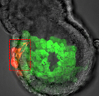
During blastocyst development, asymmetric cell divisions generate polar and apolar daughter cells, which organise into outer and inner positions, respectively, to form the trophectoderm (TE) and inner cell mass (ICM) lineages. The Hippo signaling pathway is crucial for setting up this early lineage specification, but how Hippo signaling relates to cell position and polarity remains unclear. In this issue (p. 2813), Yojiro Yamanaka and colleagues carefully analyze the initiation process of the first lineage specifications in the 16-cell mouse embryo. The authors count the number of polar/apolar cells and outer/inner cells in intact embryos and find that many apolar cells are located in outer positions with only one or two cells fully internalized. Notably, many of these outer apolar cells have high cytoplasmic phosphorylated YAP, an early marker for inner cells. Further analysis of isolated 8-cell blastomeres confirms that polar and apolar cells have intrinsic differences in the regulation of phosphorylated YAP prior to setting up the outer/inner configurations in the embryo. The authors suggest that polarity regulates the outer/inner cell positioning, as well as Hippo signaling, in order to activate TE and ICM lineage specification.

Scribble gets done for intracellular trafficking

FigureCell polarity is fundamental for biological activity across many varied cell types within different animal species. Intracellular trafficking regulates the differential distribution of proteins that is fundamental to establishing cell polarity, but how cell polarity regulators exert their effects on trafficking machinery is largely unknown. Now, on p. 2796, David Bilder and colleagues identify a specific and unexpected role for Scribble, a conserved core polarity protein, in controlling cargo sorting during intracellular trafficking in several Drosophila epithelial tissues. The authors show that Scribble mutants phenocopy endocytic internalization mutants but that they themselves are not defective in endolysosomal trafficking. Instead, Scribble controls cargo sorting within the retromer pathway, a system used to recycle proteins from endosomes to the trans-Golgi network. Depletion of the Scribble module affects the localisation of canonical retromer-dependent cargo, such as Wntless and Crumbs, but does not affect cargos that are retromer independent. This work brings a new aspect to the interplay between membrane traffic and polarity, and will help to decipher how these two essential pathways interact to establish and maintain epithelial polarity.


PLUS…


Regeneration, morphogenesis and self-organization

Dev2745In March 2014, the RIKEN Center for Developmental Biology in Kobe, Japan, hosted a meeting entitled ‘Regeneration of Organs: Programming and Self-Organization’. Scientists from across the globe met to discuss current research on regeneration, organ morphogenesis and self-organization – and the links between these fields. As discussed by Daniel Goldman, a diverse range of experimental models and organ systems was presented at the meeting, and the speakers aptly illustrated the unique power of each. See the Meeting Review on p 2745.


Cellular and physical mechanisms of branching morphogenesis

nelsoncropppedBranching morphogenesis is the developmental program that builds the epithelial trees of various organs, including the airways of the lung, the collecting ducts of the kidney, and the ducts of the mammary and salivary glands. Recent advances in cell lineage analysis and real-time imaging have uncovered surprising differences in the mechanisms that build these diverse tissues. Victor Varner and Celeste Nelson review these studies and discuss the cellular and physical mechanisms that can contribute to branching morphogenesis. See the Review on p. 2750.


Harnessing developmental processes for vascular engineering and regeneration

DEV2760The formation of the vasculature is essential for tissue maintenance and regeneration, and understanding how vascular formation is coordinated in vivo can offer valuable insights into engineering approaches for therapeutic vascularization and angiogenesis. Here, Kyung Min Park and Sharon Gerecht discuss how the process of vascular development can be used to guide approaches to engineering vasculature.  See the Review on p. 2760.


Thumbs up (No Ratings Yet)
Loading...

Categories: Research

Navigate the archive

Use our Advanced Search tool to search and filter posts by date, category, tags and authors.

The Embryology Course at the MBL

Posted by , on 7 July 2014

Right now I am attending the Embryology Course at the Marine Biological Laboratory (MBL). I am a graduate school student from Osaka University in Japan and I am looking for what I will do during my postdoc next year. This course supplies both lectures and experiments for various modules, including C. elegans, Drosophila, planaria, hydra and frog and many other organisms. Previously, I have only worked with mouse, so this course is a very good opportunity for me to learn many kinds of animal development.
I have already spent a few weeks here, and they have been more wonderful than I had expected. Today I want to talk about the lectures, experiments and friends I have met here.

Lectures
Every morning we have a lecture from a different expert. They teach not only basic knowledge of each animal, but also discuss frontier research in their field. After the lecture, we have a research discussion. During this time, the students get to discuss the lecture with the expert and almost all students ask questions. The students have a positive attitude and this helps to further stimulate my interest. Unfortunately, one hour is not enough time for us to discuss everything. However, we have a chance to go to dinner with the lecturer and engage in further discussion of the topic. At that time we talk not only about science but our life plans, hobbies, and so on. This is a good opportunity to form a more personal and meaningful relationship.

Experiments
In the afternoon, we perform experiments. To my surprise, we are able to perform whatever experiments we want. There is a protocol of course, but there is a wide variety of different experiments to perform and no restrictions. Therefore, we can make a plan of experiments ourselves, enabling us to arrange and possibly improve the experiment.
The analyzing facility is very substantial. For example, there are more than 3 conforcal microscopes, a Lightsheet fluorescence microscope, a laser ablation system and an electroporator.
In the 2nd week of the course I succeeded in the time-lapse imaging of follicle cell rotation in Drosophila eggs. Follicle cells rotate during their development. I have never worked with Drosophila so it was a little hard time to take the egg from the ovary and perform the time-lapse imaging. However, after many tries I eventually got the result. I was very happy and had a great experience.

Friends
The people in the course are from various countries, including the US, Germany, Croatia, Argentina, Taiwan and so on. We do not all study the same organism, but we all are interested in developmental biology. The students are together almost every day from morning to midnight, so we have plenty of opportunities to interact with each other and make friends. This allows me to know the research techniques and research goals of the same generation in other countries, which helps to expand my views.

I really look forward to spending the rest of the course with these students!!

Thumbs up (3 votes)
Loading...

Tags: ,
Categories: Events

Announcing DMM’s special issue ‘Spotlight on Zebrafish: Translational Impact’

Posted by , on 6 July 2014

In recent years, the zebrafish has emerged as an increasingly prominent model in biomedical research. Its optical transparency for the first few weeks, high fecundity and ex vivo fertilization have made it traditionally suitable to study developmental biology. However, over the past decade these same features have enabled the zebrafish to become a preeminent disease model and tool for studying disease mechanisms. Discoveries in zebrafish disease models are leading to new perspectives on human disease and new drugs that are entering the clinic in diverse areas from cancer to tuberculosis.

To showcase the translational impact of the model across multiple disease areas, Disease Models & Mechanisms has compiled a Special Issue packed with reviews, research and resource articles from researchers at the cutting-edge of their respective disease area of interest.

F1.medium

In the front section of the issue, Jennifer Phillips and Monte Westerfield provide an overview of the use of zebrafish in translational research in an ‘At a Glance’ poster article, illustrating recent examples of successful clinical translation of zebrafish studies. In the other Review articles, important insights from the zebrafish model into cancer, cardiovascular disorders, infectious diseases and more are discussed, with contributions from Jason Berman, Randal Peterson, David Langenau, Annemarie Meijer, Wolfram Goessling and others.

In the research section of the issue, striking examples of how the zebrafish can advance our understanding of disease mechanisms and contribute to the development of new disease models and therapeutic tools are presented. For example, Ana Vacaru, Kirsten Sadler and colleagues analyze zebrafish liver steatosis, protein markers and target gene expression to provide a new perspective on the unfolded protein response (UPR) and its role in fatty liver disease (FLD). In another study, Tamara Stawicki, David Raible and colleagues use forward genetics to identify new genes involved in resistance to hearing loss, a disorder that affects over 35% of people over 65. Alexa Burger, Daniel Haber and colleagues provide a new zebrafish model of chordoma, a rare type of bone cancer that is thought to arise from the remnants of embryonic notochord cells and for which, until now, there has been no animal model, few cell lines and limited treatment options. Nadia Danilova, Shuo Lin and colleagues use two genetic zebrafish models of ribosomal protein (RP) deficiency and find that treatment with exogenous nucleosides leads to improved survival, increased blood count and decreased morphological defects, suggesting the use of these compounds for novel therapeutic purpose in humans with RP deficiency, such as Diamond Blackfan anemia patients. Exquisite live imaging in zebrafish to visualize immune response is used by Tjakko Van Ham, Ben Giepmans and colleagues to study the resolution of neuroinflammation in vivo and by Mai Nguyen-Chi, Georges Lutfalla and colleagues to model chronic inflammation during transient Escherichia coli infection in the notochord.

For a more detailed introduction to our special issue read our Editorial by E. Elizabeth Patton, Paraminder Dhillon, James F. Amatruda and Lalita Ramakrishnan and to read all the articles in the issue, for free, visit http://dmm.biologists.org/content/7/7.toc

Don’t forget our special ‘Model for Life’ article in which the DMM Editor-in-Chief, Ross Cagan, interviews Leonard Zon, pioneer in the zebrafish disease models community. In this article, Zon recalls the evolution of his career from developmental biologist to physician-scientist and the stories behind some of his major research accomplishments. He also discusses challenges and opportunities in zebrafish research and provides advice on translating basic research findings to the clinic.

F1.medium (1)


 “I think you could easily argue that zebrafish is the best chemical system. Access to the embryos is so easy, and the field really blossomed with interesting screens into all sorts of phenotypes”.


Read the full interview here: http://dmm.biologists.org/content/7/7/735.full


Thumbs up (1 votes)
Loading...

Tags: , ,
Categories: News

Goalward-bound: why biological research is like football

Posted by , on 3 July 2014

june 2014

The 2014 FIFA World Cup has mesmerised football fans all around the world over the past weeks, but besides just the fancy footwork on display, we’ve also seen some amazing athleticism. Many of the matches have taken place under scorching, highly humid conditions! Though this might not be foremost in our minds as marvel at the ball-mastery and cheer on our favorite teams, at the same time we are witnessing the marvel of muscle tissue too.

Muscle consists in organized patterns of muscle fibers that contain multiple nuclei. During development, these fibers are assembled from fusion of muscle cells. In the fruitfly Drosophila, founder cells fuse with multiple fusion-competent myoblasts (FCM) to form the muscle fiber. In a recent study published in Development, Ciglar and colleagues attempted to understand a bit more how different muscle cell fates (ie: founder cells and FCMs) are determined during development.

They showed that the proteins tramtrack69 (Ttk69) and lame duck (Lmd) were essential for the normal development of Drosophila muscles. Interestingly, they showed that the acquisition of FCM identity resulted from the combined actions of Ttk69 and lame duck: Ttk69 repressing the expression of founder cell genes whereas Lmd activating the expression of FCM genes.

On this picture you can observe (in white) the expression of β-3 tubulin, a marker of cell “skeleton”, in normal embryos (left picture) and in embryos in which Ttk69 has been deleted by genetic engineering (right picture). Since the deletion of Ttk69 leads to the formation of abnormal muscle fibers, the authors conclude that Ttk69 is mandatory for normal muscle development.

This study is a step toward the understanding of the complex machinery involved in muscle development. Robust understanding of what factors direct cell fates during development is key for stem cell biologists. As we try to direct stem cells towards a specific fate, we try to recapitulate development, and we base our experimental strategies on what we know from developmental biology. In this sense, biological research is like football: scoring a beautiful goal involves patience, hard work, and usually a long skillful build-up involving players with different expertise. And as we focus on the strikers that reach the scoring line, we must remember to admire the entire team!


Picture credit:

Ciglar, L., Girardot, C., Wilczy ski, B., Braun, M., & Furlong, E. (2014). Coordinated repression and activation of two transcriptional programs stabilizes cell fate during myogenesis Development, 141 (13), 2633-2643 DOI: 10.1242/dev.101956
 

Thumbs up (1 votes)
Loading...

Tags: , ,
Categories: Research

This month on the Node- June 2014

Posted by , on 1 July 2014

Node 4th birthday


This month the Node celebrated its fourth birthday! Here are some of the other highlights:


Research:

Embryo electroporation2– Kevin Chalut wrote about his recent paper  at the crossroads of biology and physics, where he shows that the nucleus of ES cells exiting pluripotency have an unusual physical property.

– How is the primitive streak formed? Octavian discussed his eLife paper

– Elsa and Aitana described a new embryo electroporation method that allows higher efficiency rates and reduces cell damage, published in Development

– And Thomas gave his perspective on past and current work on the evolution and mechanisms of segmentation.


Meeting reports:

– Juan attended the Annual Meeting of the Japanese Society of Developmental Biologists, which took place in Nagoya last month.

– A mix of lectures, practicals and community building featured at the latest Master Course on Bioimage Data Analysis.

– We had the pleasure to host two posts by high school bloggers who attended this year’s ISSCR meeting in Vancouver (here and here).

– And Angelo shared his reflections on the Woods Hole Embryology Course, where he has taught for the last 6 years.


Photo Cred: Stem Cell Network

 Photo Cred: Stem Cell Network


 Also on the Node:

P Ingham– From trithorax to hedgehog, from Drosophila to zebrafish: read our interview with this year’s Waddington Medal winner Phil Ingham.

– Did you know that there is a model organism used in developmental biology that needs artificial moonlight? Read ‘A day in the life of a Platynereis dumerilii lab‘ to find out more!

– Developmental biologist Julian Lewis sadly passed away last April. We reposted an obituary by Paul Martin and David Ish-Horowicz looking back on his life and work.

– And Megan shared her tips on how you should prepare your application for a postdoc or lectureship position.


Happy reading!


Thumbs up (1 votes)
Loading...

Categories: Highlights

A key component of cell division comes to light

Posted by , on 30 June 2014

Jens Lüders leads the Microtubule Organization laboratory (Photo: Battista/Minocri, IRB Barcelona)

 “la Caixa” PhD student Nicolas Lecland is the first author of the study published in Nature Cell Biology (Photo: Battista/Minocri, IRB Barcelona)

A breakthrough at IRB Barcelona fills a knowledge gap in understanding how the cell division apparatus, the mitotic spindle, is formed.

The in vivo visualization and monitoring of the starting points of microtubules — filaments responsible for organising the mitotic spindle — provides novel insight into the dynamic architecture of this structure.

The findings will also contribute to understanding how the mitotic spindle is perturbed by drugs that target microtubules and that are used in chemotherapy.


The division of a cell in two requires the assembly of the mitotic spindle, an extremely complex structure, which is the result of the coordinated action of a multitude of proteins and a finely tuned balance of their activities. A large part of the time that a cell requires to divide is devoted to assembling the mitotic spindle, which, superficially, resembles a ball of thread with the shape of a rugby ball.

The most abundant components of the spindle are the microtubules. “By labelling the ends of thousands of these fine filaments, which are indispensable and extremely dynamic and variable, we have finally been able to follow their distribution and movement during the assembly of the mitotic spindle,” explains Jens Lüders, a cell biologist from the Institute for Research in Biomedicine (IRB Barcelona). The breakthrough appeared yesterday in the advanced online edition of the journal Nature Cell Biology.

“For more than 10 years we have been able to track only the growing ends of microtubules but not the starting points. As a result, we lacked essential information in order to understand the dynamic architecture of the mitotic spindle and how it contributes to cell division,” says Lüders. Headed by the German scientist who runs the Microtubule Organisation group at IRB Barcelona, the study carries only two names, his own and that of the French researcher Nicolas Lecland, first author, who completed his PhD at IRB Barcelona through a “la Caixa” fellowship.

The scientists have demonstrated that the protein γ-tubulin localizes at the starting points of the microtubule filaments and is relatively stably associated with these structures. Using a version of γ-tubulin that carries a fluorescent label activated by laser light, the researchers were able to follow the movement of the starting points of microtubules within mitotic spindles by filming dividing human cells.

The Advanced Digital Microscopy Facility, a joint IRB Barcelona-Barcelona Science Park Facility run by the IRB physicist Julien Colombelli, has been crucial for setting up the technology required. “The success of this study is also the result of the technical know-how and cutting-edge technology available, without which we would never have been able to tackle this project,” emphasizes Lüders.

The researchers describe for the first time where most microtubules form inside the mitotic spindle, how they develop, and how their starting points are transported—with the help of three motor proteins—to opposite poles of the spindle, where they attach. Simultaneous to this process, the opposite ends of the filaments extend towards the cell centre, where they interact with chromosomes.

When the spindle is finally assembled, the microtubules pull the chromosomes to opposite poles and initiate the physical division of the cell. “We now have a more complete understanding of how the spindle assembles and functions and can use our novel marker for testing old and new hypotheses about underlying mechanisms,” says the scientist.


A new tool to study cancer

In addition, the breakthrough paves the way to “better” understanding the mode of action of drugs that inhibit microtubules and that are used in chemotherapy. These kinds of drugs impede the mitotic spindle, thus preventing cell division and interfering with tumour growth.

In spite of the many years of clinical success of these treatments against cancer, little is known about how they impair spindle architecture and function. Although these drugs are highly efficient, they do not show the specificity desirable as they also affect healthy dividing cells. In addition, they affect non-dividing cells such as neurons, in which microtubules also have important functions.

“A better understanding of the differences in spindle organisation between cancer and healthy cells and how they respond to microtubule-targeted drugs is essential in order to optimise treatments, for example by identifying more specific drugs or new targets. This tool could be useful to achieve these objectives,” states the researcher.

The study has been supported by structural funds from the Generalitat de Catalunya, a Marie Curie grant from the European Union, and the Plan Nacional, of the Ministry of Economy and Competitiveness.


Reference article:
The dynamics of microtubule minus ends in the human mitotic spindle
Nicolas Lecland and Jens Lüders
Nature Cell Biology (2014) Doi: http://dx.doi.org/10.1038/ncb2996

Video: gtubpaGFPmerge copy


This article was first published on the 30th of June 2014 in the news section of the IRB Barcelona website


Thumbs up (No Ratings Yet)
Loading...

Tags: , , , , ,
Categories: Research

StemCellTalks sends high school bloggers to the ISSCR pt. 2

Posted by , on 26 June 2014

StemCellTalks is a Canadian high school stem cell outreach initiative that has been running in 7 cities in Canada since 2010. The program has featured over 50 stem cell “experts” during this time, involved the participation of over 500 gradute student volunteers and reached over 5000 grade 11/12 students. This year, sponsored by Stem Cell Network and Let’s Talk Science, the Vancouver chapter was able to partner with the International Society for Stem Cell Research and send five talented student bloggers – Lauren Dobishok, Tanner Jones, Mindy Lin, Vivian Tsang and Michelle Tse –  to its Annual Meeting, which was hosted in Vancouver last week from June 18-21th. Three of these blog posts (herehere and here!) have been featured on another excellent stem cell blog – Signals – and we are happy to be able to share the final two posts here on The Node!

What is stem cell tourism? Narrated by Professor Timothy Caulfield from Stem Cell Network on Vimeo.


By Tanner Jones (Dr. Charles Best Secondary, Vancouver, British Columbia, Canada)

With the promising restorative properties of stem cells, the hopes for treating a variety of diseases are close at hand; however, are these discoveries being accurately conveyed to the public? What are the repercussions of showing diseased patients a treatment that may not be available to them? As patients search for these therapies, many will travel to other countries where their regulatory laws are not in compliance with western standards. This hazardous phenomenon has been classified as stem cell tourism, and it poses an immense risk to patients who seek treatment in illegitimate clinics.

While attending StemCellTalks Vancouver, a conference where youth are educated on the capabilities of stem cells, I was captivated by Dr. Tania Bubela’s speech. Her account of how media can exaggerate research to the general public resonated with me. Dr. Bubela explained that while many clinical trials using stem cells are being run, these trials usually take as long as fourteen years to complete. As these clinical trials are being performed, the media often overstates the work that is being done, creating a certain amount of hype towards the general public. Although this ripple effect seems positive, it leaves many desperate patients confused as to why these treatments are not accessible to them. While seeking treatments that have been reported by the media, many individuals will stumble upon clinics in other countries who have promised successful outcomes for clinical therapy. Using patient testimonials, perspective applicants for these clinics are drawn in, hoping that there experience will be positive as well. In reality, most of these clinics have little evidence or research that supports their claims, yet patients will travel great lengths to visit them as they feel it is their only option. Not only are these clinics extremely expensive, in some cases, treatments my result in harmful side effects for the patient.

The 2014 ISSCR Annual Meeting, held at the Vancouver Convention Centre, provided some of the attendees a rare opportunity to gain a greater understanding of the ethical issues associated with stem cell research. As part of this conference, I was quickly introduced to an issue surrounding stem cell tourism during the Presidential Symposium where Dr. Paolo Bianco, Dr. Elena Cattaneo, and Dr. Michele De Luca, were being presented with the ISSCR Public Service Award. These phenomenal scientists have been championing the cause to halt the introduction of a new stem cell treatment in Italy. The Stamina Foundation in Italy has been treating patients with unproven stem cell therapies that have not been tested in rigorous clinical trials. The Foundation claims that by using mesenchymal stem cells, they can treat Parkinson’s disease as well as Spinal Muscular Atrophy; however, there is no evidence that mesenchymal stem cells can aid in the treatment of either of these diseases. One of the potential dangers of this therapy is the possible generation of bone or fat in organs. These public figures have been tirelessly debating the medical standards and regulatory oversights associated with the Stamina Foundation. As Dr. Bianco humbly accepted his award, he stated that researchers and physicians should be protecting patients from the physical harm, the financial exploitation and the moral illusion that can be produced by these illegitimate clinics.

With the daunting task of ending stem cell tourism, some wonder if it will ever be accomplished. Despite the challenges, Dr. Zubin Master, a Professor at the Albany Medical College, has proposed a few ideas that may lead to the extinction of this problem.  He suggests that physicians, patients and the public should be educated on the danger of these unproven therapies. If a greater understanding is developed within the population, many people will be less likely to engage in stem cell tourism. Dr. Master also believes that the most powerful initiative that can be taken to end stem cell tourism would be the involvement of patient advocacy groups. As ambassadors for their disease, patient advocacy groups disseminate information and educate individuals who are suffering from the same disease.  These trusted organizations are perceived as a neutral party with the patient’s best interests in mind, while some individuals may view scientists and clinicians as a barrier to certain treatments due to scientific protocols and regulations to clinical trials. With the sharing of information and by releasing statements on the potential risk of illegitimate stem cell clinics and the need for strict regulations, patient advocacy groups can generate an influential effect on patients currently thinking of participating in stem cell tourism.

Will stem cell tourism continue to be a problem in the future? With the prospective advancements in regenerative medicine and other treatments, many hope that patients will remain in their country to seek therapy. Until that time, it is possible that patients may continue to expose themselves to the possible physical harm and financial exploitation associated with these unproven therapies. Unless they are educated on the potential hazards of these illegitimate clinics, stem cell tourism will continue to attract those who are desperate and feel they have no alternatives.

Thumbs up (1 votes)
Loading...

Tags: , ,
Categories: Events, Outreach

PhD position on Drosophila intestinal homeostasis in Cardiff

Posted by , on 25 June 2014

Closing Date: 15 March 2021

the JQ lab is looking for candidates for one 3-year PhD studentship to work on the regulation of intestinal stem cell neutral competition during the homeostasis of the Drosophila adult intestine, at the European Cancer Stem Cell Research Institute, Cardiff University.

Please note deadline is 11 July 2014.

Details, application link and poster:

http://www.findaphd.com/search/ProjectDetails.aspx?PJID=55561


PhD_JQ_Cardiff_Oct2014

Thumbs up (No Ratings Yet)
Loading...

Categories: Jobs

Obituary: Julian Hart Lewis (1946-2014)

Posted by , on 25 June 2014

This obituary first appeared in Development.


Paul Martin and David Ish-Horowicz look back on the life and work of their long-time friend and colleague Julian Lewis, who passed away on April 30th 2014.


Julian LewisJulian Lewis made unique contributions to several areas of cell, developmental and theoretical biology. He combined a formidable intellect and mathematical training with experimental dexterity and deep biological insight, and used these to great effect to study key questions in early embryonic patterning, neurogenesis and, most recently, Notch signalling and somitogenesis. His fluent prose was evident in his publications and also in the textbook Molecular Biology of the Cell, to which he was a major, founding contributor. His kindness and gentleness endeared him to all, especially to the many people whom he mentored as they passed through his lab. He inspired them and his numerous collaborators and colleagues, all of whom will be bereft at the loss of an irreplaceable colleague.

Julian grew up in West London, attending St Paul’s School, where he was an outstanding linguist. He studied physics at Oxford University, remaining there for his DPhil in theoretical physics. He then spent eighteen months as a postdoc at the Institute for Physical Problems in Moscow, helped by his fluent spoken and written Russian – just one of his non-English languages.

Julian’s long-standing fascination with natural history led him to accept a postdoc position in Lewis Wolpert’s lab at the Middlesex Hospital, which soon became the epicentre of studies on chick limb patterning – the system that triggered many influential theories of vertebrate embryonic patterning that we now take for granted. Julian began working closely with Denis Summerbell, a doctoral student in the lab and, together, they provided the theoretical and experimental basis for the progress zone model of limb patterning, which still influences our ideas of how spatial organisation arises during vertebrate development (Summerbell et al., 1973).

Other colleagues in the Wolpert lab at the time included Jonathan Slack, Cheryl Tickle, Jim Smith and Nigel Holder. This team unearthed many important mechanistic insights from cut-and-paste surgical experiments, thereby laying the foundation for subsequent molecular advances in understanding limb patterning. Julian’s big contributions were in figuring out how cells in the progress zone might measure ‘time’, and how this in turn imparts proximodistal positional values on cells. Several papers involved a combination of experimental and mathematical analysis; they included Julian’s first exploration of how timing mechanisms might pattern tissues – a much understudied problem at the time.

In 1978, Julian set up his own lab at the Anatomy Department of King’s College, London on the Strand,where he taught histology, with a healthy dose of cell biology, and also met his American future-wife Sherry, who was in London doing a PhD in neuroanatomy. His lab there began to examine how the various tissue components of the limb – skeletal elements, muscles, nerves and blood vessels – arrange themselves appropriately in relation to one another. The experiments revealed a hierarchy of interactions such that, for example, connective tissue defined ‘trunk roads’ taken by all limb nerves, which then followed individual paths in response to chemoattractants released by skin or muscle targets (Lewis et al., 1981).

While still at King’s, Julian and his postdoc Gavin Swanson switched models and began to study development of the otic vesicle, which gives rise to the sensory epithelium of the inner ear. One transition paper had them peeling open the otic vesicle and grafting it onto a host chick limb bud, where it differentiated to form the exquisite normal patterning of hair cells, making the process accessible to experimental manipulation (Swanson et al., 1990). Julian’s innovative idea of injecting white paint into the ear to visualise its morphology was a further example of his ingenuity.

In 1986, Julian jumped at an offer to move his lab to the new Imperial CancerResearch Fund (ICRF) Developmental Biology Unit, which had just been set up in Oxford under the directorship of Richard Gardner. Here was a thriving community of interactive groups with most of the major model systems represented along a compact floor at the top of the Zoology Building: flies (Ish-Horowicz and Ingham), chick (Lewis), frogs (Slack), mice (Gardner, Copp and Beddington) and, later, zebrafish (Ingham). These were very exciting times in developmental biology, as striped gene expression patterns in Drosophila competed with new mesoderm-inducing factors in Xenopus for pages in Nature. Julian’s interactions with everyone in the Unit were key to its success and to that of its students and postdocs.

Julian’s major interest remained the inner ear, and he uncovered clues about its morphogenesis, the inductive signals that kick-start its invagination from head ectoderm and how the semicircular canals are formed. He also made a foray into wound healing; in the early 1990s, he published a paper showing that wounds in the simple embryonic epidermis closed by means of an actomyosin cable that assembled in the leading edge cells (Martin and Lewis, 1992). This cable turned out to be a general feature of wound healing in many systems, and is also present during normal morphogenesis, e.g. during dorsal closure in fly embryos.

Julian also became increasingly fascinated by the problem of how local patterning was established in the otic epithelium; in particular, with the beautiful hexagonal arrays of individual hair cells, each hair being surrounded by separating support cells. This patterning smelled of ‘lateral inhibition’, the process mediated by Delta-Notch signalling that had been shown to generate regularly spaced neural precursors in Drosophila – an idea that led him to embark on a series of landmark studies of Notch signalling in vertebrates.

In collaborationwith the Ish-Horowicz lab down the corridor and the Kintner lab in San Diego, Julian’s team showed using Xenopus and chick that a cell’s decision whether to become neural is indeed regulated by Delta-Notch signalling and lateral inhibition – individual differentiating neural cells express the ligand Delta, which inhibits direct neighbours from also differentiating and preserves them as progenitor cells (Henrique et al.,1995; Chitnis et al., 1995). Subsequent work in the chick retina confirmed this model, showing that Delta- Notch signalling regulates the timing and progression of differentiation, but not the type of neuron formed (Henrique et al., 1997).

The Oxford Unit was closed in 1996, and several groups, including the Ish-Horowicz and Lewis labs, transferred to the main ICRF (now Cancer Research UK) institute at Lincoln’s Inn Fields in London. There, Julian began to switch his research to zebrafish, taking advantage of its genetics and suitability for advanced microscopy (the physics of which he understood well) to study the molecular processes involved in cell fate diversification in vivo.

The Delta-Notch story continued with studies of its role in patterning in the developing zebrafish gut and vasculature, but Julian also began a series of incisive experiments on vertebrate segmentation, studying in particular how the regular production of somites (the precursors of our axial skeleton and muscles) is controlled by a molecular oscillator (‘clock’). Here too, he showed that Notch signalling was crucial, acting to synchronise the clocks of neighbouring cells (Jiang et al., 2000). Equally ground-breaking, he modelled the zebrafish clock as a simple, delayed negative-feedback loop based on transcriptional autorepression. He showed that the period of such a clock would depend on the kinetics of synthesis and breakdown of the mRNAs and proteins of the clock, rather than on their absolute protein concentrations (Lewis, 2003). Crucially, he showed that noise in the circuit, far from disrupting the oscillations, contributes to the circuit’s robustness. This single-author paper has formed the basis for most of the subsequent work in the field.

For his collective work, Julian was awarded the British Society of Developmental Biology’s highest accolade, the Waddington Medal, in 2003, and was elected to EMBO membership in 2005 and as a Fellow of  The Royal Society (FRS) in 2012. He closed his lab in 2012 but continued working; his most recent paper was published this April in Development (Soza-Ried et al., 2014) and was a perfect example of what the funders now call ‘predictive science’. His lab showed that the development of normally sized somites could be rescued in embryos lacking endogenous Delta expression by delivering regular pulses of Delta activity, thereby confirming his mathematical prediction of Notch-mediated clock synchronisation. It was particularly appropriate that this last paper was published in Development – his 24th in this journal (and its predecessor, JEEM) – on whose editorial board he served between 1988 and 1998.

Julian was a wonderful mentor who gently nudged the best out of all of his graduate students and postdocs, showing immense patience with those of us who were slower than he was in grasping inferences, concepts and equations. He wrote beautifully; his prose was almost lyrical, as if it had been ‘sprinkled with magic’ as described by one journal editor.

For the last ten years of his life Julian suffered from prostate cancer, which led to his making an important contribution to the cancer research community. He talked widely on the biology and treatment of cancers to general scientific and lay audiences. These were especially powerful and important talks; near the end of each, he would show data from a drug trial (Fong et al., 2009) and a bone scan showing metastases, at which point he indicated that he himself was a participant in the trial and that the bone scan and metastases were his. The bravery of a patient advocate talking so lucidly and in such clear scientific language about his personal experience taking a novel cancer drug had tremendous scientific and emotive impact. His tremendous courage and stoicism were also evident in his continued productivity despite his pain and discomfort. In the weeks before he died, Julian also delivered final drafts of two of his chapters for the 6th edition of Molecular Biology of the Cell, as well as a new section on computational biology.

Julian was a scientist’s scientist, with a legendary ability to ask the gentle ‘killer’ question at a seminar despite sleeping through most of it. He could be slightly ‘over the top’ on matters of clarity and precision, once walking out of a seminar to check a controversial but critical fact from a journal article in the library – and he was the speaker! He also had an idiosyncratic dress style, classifying his sweaters into two categories: ‘with holes’ and ‘without holes’. But he was also a lovely human being and a ‘renaissance man’ whose wide interests incorporated music, artistic prints, ceramics (which both he and Sherry collected avidly) and second-hand books, which were usually read in the original Russian or French. He was a devoted husband and father – with Sherry he raised three science-orientated daughters. He is sorely missed by them, and by all of us who knew and loved him.


References:

Chitnis, A., Henrique, D., Lewis, J., Ish-Horowicz, D. and Kintner, C. (1995). Primary neurogenesis in Xenopus embryos regulated by a homologue of the Drosophila neurogenic gene Delta. Nature 375, 761-766.

Fong, P. C., Boss, D. S., Yap, T. A., Tutt, A., Wu, P., Mergui-Roelvink, M.,Mortimer, P., Swaisland, H., Lau, A., O’Connor, M. J. et al. (2009). Inhibition of poly(ADP-ribose) polymerase in tumors from BRCA mutation carriers. N. Engl. J. Med. 361, 123-134.

Henrique, D., Adam, J., Myat, A., Chitnis, A., Lewis, J. and Ish-Horowicz, D.(1995). Expression of a Delta homologue in prospective neurons in the chick. Nature 375, 787-790.

Henrique, D., Hirsinger, E., Adam, J., Le Roux, I., Pourquié, O., Ish-Horowicz, D. and Lewis, J. (1997). Maintenance of neuroepithelial progenitor cells by Delta- Notch signalling in the embryonic chick retina. Curr. Biol. 7, 661-670.

Jiang, Y.-J., Aerne, B. L.,Smithers, L., Haddon, C., Ish-Horowicz, D. and Lewis, J. (2000). Notch signalling and the synchronisation of the somite segmentation clock. Nature 408, 475-479.

Lewis, J. (2003). Autoinhibition with transcriptional delay. A simple mechanism for the zebrafish somitogenesis oscillator. Curr. Biol. 13, 1398-1408.

Lewis, J.,Chevallier, A., Kieny, M. and Wolpert, L. (1981). Muscle nerve branches do not develop in chick wings devoid of muscle. J.Embryol. Exp. Morphol. 64, 211-232.

Martin, P. and Lewis, J. (1992). Actin cables and epidermal movement in embryonic wound healing. Nature 360, 179-183.

Soza-Ried, C.,Ö ztü rk, E., Ish-Horowicz, D. and Lewis, J. (2014). Pulses of Notch activation synchronise oscillating somite cells and entrain the zebrafish segmentation clock. Development 141, 1780-1788.

Summerbell, D., Lewis, J. H. and Wolpert, L. (1973). Positional information in chick limb morphogenesis. Nature 244, 492-496.

Swanson, G. J., Howard, M. and Lewis, J. (1990). Epithelial autonomy in the development of the inner ear of a bird embryo. Dev. Biol. 137, 243-257.

Thumbs up (1 votes)
Loading...

Tags:
Categories: Research

A day in the life of a Platynereis dumerilii lab

Posted by , on 25 June 2014

Hello! I am Maggie Pruitt, a postdoctoral researcher in the Department of Genetics, Development, and Cell Biology at Iowa State University (Ames, Iowa, USA – think middle America or fields upon fields). I work in Dr. Stephan Schneider’s evo-devo laboratory, and my work mostly focuses on studying components of the Wnt/β-catenin signaling pathway during early Platynereis dumerilii development.

The Schneider lab is part of the small, but growing, Platynereis community. Many of the Platynereis labs seem to study developmental biology or evolutionary developmental biology. While the Schneider lab is focusing on early development and the early gene regulatory networks present in Platynereis, other labs work on eye and brain development, gametogenesis, the molecular mechanisms of how the moon affects the life cycle of Platynereis and how neuropeptides control swimming depth of Platynereis larvae within the water column, to name a few.


Platynereis dumerilii as a model organism

Platynereis is a polychaete annelid and belongs to the clade Lophotrochozoa, which is the third but understudied branch of bilaterally symmetric animals (understudied compared to ecdysozoans like the nematode C. elegans, and deuterostomes like the vertebrate mouse). These worms can be collected in the wild from the coasts of the Mediterranean or coasts of Western Europe; however, they can also be easily cultured in a lab.

 
Culture_room

Our culture room. Notable features include boxes of adult or young worms, our artificial moonlight, and an injection microscope (yes, we can inject Platynereis embryos!).


As Platynereis is a marine worm, the worms need seawater. Filtered natural seawater (NSW) is by far the best for the worms. But, if your lab is not located close to a seawater source (like us in the middle of the USA) or you want to avoid steep costs for shipping NSW to your lab, you need to get a little creative. Artificial seawater (ASW) can be made using a salt called Instant Ocean. While this can be technically called seawater, the worms (or more specifically the embryos) do not really survive well in it. So…. since the Schneider lab is far from the sea and the worms do not like ASW, what do we use? This is where creativity comes in. Actually, it is where Dr. Dennis Lavrov’s sponge lab (also at Iowa State University) comes in. We provide the salt to make ASW to the Lavrov lab and their sponges. Then, once this seawater has been “conditioned” by the sponges (i.e. when the Lavrov lab needs to change the water in their tanks), we take their wastewater, filter it, sterilize it, and voila! Seawater that our embryos like, cleverly named DSW (Dennis seawater). We use 100% DSW for all of the fertilizations. Once the worms are juveniles or adults, we keep them in a 50/50 mixture of DSW and ASW.

The Schneider lab Platynereis culture is not huge, so optimal breeding success is key. One way to maximize this is to keep the culture on a fairly tight routine. The culture should be maintained at 18oC or 64.4oF, a standard for Platynereis labs around the globe, as the speed of early development is very temperature sensitive. In addition to constant temperature, it is good to have the worms on a regular feeding and water change schedule. Our young and adult worms receive minced organic spinach (only the best, pesticide-free spinach is good enough) on Mondays and ground fish food flakes on Fridays. The last incredibly important thing is to have tight control of the daily light and dark periods.

Sexual maturity and spawning in Platynereis dumerilii are synchronized by a lunar cycle. In the lab, the culture is on an artificial light cycle. The worms experience summer all year round, and so have 16 hours of light and 8 hours of total darkness each day. In addition, they are exposed to a 28-day night cycle in which three weeks of pitch-dark nights are followed by 7-days of full moon. Our high-tech recreation of this lunar cycle comes in the form of a $1.99 night light from Walmart, and a very reliable undergraduate researcher as a timer to turn the light on and off on the correct days. Sexually mature worms can be collected 3-6 days after our “moon” phase. Any disruption in this light cycle (exposing the worms to light during the dark phase), like leaving a digital clock on that has a bright backlight, has detrimental effects on the breeding success.

Sexual maturity can occur in worms as young as three months old, but sometimes we have “geriatric” 18-month-old worms finally maturing. In Platynereis, completed sexual maturity lasts one day/night, so an artificial cycle with artificial light allows for collective sexual maturity in the culture. Maturing worms are easily distinguished from non-mature worms by their change in appearance. Maturing worms have an empty gut and become more opaque. As the worms continue to mature, the females become yellow (lots of eggs) and the males have a cream anterior and a red posterior (lots of sperm and blood vessels).

 
male_female_anterior

Anterior halves of a female worm (left) and a male worm (right). The distinctive colors, yellow versus cream/red, are noticeable.


The awesome power of the Platynereis mating system

In the wild, Platynereis will swarm at dusk. However, in the lab we keep the sexually maturing male and female worms in separate bowls so the worms can last until the morning. Matings are easy to set-up; all you need is a glass bowl, some DSW, and a sexually mature male and female. After two worms are placed together in a bowl, they will exchange pheromones, which triggers their nuptial dancing and mating ritual:


The female is swimming in small circles, and the male is swimming in large circles.  Pheromones are exchanged, the male first releases his cloud of sperm, and then the female releases her eggs (courtesy of Albrecht Fischer, University of Mainz, Germany).


Go ahead; watch the movie again. It is awesome and is partially responsible for getting me hooked on Platynereis. And at the end of this nuptial dance you are left with thousands of synchronously developing embryos and two dead worms. What a way to end your life, right? Perhaps before we pass judgment on how and when these worms die, I should give a bit more information on their life before the fatal spawning event.


Crash course to the life cycle of Platynereis

One of the many reasons why Platynereis is a good model system for developmental biology is that Platynereis develops rapidly. Platynereis begins as a 160μm egg and after instant fertilization of many eggs at once Platynereis embryos develop synchronously. In fact, the thousands of embryos from a single mating develop synchronously through the embryonic and larval stages. During early development, Platynereis embryos exhibit spiral cleavage. Spiral-cleaving embryos develop by a series of stereotyped asymmetric cell divisions that allows for the identification of individual cells by their positions and size. By 24h, the embryos have developed into a free-swimming larval form, a trochophore that swims freely within the water column, and after just 96h, Platynereis is a three-segmented juvenile worm that stays on the bottom of the dish. Segments are added throughout the lifetime of Platynereis at the tail end from a posterior growth zone, and Platynereis maintains a lifelong capability to grow and regenerate posterior segments after loss. The size of an adult worm varies tremendously, but on average they are about 35mm. During segment proliferation, the sexually immature worms live in self-spun silk tubes. The tubes are open at both ends and allow the worm to “check out” their environment, attack passing worms and prey, and eat their food. In their own world they are mostly holistic vegetarians that turn into fierce cannibalistic predators if the opportunity arises.
 

Here is one of approximately 90 boxes where we house our young and adult worms. In this box, you can see the many worms, each with their own silk tubes. If you look closely, you can even see one worm peeking out of its tube (right of center).


To sexually mature, Platynereis goes through a process called epitoky. Essentially, the whole body of the worm is irreversibly modified for reproduction, and the worm is transitioning from a sexually immature “atoke” form, to a sexually mature “epitoke” form. Some of the radical body modifications during this process include upgrades of motor and sensory organs, tune-ups of the muscular system for speedy swimming, resorbing of some muscles and all of the gut tissues, and the growing of thousands of gametes within their central body cavity. Indeed, the female and male worms are mostly reduced to growth chambers for gametes, bags of eggs and sperm, respectively. Once epitoky is complete, and the worm has reached its climax — one-day of sexual maturity, the worm will leave its silk tube home and swim into the pelagic zone (open water column). Here they will find like-minded partners (the good) or end up as a healthy protein shake for some higher ups in the food chain (the bad). After reproduction, the worms die. Maybe this death is still shocking to you, but the entire body of the worm changed for this one event…. the worm dies after completing a final task. From an evolutionary standpoint, the only task that matters.


A typical day in our laboratory

An ideal day in the lab is one in which everything is working correctly. Since this seems to be a mythical creature, I’ll stick to a typical day in our lab. Our typical days come in two flavors, depending on whether or not we have mature worms for fertilizations.

In the absence of sexually mature worms, a typical day in the lab consists of cloning, cloning, cloning, and maybe some more cloning. The goal of most of the cloning work is to make an in situ probe, and determine the expression pattern of the gene over the first 24h of development. The lab has an ever-growing list of interesting genes to clone, so this is a lab effort. I seem to spend most of my time working with all of the hard-earned embryos I collected in previous weeks (it can be hard to share embryos from late night fixations or embryos treated with pharmacological inhibitors). My favorite, and most frequent, activity is performing in situs. Cloning a new gene can be quite satisfying, but not nearly as much as uncovering a beautiful expression pattern that perfectly fits your hypothesis.

In the presence of sexually mature worms, I can typically be found running between our molecular lab (located on the 5th floor) and our culture room (located on the 7th floor). Maybe I’m being silly, but I feel extremely lazy taking the elevator up two flights of stairs. Since we only have sexually mature worms for less than two weeks each month, I try to take advantage of these days. Some of the fertilizations need to be saved to propagate the culture, but I try to use the rest. So, these days begin with a short run up to the culture room to set-up fertilizations. I’ll try to have a plan-of-action for the day, like how I plan to use the embryos, but sometimes, the worms just do not cooperate. Females weren’t feeling the males, all males and no females, etc. Pending total failure with the fertilizations, the embryos will be 1) used for microinjection or a pharmacological inhibitor experiment, 2) fixed for in situ hybridization or immunohistochemistry, or 3) used for RNA isolation. These days can get pretty hectic, but being busy means you and the lab will be set, sample-wise, for the weeks without the matures. It is only when slippery fingers make an appearance and multiple tubes of embryos or entire plates of treated embryos are dropped that these days are the worst. When this happens (yes, it happens), I usually get a coffee, then finish the day pretending the lab is in the absence of matures…. until the next morning.


Node day in the life new doodle square
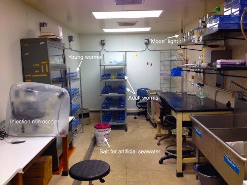
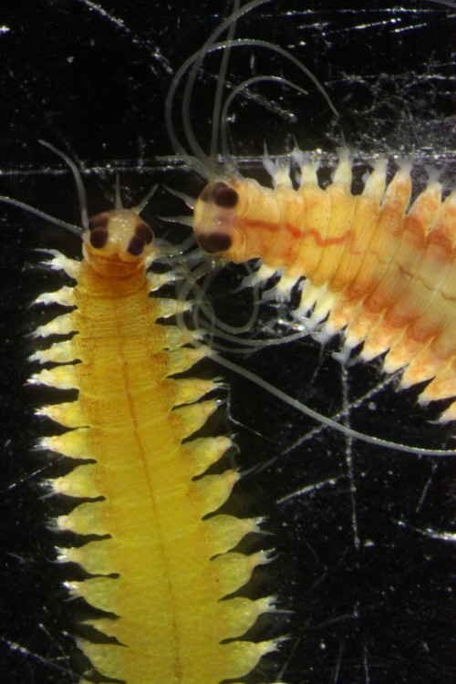
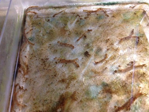
This post is part of a series on a day in the life of developmental biology labs working on different model organisms. You can read the introduction to the series here and read other posts in this series here.


Thumbs up (8 votes)
Loading...

Tags: , ,
Categories: Lab Life