The community site for and by
developmental and stem cell biologists

Literary Paper Openings

Posted by , on 24 May 2013

What would a paper be like if it started with the opening line of a famous book? Since yesterday Twitter has been buzzing with answers to that question! We have storified some of the tweets below, but the discussion is still up and running, so why not follow the hashtag #LiteraryPaperOpenings?

Thumbs up (2 votes)
Loading...

Tags: , ,
Categories: News

Navigate the archive

Use our Advanced Search tool to search and filter posts by date, category, tags and authors.

From science to art- an interview with sculptor Briony Marshall

Posted by , on 23 May 2013

Briony Marshall is a London-based sculptor and installation artist. Initially interested in science, Briony studied Biochemistry at the University of Oxford. After her undergraduate studies she moved on to art and sculpture, but her beautiful pieces have a strong scientific inspiration. Her first solo exhibition is currently at the Pangolin London Sculpture Gallery, and includes several developmental-biology inspired pieces. The Node interviewed Briony and asked her about her work.

Sculptor Briony MarshallThe Node: You initially trained as a biochemist. What made you decide to leave science and pursue an artistic career?

Briony: It took me a while to get there. I think I got a little bit put off by the idea of doing a PhD and research… the long process of being a researcher. If someone could have made me a professor straight away I would have been very happy! I really studied biochemistry because I thought it was the most interesting cutting edge bit of science, I don’t think I ever really saw myself as being a biochemist. It was purely because I enjoyed it and I thought it was a fascinating subject that holds quite a lot of answers to what goes on in the world.

The Node: A lot of your work, including the pieces in this exhibition, are inspired by science. Is science only a source of inspiration, or does your scientific training also instruct your creative process?

Briony: I am a scientist inherently, the way I view the world. I like to unpick how it works. So when I initially turned to art, I felt like I was turning my back on science and my previous career. I was trying to launch myself fully into the art world, which is perceived as a very different environment, and a different process. But as an artist you have to be true to yourself. The unique thing about an artist is that they have a particular point of view on the world, which is related to the whole of the experience that they have had, the times they live in, and their life. So, as I developed as a student (and I never intended it) I just started using science as metaphors. My molecular work looks at parallels between micro world and macro world- the way atoms interact with each other can give you insights into the way people interact with each other, and the way society works. That’s the lens through which I view the world, and it comes out in the work.

I’ve always been a practical person. I think that’s why I’m a sculptor, not a painter. 2D art can be very virtual… it’s creating illusions of other worlds, while as a sculptor you are dealing with materials, and gravity… I think a lot of sculptors are quite practical people- you have to be- and I think the sort of people who are drawn to sculpture are people who like problem-solving, which is very similar to people who are drawn to science.

You also asked about the creative process. It’s similar, in a way, to the scientific process, where you have to accumulate knowledge, you have to work very hard for a long time. But often the important bit is an intuitive leap, when you suddenly view things in a different way. I think that is very common to both science and art.

The Node: Could you explain your creative process a little more? How do you find the scientific themes that inspire your work, and how do they then evolve into an art piece?

Briony: It varies a little bit, but it often starts with reading. Sometimes it is just popular science books, sometimes more detailed research. The internet is a wonderful thing because you can browse and connect, and there are amazing departments that put a lot of scientific knowledge and lectures and information online now. Even 10 years ago, I would have had to be going to scientific libraries! I do a certain amount of research, and then there is a moment of faith and waiting for the idea to pop in.

Often there are themes that I’m interested in. As a sculptor I’m quite interested in how form develops. How you go from nothing to something, which is a creative process as well.  There are creative processes in nature: the birth of the universe in physics, or in developmental biology the development of complex forms. That relates to me as a sculptor- how you create form and complexity.

I have also developed my own methodology where I work really small, in little wax maquetes. I can work really fast on a piece, complete it in a single sitting, and then I will spend longer deciding what scale the final piece should be, and then enlarging it. For each of my Carnegie stages* in the exhibition, you can actually see 3 different scales. Each of them [the stages] has a little maquette that you can pick up and hold in your hand, which really gives you a sense of this little precious thing. And then there’s a series where I was really working out their forms in plaster, and they are almost portrait size. And then the final installation. It took me a while to decide what size to do them. They have a presence, and I really want people to go up and interact with them. They are almost equivalent volumes to a human body, hanging in the space.

Carnegie stages 6 - 10

The Node: You mentioned your Carnegie stages piece. What inspired you to create this piece, and what is the message that you are trying to convey with it?

Briony: I was doing some research for another piece in the exhibition, and I came across the charts of Carnegie stages. I was so struck at just looking at the particular ones I picked, stage 6 to 10. They are reminiscent of African masks, minimalist sculptures, of early greek Cycladic art… They [the biological representations] jumped out at me as they seemed already part of the cannon of sculpture. But then they are not, they are part of our own development. I thought it was fascinating. Why is there that link? Do people naturally tune in to these forms because there is some ancient link? And they do: people that come in to the exhibition have made links to various things they connect to in the canon of art.

The Node: Another developmental-biology inspired piece is Embryonic growth, which shows a young human embryo at the centre of a spiral. What was your inspiration for this piece?

Briony: Well, it is slightly embarrassing, because one of the inspirations is a scientist called Ruppert Sheldrake, who had the controversial idea of morphic fields. They are such radical ideas that people baulk at the idea of them. But as a sculptor it’s a fascinating idea: he thinks that some of the reasons why we don’t understand exactly how different parts of the embryo know where they are and develop form is that there is a morphic field… like the gravitational field, that we don’t necessarily fully understand but that we have clear evidence that exists.

In his hypothesis, the developing embryo might be tuning in to the fields of other shapes that have previously existed. As a sculptor that is an amazing idea, that by creating a shape in space you are not just creating it, but creating future resonances. It is an interesting idea, but I think it is more of a metaphor for things, rather than a real scientific thing. But it did make me think about all sorts of things influencing the embryo as it is developing. In the piece, the spiral around the embryo is a Fibonacci spiral, giving the idea of order, mathematical beauty and the patterns that exist. There is genetic inheritance, you have environmental factors, all these things around the developing embryo influencing how it grows, giving it energy, a pattern and a blueprint for its development.

Embryonic Growth

The Node: In addition to creating art you also teach, and you have tutored and lectured in the past on art and science at the Art Academy. I’m curious as to what message you try to get across to your students who are looking for inspiration for their work in science.

Briony: To be honest I haven’t necessarily given advice. The courses have been more showing the breadth of work that people do- there is quite a breadth of art and science, from philosophical to illustrative works. But I have told people that it needs to be a genuine mesh to what they are about as a person, rather than being a gimmick. Some people almost like the aesthetics of test tubes and scientific process, and scratch the surface of it. I think it is such an important part of society. If you are going into that realm you need to go further than the clichés for it to have value. Otherwise it’s a very superficial look.

Briony Marshall’s solo exhibition Life Forming is at the Pangolin London Sculpture Gallery until the 15th of June. You can find more information about the exhibition at the Pangolin London website and explore some of Briony’s past science-inspired work on her website.

*Carnegie stages are a standardized system of stages that chronologically describe the development of the vertebrate embryo

All images courtesy of Pangolin London; Photography Steve Russell

Thumbs up (7 votes)
Loading...

Tags: , , ,
Categories: Events, Interview, Science Art

Woods Hole Images Round 2- the winner

Posted by , on 21 May 2013

The polls have closed for the second round of beautiful images from last year’s Woods Hole course. Making an unexpected dash to victory in the last hours of voting, the two-headed regenerated planaria is the winner of this round!

2012Round2_C_large


This picture was taken by Chang Liu of the Shanghai Institute of Biochemistry and Cell Biology. It shows a regenerated Dugesia sanchezi planarian immunostained for acetylated tubulin (green) and phospho-histone-H3 (dividing cells, purple).


The runners-up to this competition were an image of a slipper limpet larva by Joyce Pieretti (University of Chicago), Manuela Truebano (Plymouth University), Saori Tani (Kobe University) and Daniela Di Bella (Fundacion Instituto Leloir); the embryo of a dwarf cuttlefish by Maggie Rigney (University of Texas, Austin) and Nipam Patel (University of California); and a Longfin inshore squid by Wang Chi Lau (Chinese University of Hong Kong).


Look out for this winning image in the cover of a coming issue of Development, and stay tuned for round 3 of the Woods Hole image competition!

Thumbs up (1 votes)
Loading...

Tags: ,
Categories: Images, News

San Francisco Declaration on Research Assessment

Posted by , on 17 May 2013

In December last year, a group of editors and publishers, including editors from our sister journal Journal of Cell Science, got together at the annual ASCB meeting to discuss ways in which we could improve the way in which scientific output is evaluated. There is much discontent in the community with the all-pervasive importance of the Impact Factor, and this meeting looked at how we might find a more balanced way to assess individual researchers and their work.

DORAToday, the results of this discussion are being made public, with the release of the “San Francisco Declaration on Research Assessment” website, and accompanying editorials in Science, eLife and other journals. As initial signatories to the Declaration, The Company of Biologists and its journals fully support, and comply with, the proposals for Publishers stated in the Declaration – look out for more information on this in an upcoming editorial in Development.

I’d encourage you to read the Declaration, and – if you agree with its principles – to sign it. In my personal opinion, reflected in what I frequently hear from members of the community, the prevalent use of the Impact Factor as a mechanism to judge a scientist’s worth (and hence future job prospects) isn’t healthy. I also think that developmental biologists may suffer more than some in other fields: the very nature of our work often makes for long-term projects, and so the two-year window of the Impact Factor does not reflect the speed of our field. So I hope that this Declaration will help to change the culture of research assessment, and to ensure that research and the researchers behind it are assessed on their individual merits and not on the number associated with the journal in which they happen to have published.

We’d love to hear your thoughts on this Declaration, so please comment below if you’ve got anything you’d like to discuss with the journal and with the developmental biology community at large.


Thumbs up (6 votes)
Loading...

Categories: Discussion, News

From Sequence to Function

Posted by , on 17 May 2013

DSC_0283_cleaned_small


It took longer than the human genome, if by only a few years, but it has finally arrived. The sequencing of the zebrafish (Danio rerio) genome reported in Howe et al. is one of two zebrafish publications to recently appear in  the journal Nature.  The second article, Kettleborough et al., makes use of this high quality genome sequence and not only creates the tools required for the functional annotation of all zebrafish protein coding genes, but describes the active pursuit of this goal.

About a decade ago, when the human genome was first published, there was a lot of hope and expectations that this would lead to an immediate advance in the treatment of many diseases and the understanding of ourselves. As these things often are, it turned out there is a lot more to understanding our genomes than just the decoding of a reference sequence. Now with the zebrafish genome in hand it is possible to see that 70% of human protein coding genes have a direct zebrafish ortholog. This at the same time represents 84% of all human genes with a disease association in OMIM.  Although both vertebrate organisms it still remains striking, almost humbling at just how close we as humans are to our aquatic relatives. It will be the continuation of detailed investigations involving model organisms which will play a fundamental role in connecting genotype to phenotype.

It is exactly this similarity which forms the basis of Kettleborough et al.’s  approach in actively knocking out all 26,000 zebrafish protein coding genes.  We still do not understand the function of a large proportion of our own genes but by providing loss of function alleles as a resource to the greater community and also functionally annotating these alleles we will hopefully gain greater insight into our own genomes.

Investigating the phenotypic outcomes of these alleles is now well under way at the Wellcome Trust Sanger Institute. The phenotypic consequences within the first 5 days of development are evaluated and annotated as part of a multi-allelic phenotyping approach explained in detail in Dooley CM et al.


All alleles, availability, phenotyping information and much more is available at: http://www.sanger.ac.uk/Projects/D_rerio/zmp/ so stop by and have a look!

Thumbs up (3 votes)
Loading...

Tags: , , , , ,
Categories: News, Research, Resources

Stem cells crossing boundaries

Posted by , on 16 May 2013

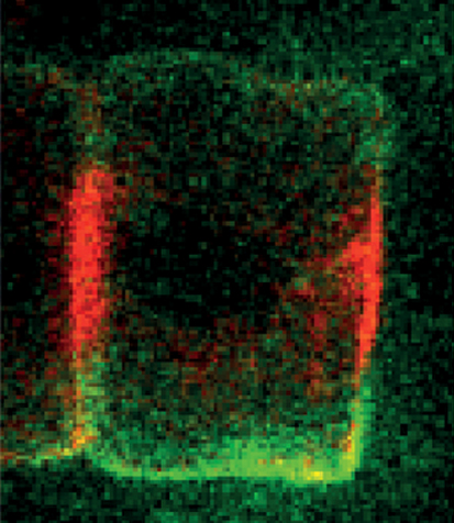
drosophila midgut hindgutFor most of us, we don’t all end up settled as adults in the same town where we were born.  The same is true for many cells, including some stem cells in the fruit fly intestine.  A recent paper describes the migration of progenitor cells, some of which will later become stem cells, in the intestine of developing flies, with the help of stunning images of these boundary-crossing cells.

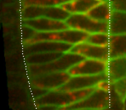
Adult stem cells divide and differentiate to compensate for cell loss or cell injury in adult tissue.  Understanding where these adult stem cells originate during development and how they migrate to their final position in adult tissue is an important question in stem cell biology.  A recent paper in Development describes the migration of Drosophila intestinal stem cells during metamorphosis.  Takashima and colleagues traced the migration of progenitor cells in the intestine—in the midgut, hindgut, and the excretory Malpighian tubules.  A subset of adult midgut progenitors migrates posteriorly to form the adult ureters, and in later pupal stages these progenitors migrate to the Malpighian tubules to give rise to renal stem cells.  These results establish, for the first time, the origin of the renal stem cells in Malpighian tubules.  Conversely, during early pupal development a subset of hindgut progenitor cells migrates anteriorly, with these presumptive stem cells later differentiating into midgut enterocytes.  Takashima and colleagues found that Wingless helps regulate the balance of hindgut progenitors that differentiate into midgut or hindgut enterocytes.  These results show that the boundary between the midgut and hindgut regions is not stable until later in development.  Pluripotent progenitor cells are able to cross this boundary and adopt the fate of their new domain.  In the image above, hindgut progenitor cells (green) are found in the hindgut-midgut boundary (the hindgut proliferation zone, HPZ).  Early in development, hindgut progenitor cells were lineage traced and later show their migration across the HPZ and toward the midgut (lineage traced cells in red).

For a more general description of this image, see my imaging blog within EuroStemCell, the European stem cell portal.
ResearchBlogging.orgTakashima, S., Paul, M., Aghajanian, P., Younossi-Hartenstein, A., & Hartenstein, V. (2013). Migration of Drosophila intestinal stem cells across organ boundaries Development, 140 (9), 1903-1911 DOI: 10.1242/dev.082933

Thumbs up (1 votes)
Loading...

Tags: , ,
Categories: Images, Research

A Taste of Stem Cells

Posted by , on 16 May 2013

Continuous supply of mature differentiated cells by adult stem cells is required in most of adult tissues especially those with rapid turnover rates. In recent years, using advanced cell biological methods, many studies have uncovered homeostatic mechanisms that are driven by specific tissue resident stem cells. Mammalian lingual epithelium (tongue) always had been a focus for identifying diverse taste receptors, cells and their mechanism of action. However, stem cells for this high turnover tissue remained largely uncharacterized. In a recent study published in Nature Cell Biology, Tanaka et al., show how homeostasis and regeneration of lingual epithelium are maintained by distinct stem cell population.

Mammalian tongue is composed of taste buds and keratinized epithelial cells, the later providing rigidity for the organ. The authors of this study attempted to identify stem cells that generate these differentiated epithelial cells. They combine the knowledge of classical thymidine labeling studies and state-of-the-art multicolour cell lineage techniques. It has been proposed previously that the keratin 5/14 positive lingual epithelial cell population possesses characteristics of stem cells. Authors of this work proved that these cells were not a distinct population of cells that gives rise to differentiated cells, however the stem cells they have identified do express these markers. To identify the individual population origin of differentiated cells, they labeled cells with CreERT2-inducible multicolour fluorescent reporters (green, blue, orange, red). They identified few label retaining cells after 28 days at the interpapillary pit (IPP) of the lingual epithelium. To identify a specific marker within this small population of cells, they have crossed various stem cell marker-CreERT2 knock-in mice with the multicolour expressing rainbow mice and confirmed Bmi1 (Bmi1 polycomb ring finger oncogene) as a lingual stem cell marker! Previously, Bmi1 has been reported as intestinal stem cell marker. There is no obvious explanation on how they chose Bmi1 as a likely candidate (cherry pick?!). However, further experiments with RNA in-situ hybridization proved the highest expression of this gene in lingual epithelial stem cells. Taste bud cells were not labeled long-term in Bmi1CreER/+/Rosa26rbw/+ mice confirming that the Bmi1-positive cells identified are unipotent stem cells giving rise to keratinized epithelial cells.

Furthermore, to examine the regenerative capacity of Bmi1-positive stem cells, the authors injured the lingual epithelial cells with different doses of irradiation. Progeny of Bmi1-positive cells were detected on the surface of keratinized epithelial cells at day 7 after irradiation and regenerated the injured tissue. To analyze the regenerative ability in the absence of Bmi1-positive cells, they have effectively deleted Bmi1-positive cells by crossing Bmi1CreER/+ mice with Rosa26loxp–stop–loxp–dta/+ mice. In this system, Tamoxifen induction of CreERT2 trigger the expression of Diphtheria toxin (DTA) which kills the cells. Removal of Bmi1-positive cells leads to decreased proliferation of basal cells, proving the hypothesis that the Bmi1-positive cells play a role in tissue regeneration.

The authors conclude that the Bmi1-positive cells are slow cycling long-term stem cells, giving a hint to hunt for rapid proliferating stem cell population in lingual epithelium, which is not identified yet. Also, further investigations on the role of Bmi1-positive cells in lingual origin of squamous cell carcinoma would allow targeting this gene for therapeutic interventions.

 
Tanaka T, et al., Identification of stem cells that maintain and regenerate lingual keratinized epithelial cells. Nature Cell Biology. 2013 May;15(5):511-8.

Thumbs up (3 votes)
Loading...

Tags: , ,
Categories: News, Research

Enter the EuroStemCell non-fiction writing competition!

Posted by , on 15 May 2013

graphic_non-fiction_example_simpleSpring has sprung! The sun is out at last (sort of), but that’s not the only great news we’ve got from EuroStemCell: we’ve launched our first ever stem cell non-fiction writing competition.

This is your chance tell a stem cell story with your imaginative science writing, by creating a comic or by writing a poem. And you could win 300 Euros and see your work live on eurostemcell.org.

Judges for the competition include CellStemCell Editor Debbie Sweet and well-known science fiction author Ken McLeod, amongst others.

Go on, give it a go! Full details at http://www.eurostemcell.org/stem-cell-competition


In other news from EuroStemCell, there’s lots of new material on our website:

Our latest newsletter fills you in on this and more.

Sign up to receive our monthly e-newsletter at http://www.eurostemcell.org/newsletter. You can also keep in touch by following @eurostemcell on Twitter or liking us on Facebook. Your feedback is always very welcome – via these channels or use our website contact form to get in touch.

Thumbs up (No Ratings Yet)
Loading...

Tags: ,
Categories: Events, News, Research

Bullying in the lab

Posted by , on 14 May 2013

The lab can be one of the greatest places in the world to make live long friends. Spending countless hours in a tissue culture room late into the middle of the night, sharing similar frustrations when experiments don’t work or talking about how your PI does not have a clue what they are talking about can really bring people together. This is also compounded by the fact that you will spend some of the most formative years of your life with these people. They’ll be the first people you see in the morning, the last people you see at night, and the people that you will spend most of your time socialising outside of the lab with.

However, pressure in every profession can push people to the edge and increase tensions between lab members and in some cases result in bullying. Unfortunately, education does not prevent bullying from occurring in academia and is sadly somewhat a regular occurrence within labs and surprisingly is still found to occur at the higher levels among faculty members.

For PhDs and Post-Docs the constant pressure to perform both technically and mentally can be a lot to handle. In some cases this can get the best of people, with envy being a consuming emotion that results in tension between lab members. Resenting other people in the lab due to their success no matter how big or small can result in the abuse or coercion of others and in some extreme cases result in bully’s tampering with their victims work and stealing their supplies.

Intimidation from other lab members is not the only place where this mental torture comes from, with many PI’s being some of the biggest bully’s within academia. On many occasions I have watched group leaders shout openly at their students, berate their work and on more than one occasion throw their lab book out the window.

One post-doc that I use to work with would go MIA for a few days after meeting with the PI, just to relieve the tension within the lab.  Another lab beside me would orientate a magnet on their freezer in a certain way to represent whether the PI was in or not, so much was the fear of dealing with her. On another occasion a PhD student friend of mine suffered intimidation for months from her PI and eventually was fired without cause. When they approached the HR department within the college they were told that the Professor was ‘too powerful’ and the college could do nothing to resolve their cause, leaving them with no choice but to leave quietly.

If you are suffering from bullying within the lab or know someone who is, directing them towards your university counselling service can always help. If not, lending a helpful ear or discussing your problems with a member outside of the lab might help them/you see the bigger picture and allow you get confront the problem.

www.postpostdoc.com

Thumbs up (5 votes)
Loading...

Categories: Careers, Discussion, Lab Life, Research

The Node new community manager!

Posted by , on 14 May 2013

Cat The Node photoHi! My name is Cat and I am the new Community Manager here at The Node. I am originally from Portugal, but moved to the UK to study a long time ago (almost a decade!). Until very recently I was a PhD student at Jordan Raff’s lab in Oxford, using fruit flies to study how centrosomes are formed- so I like to think of myself as a cell biologist with a developmental twist! Although I had a great time in the lab, I eventually realized that I enjoyed much more interacting with scientists and hearing about their science than doing my own…

This is why I am so excited about my new position as The Node Community Manager- in its essence, The Node is a place to bring scientists (you!) together, to share ideas and opinions and the latest research. I want to play my part in facilitating that! Eva did an excellent job in setting up and developing The Node in the last three years, and that is a hard act to follow. Going forwards, I will do my best to continue making The Node the vibrant and dynamic community website that it is, and you’ll be hearing a lot more from me in the coming months. Meanwhile, please continue to contribute content and comments to The Node, and do not hesitate to contact me if you have any suggestions, questions or comments. You can drop me an email at thenode [at] biologists.com (or by clicking here) and please do say hello if you see me at a conference. I look forward to meeting you!


Thumbs up (11 votes)
Loading...

Categories: News