The community site for and by
developmental and stem cell biologists

Evolution of development and an uncommon model organism

Posted by , on 8 December 2010

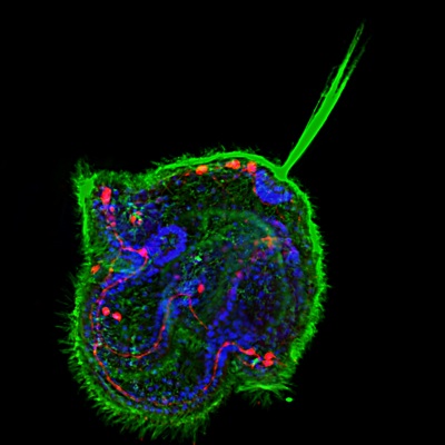
We can all articulate the importance of using model organisms to understand biology, but many of us fall short in our understanding of some of the more uncommon model organisms.  Thankfully, there are amazing biologists out there that save the day!  These researchers use some of the more atypical model organisms to understand how different organisms develop and how developmental processes have evolved.  Today’s image features the crustacean Parhyale hawaiensis.

The establishment of the dorsoventral (DV) axis in many organisms is fundamental to the proper organization of organs and tissues.  In arthropods, the organization of tissues around the DV axis is well conserved, yet how the axis is established is not.  For example, ventral midline cells play a restricted role in DV patterning in Drosophila, yet they play a prominent role in establishing the DV axis in the crustacean Parhyale hawaiensis, according to a recent paper by Vargas-Vila and colleagues published earlier this year in Development.  In addition, the Parhyale ortholog of the transcription factor gene single-minded (Ph-sim) is expressed in midline cells and is required for differentiation of midline cells.  These results suggest the importance of ventral midline cells in DV patterning in the last ancestor common to both crustaceans and insects.

Images above show Parhyale embryos at different stages, with nuclei in blue.  Throughout early development, Ph-sim is expressed in ventral midline cells, as seen in the false-color overlay (red) of expression patterns (A-C).  In Ph-sim (RNAi) embryos, midline staining of the midline marker Ph-otd-1 is absent (compare D and E), and the nice clear line of ventral midline cells (red arrows in F) is no longer visible (compare F and G).

ResearchBlogging.orgVargas-Vila, M., Hannibal, R., Parchem, R., Liu, P., & Patel, N. (2010). A prominent requirement for single-minded and the ventral midline in patterning the dorsoventral axis of the crustacean Parhyale hawaiensis Development, 137 (20), 3469-3476 DOI: 10.1242/dev.055160

Thumbs up (8 votes)
Loading...

Tags: , ,
Categories: Images, Research

Navigate the archive

Use our Advanced Search tool to search and filter posts by date, category, tags and authors.

Ernst Haeckel and the recapitulation of an “early” biological debate

Posted by , on 8 December 2010

Scientists don’t spend free time to think about the changes that made possible the birth of a new way to make research. For example, how we moved from a world driven by religious and philosophical beliefs to a world demanding explanations and mechanisms? Ernst Haeckel was one of the scientists who made that change possible and, more important to us, he accomplished it from the field of developmental biology. Although this post is not intended to be a review of Haeckel’s work (that would be a daunting and overwhelming task), it is worth to mention some interesting things about this story. As many scientists at their times, Haeckel’s work was criticized and Haeckel was, indeed, heavily attacked not only by religious and conservative people, but also by other fellow scientists. We can summarize some aspects of Heckel’s view as follows: Haeckel was impressed and inspired by the work of previous scientists (being J.F. Meckel and K.E. von Baer the most important people), showing the resemblance between embryos from different animal species at early stages of development. Although Haeckel was not the first scientist to propose a resemblance of vertebrate embryos at early stages of development, Haeckel made use of this fact, often depicting embryos in his works with some degree of abnormalities (which were used by his critics to accuse him of adulterating embryos).

Ernst Haeckel formulated the known Fundamental Biogenetic Law, in which he describes the parallelism between embryonic development and the phylogenetic history, claiming that embryonic development is a rapid recapitulation of the evolution, or “ontogeny recapitulates phylogeny”. Most conservative people viewed Haeckel’s propositions as a challenge to the more religious views about the origin of man. Haeckel made comparisons between early embryos from different species; his famous drawings, that appeared in his works, especially in Natürliche Schöpfungsgeschichte, were famous at the time, and they were criticized by other scientists. Some people at the time claimed that the only evidences for this proposition were the drawings made by Haeckel, but we have to consider that experimental biology was at a sort of “very early stage of development”. Hence, Haeckel’s work was abandoned from the main stream of science, especially between the World Wars, when chemistry and physics gained much more attention. However, Haeckel’s work likely inspired many future scientists, including his students. One of them was Hans Spemann, who later made one of the most important experiments in biology. Even, when the findings of Spemann and Mangold can be considered as opposed to Haeckel’s biogenetic law (because now the embryological development is driven by hidden forces with molecular nature, and since all organisms are different, these forces should differ in nature), the work of Spemann led in time to the discovery and (partial) understanding of the Wnt pathway, which is maybe one of the most conserved signaling pathways in nature and one of the most important driving forces in embryological development, validating Haeckel’s work: indeed, embryological development involves the expression and function of conserved genes through evolution. This realization brought Haeckel’s work one more time into the public attention, and once again, critics to his work appeared, with high press coverage at the time.

Image attributed to Ernst Haeckel, published in his work  Natürliche Schöpfungsgeschichte, and illustrates the similarities between embryos of different species (man, dog and turtle). His rivals argued that embryos compared in Haeckel’s drawings usually had abnormalities and that they corresponded to different developmental stages. Image source: Wikipedia Commons.  

Today, two papers published in Nature (vol. 468, Number 7325) “recapitulate” this classic debate: Domazet-Lošo and Tautz show in Zebrafish that the transcriptome expressed during the phylotypic stage (the stage in which species from a phylum resemble each other) is older compared with the transcriptome expressed in adult stages. They conclude that “our study provides strong molecular support for a correlate between phylogeny and ontogeny”, which agrees with the propositions of Haeckel and previous researchers (like K. von Baer). In the same issue, Kalinka and co-workers took a similar approach with six Drosophila species, observing also maximal conservation of gene expression at the phylotypic stage. Haeckel was discredited by many scientists, even in these days. He has been accused to be convinced to fraud, showing that embryos in drawings are stylized, altering embryos, and heterochrony is not considered in the drawings itself. With the available tools nowadays, we know that embryological development is variable between species. I believe that this is not the point. Haeckel’s work helped to popularize an important idea in biology, and we can discuss at which extent conserved genes and signaling pathways are integrated in the early (or late) development, validating the general concept about the relationship between evolution and development.

It should be an outstanding improvement in scientific journals, to include historical profiles and short reviews (no more than one page) about these relevant figures in biology (and science in general).

Thumbs up (5 votes)
Loading...

Tags: ,
Categories: Discussion, Research

Zebrafish transgenes go ubiquitous

Posted by , on 7 December 2010

The Node’s staff has kindly asked me to write a little “behind the scenes” on our zebrafish paper released today in Development, “Ubiquitous transgene expression and Cre-based recombination driven by the ubiquitin promoter in zebrafish” (http://dev.biologists.org/content/138/1/169).

The spark to pursue the project were the first conversations I had in spring 2008 with senior postdocs in Leonard Zon’s lab at the Children’s Hospital Boston right when I started my postdoc to study hematopoietic cell fate control using zebrafish. Coming straight out of my graduate work on Wnt and Hh signaling in Drosophila, my thinking was centered on mutants, transgenes, and recombinase-mediated manipulations. The zebrafish is still a relatively new, yet increasingly popular, model organism with compelling imaging possibilities and malleable genetics. It was no small shock however to learn how some key molecular tools were not working well or even totally missing in zebrafish. A reoccurring theme was the lack of a truly ubiquitously expressing promoter for transgenes, in particular one that is active in red blood cells and adult organs. I realized that I would need Cre/loxP tools for my project ideas, all of which depend on a ubiquitous promoter such as Rosa26 transgenes in mice, to permanently express a lineage tracer transgene in the cells we wish to track.

As one of my first practical things in the lab, I therefore assembled the zebrafish ubiquitin (ubi) locus through database searches and ordered primers to amplify the 5’ region of the gene. Why ubi? ubi:GFP are the most reliable transgenic markers in Drosophila for ubiquitous labeling of cells, and I used plenty of such strains in my past projects. After injecting zebrafish ubi promoter-driven EGFP reporter vector into zebrafish embryos, I saw ubi expressed transgenes at all developmental stages and a multitude of adult organs, including cell types such as red blood cells that have so far been missed by quasi-ubiquitous zebrafish promoters such as beta-actin or ef1alpha. The picture below is a mosaic ubi:EGFP embryo from one of these initial injections.

ubi:EGFP reporter injected mosaic zebrafish embryo at approximately 36hpf.

Shortly after these first tests, Charles Kaufman joined the lab as a postdoc. While we discussed during our very first chat his ideas to tackle melanoma formation using transgenic zebrafish, he mentioned requiring a ubiquitous promoter. “There really isn’t a good ubiquitous promoter in zebrafish”, I said. Charles originally trained in mice and was thus used to luxurious molecular genetics tools; so he replied, astonished, “So how are we supposed to do anything then?” “Well, maybe we now have a ubiquitous promoter” I replied and outlined my preliminary data. The rest unfolded as a team effort to first elucidate if ubi is truly that ubiquitous and to subsequently create a tool box for genetic lineage tracing and Cre/loxP-regulated transgenes in zebrafish.

To confirm if ubi truly expresses ubiquitously also in our lab’s favorite tissue, blood, Pulin Li and Emily Pugach successfully carried out adult zebrafish blood transplantation assays and characterized ubi expression in hematopoietic cells. As if timed for the project, Owen Tamplin brought a batch of the original CreERt2 plasmid to the lab when he joined as a postdoc. CreERt2 is a version of the Cre recombinase that is inducible by 4-hydroxytamoxifen (4-OHT). With the precise developmental staging possible in zebrafish embryos, timed addition of 4-OHT to the dish allows for precise temporal activation of a given CreERt2 driver, a principle that has already been successfully established in zebrafish. We therefore generated ubi:creERt2 transgenic zebrafish as a source of ubiquitous inducible CreERt2 recombinase activity and confirmed its sensitivity to 4-OHT at various developmental stages.

To harness the full lineage tracing potential of ubi, we also created ubi:loxP-EGFP-loxP-mCherry, or ubi:Switch as we call it. This transgene initially expresses EGFP ubiquitously, but any cell with active Cre will cut out the EGFP cassette and put mCherry under ubi control, thus indelibly marking this cell and its descendents with fluorescent red throughout development. ubi:Switch now allows simple lineage tracing experiments where ubi:Switch transgenics are crossed to any tissue-specific Cre- or CreERt2-expressing transgenic zebrafish strain of choice, many of which are currently under development in labs around the world.

Future versions of ubi:Switch can easily be cloned to express different fluorescent color combinations tailored to specific experiments. Furthermore, ubi:creERt2 facilitates the future creation of tissue-specific loxP lineage tracing transgenics, which can then be universally tested by crossing to this ubiquitous CreERt2 source. The time spent building these tools was not only great training, but now enables us – and hopefully other zebrafish researchers in the field – to perform lineage tracing experiments and to create new exciting transgenics. The lack of gene knockouts or RNAi technology remains a heavy burden on the zebrafish community. Maybe ubi as new ubiquitous transgene driver resource will now assist the development of these methods. Is ubi the final word on ubiquitous zebrafish transgene promoters? Probably not. But until we find something even more potent, ubi adds another lure to the growing tackle box of zebrafish methods to catch exciting new biology.

Thumbs up (15 votes)
Loading...

Tags: , , , , , ,
Categories: News, Research, Resources

In Development this week (Vol. 138, Issue 1)

Posted by , on 7 December 2010

The first issue of 2011 is out now…here are the highlights:

Geminin control of lineage commitment

The transition between pluripotency and multi-lineage commitment during early embryogenesis must be closely regulated to ensure correct spatial and temporal patterning of the embryo. But what regulates this crucial transition? According to Kristen Kroll and co-workers, part of the answer to this question in Xenopus embryos lies with the nuclear protein Geminin (see p. 33). The researchers show that Geminin overexpression represses many genes associated with cell commitment but increases the expression of genes that promote pluripotent and immature neuroectodermal cell fates. Geminin, they report, represses Activin-, FGF- and BMP-mediated cell commitment. Consistent with this finding, Geminin knockdown enhances commitment responses to growth factor signalling and results in ectopic mesodermal, endodermal and epidermal fate commitment in the embryo. The researchers also report that repression of commitment by Geminin depends on Polycomb repressor function, and show that Geminin promotes Polycomb-mediated repressive histone modifications of mesodermal genes. The researchers propose, therefore, that cooperativity between Geminin and Polycomb plays an essential role in controlling spatial and temporal patterning in early embryos.

Rock-ing between AP and LR axes

The vertebrate body plan features a left-right (LR) asymmetry, but how the LR axis is orientated correctly with respect to the anteroposterior (AP) and dorsoventral (DV) axes is not known. Here, Jeffrey Amack and co-workers (p. 45) report that the Rho kinase Rock2b links AP patterning to LR patterning in zebrafish embryos. During development, Kupffer’s vesicle (KV) generates a cilia-driven leftward fluid flow that directs LR patterning. The authors demonstrate that depletion of rock2b in whole embryos or in the KV cell lineage alone disrupts asymmetric gene expression during development and perturbs organ asymmetries. They show that, in control embryos, ciliated cells are distributed asymmetrically along the AP axis of the KV and generate asymmetric fluid flow. By contrast, rock2b knockdown embryos show defective KV patterning and cell morphology, and a loss of directional flow. Based on their studies, the authors propose that Rock2b is required for the AP positioning of ciliated cells within the KV and for subsequent LR patterning in zebrafish embryos.

Mesp2 Notches up somite polarity

Somites, the most obviously segmented structures in vertebrate embryos, are subdivided into anterior (rostral) and posterior (caudal) compartments. Repression and activation of Notch signalling are essential for the establishment of the rostral and caudal compartments of the somite, respectively. The mechanism by which Notch is repressed has remained elusive but, on p. 55, Yumiko Saga and colleagues identify the bHLH transcription factor Mesp2 as a novel negative regulator of Notch signalling in mouse somites. In the absence of Mesp2, somites are completely caudalised but, intriguingly, the researchers now show that the introduction of a dominant-negative form of Rbpj (a downstream effector of Notch signalling) into the Mesp2 locus largely rescues the segmental defects of Mesp2-null mice. They also report that Mesp2 represses Notch signalling independently of its function as a transcription factor by inducing the destabilisation of mastermind-like 1, a core regulator of the Notch signalling pathway. These new findings shed light on the molecular mechanisms that control the rostrocaudal patterning of somites.

Cdx1: refining the hindbrain

During embryogenesis, the vertebrate hindbrain is segmented along its anteroposterior axis into lineage-restricted compartments, known as rhombomeres (r1-r8), that dictate subsequent neural patterning. The signals that pattern the hindbrain are known, but how each rhombomere-specific gene expression pattern is established is unclear. On p. 65, Sabine Cordes and colleagues reveal that the homeobox protein Cdx1 patterns the mouse hindbrain by spatially restricting the expression of the transcription factor MafB. Mafb is required for r5 and r6 development, and its expression is restricted to these segments. The authors report that the Mafb enhancer contains candidate Cdx-binding sites, and that Cdx1 binds to these sites both in vitro and in vivo. They show that Cdx1 is expressed at the r6/r7 boundary, at the posterior limit of the Mafb-expressing domain. Importantly, in the absence of Cdx1, MafB expression extends beyond its normal r6/r7 boundary. The authors propose that Cdx1 acts as an early and transient repressor of Mafb, and thus plays a role in refining hindbrain identity.

Boc: novel roles in Shh regulation

Hedgehog (Hh) signalling gradients control many developmental processes and are influenced by numerous positive and negative regulators. The transmembrane protein Brother of Cdo (Boc) has been implicated in Sonic hedgehog (Shh)-mediated commissural axon guidance in the CNS, but how Boc affects the cellular Hh response in vivo is unclear. Here, Rolf Karlstrom and colleagues reveal that Boc is cell-autonomously required for Hh-mediated ventral CNS patterning in zebrafish (see p. 75). The umleitung (uml) zebrafish mutant is characterised by defects in retinotectal projections. The researchers show first that uml encodes Boc. Then, by analysing the phenotypes of uml mutants, they show that Boc is a positive regulator of Hh signalling in the spinal cord, hypothalamus, pituitary, somites and upper jaw, but that Boc might be a negative regulator of Hh signalling in the lower jaw. Overall, these results reveal a role for Boc in ventral CNS cells that receive high levels of Hh, and uncover novel roles for Boc in vertebrate development.

Expanding the zebrafish toolkit

The zebrafish genetics toolkit has been missing a particularly handy piece of kit: a promoter to drive ubiquitous transgene expression throughout development, equivalent to the Rosa26 locus used in mouse genetics. But no longer, for in one of Development‘s inaugural Technical papers (p. 169), Leonard Zon and co-workers report that the zebrafish ubiquitin (ubi) promoter can drive constitutive transgene expression throughout development. The authors initially identified ubi in BLAST searches using human ubiquitin. They then tested a 3.5 kb 5′ region upstream of its translational start site for transcriptional regulatory sequences and found that it drives strong and ubiquitous EGFP expression within 4 hours of injection into a single-cell embryo. Moreover, in stable ubi-EGFP transgenic lines, EGFP is strongly expressed in all external and internal organs they analysed, in all blood cell types, and from embryo to adulthood. The authors also created inducible ubi-driven CreERt2 transgenes and loxP lineage-tracer transgenes that give strong reporter activity upon Cre exposure, which further enhances and expands the zebrafish transgenesis toolkit.

To find out more, and to read the first author’s “behind the scenes” account of this work, see the related post on the Node

Plus…

The origin of ES cells has been debated in recent years. Jenny Nichols and Austin Smith now propose that there are, in fact, two possible routes by which ES cells can arise that are dictated by culture conditions.

See the Hypothesis article on p. 3

The Hippo pathway regulates growth in Drosophila and vertebrates, and, as Georg Halder and Randy Johnson now discuss, recent studies have shed light on how it governs organ size control and regeneration, and on how it is dynamically regulated during development.

See the Review article on p. 9.

Thumbs up (No Ratings Yet)
Loading...

Tags: , , , , , , , , , , , , , , , , ,
Categories: Research

Echinoderm development on film

Posted by , on 6 December 2010

“I also here salute the echinoderms as a noble group especially designed to puzzle the zoologist.”

Libbie Hyman, 1955

Echinoderms are fascinating creatures. They have extensive regenerative capabilities, a mutable connective tissue that dynamically (and deliberately) changes its stiffness, and a complex system of hydraulic canals involved in the circulation of internal fluids and locomotion.

However, the most notable feature of echinoderms is the pentamerous symmetry of their bodies, derived from a bilateral ancestor. These exclusively marine deuterostomes are mostly bottom dwellers with a biphasic life cycle, where the adult tissues develop inside a bilateral planktonic larva (swimming in the water column) and metamorphose into a benthic juvenile.

Pluteus larva
A planktonic pluteus larva of a sea biscuit.

A planktonic pluteus larva of a sea biscuit.

During my master’s project at University of São Paulo, Brazil, I studied the development of a different kind of sea urchin, a sea biscuit. Sand dollars and sea biscuits belong to a lineage of urchins that developed a secondary bilateral symmetry. Also, during their evolution, around 55 million years ago, the adult morphology changed in association with the occupation of sand beds; more specifically, the body flattened, the spines got shorter, the number of tube feet increased, and their feeding apparatus (lantern of Aristotle), which was absent in other adult irregular urchins, was retained into adulthood.

Since I was interested in the developmental origins of such changes in morphology I documented the embryonic, larval, and juvenile development of a sea biscuit species, Clypeaster subdepressus. After gathering all data, I compiled it into a science outreach video showing a resumé of the life cycle of this species, from fertilization to the first steps of the juveniles. Hope you enjoy it:

We collected adults from sand beds of São Sebastião Channel (São Sebastião, SP, Brazil) and induced gamete release (eggs and sperm). We did the fertilization in vitro and followed the embryonic development in the laboratory, under light microscopy. Embryos become swimming larvae, approximately 0.2 mm wide, which we fed with microalgae until metamorphosis. A diminute sea biscuit grows inside the larva. When the minuscule podia and spines are formed the larva sinks and undergoes metamorphosis. The juvenile sea biscuit reabsorbs the larval tissue and begins to explore its new habitat, between sand grains. [Numbers on the upper right corner show how much the scene was accelerated.]

The video is available to download here, feel free to reuse it and share it around! If you want further details on sea biscuit development (including images and video footage) the official description was published earlier this year.

Thumbs up (9 votes)
Loading...

Tags: ,
Categories: Images, Video

Keeping up with the Node

Posted by , on 6 December 2010

Like more than 500 million people in the world, the Node is now on Facebook . Our foray into Facebook was slightly overshadowed by the British royal family doing exactly the same thing a few weeks earlier, but we can guarantee you that our page will contain far more developmental biology.

We’re using our Facebook page much like our Twitter account: to notify you of new Node posts, and other brief bits of community news. Have a look , “like” us, and invite your friends and labmates to do the same. (We’ll still be updating Twitter as well, so don’t worry if you’re used to seeing us on there.)

There’s a link to our new Facebook page in the left sidebar, and the eagle-eyed among you may have spotted another new thing over there. Several people have told us that they couldn’t immediately figure out what the Node was when they arrived on the page for the first time, so we’ve tried to explain it very briefly over there.

Finally, some things are better left unchanged, it seems. We asked you whether you would like a change in the format of the e-mail notifications, and as it turns out, almost half of those that took the poll were satisfied with the current format. Good to know! Needless to say, we didn’t change the e-mail format.

Thumbs up (No Ratings Yet)
Loading...

Tags: , ,
Categories: News

Developmental biology art from Japan

Posted by , on 1 December 2010

The RIKEN Center for Developmental Biology has released the images for a series of postcards under a creative commons license. The images picture a wide range of both common and uncommon model organisms, all in a Japanese paper art style.

You can download the full set of high quality images, designed by Yukiko Fujiwara, as a zip file from their site. This axolotl is now my desktop background:

Earlier this year, I came across another artistic project out of Japan: the plates that were featured on the announcement posters of the SDB/JSDB joint meeting in Albuquerque were also physically present at the meeting:

The artwork on the plates, by design company TRAIS K.K., brilliantly features developmental biology images in the styles of traditional decorations from Japan and New Mexico, and was an absolutely perfect summary of that conference.

Thumbs up (4 votes)
Loading...

Tags: , ,
Categories: Images

Travelling Fellowships

Posted by , on 29 November 2010

Over the past months, we’ve seen a few posts on the Node from people who spent a few months working in labs abroad. All of them were funded by a Development travelling fellowship. The next deadline for these fellowships is coming up on December 31st, and Development would like to encourage you to apply.

To qualify, you must be a graduate student or postdoc planning to work for a few months in a distant lab in 2011. Have a look at the fellowship site for the full requirements, and read these stories from previous recipients on the Node.

Tetyana (from the Ukraine) went to India:
Research Snippets from the Land of the Tiger
The Maggot Meeting 2010

Cristian (from Chile) went to Germany:
Developing Science in a Far Country: The Paradoxes of Life

Shreeharsha (from India) went to Japan:
Research in the Land of the Rising Sun

Dávid (from Hungary) went to Japan:
Nippon

Terry (from the US) is currently in Israel:
International Experience

Will your story be next? It might, if you apply for one of the travelling fellowships before December 31st. Good luck!

Thumbs up (No Ratings Yet)
Loading...

Tags:
Categories: Funding, Research

International experience

Posted by , on 26 November 2010

Hello, I am Terry Jackson, a 6th year PhD student in Genetics and Genomics at Duke University which is located in Durham, North Carolina, USA. I am working on my degree in the lab of Dr. Philip Benfey whose research focuses on identifying transcription factors in the root of Arabidopsis thaliana. I am pleased to have received a travel award from the journal Development for an international collaboration with Dr. Asaph Aharoni at the Weizmann Institute of Science in Israel. His work includes investigating and identifying glucosinolates, a secondary metabolite that plants produce as a defense mechanism. Together we plan to determine the glucosinolates that are produced in many of the individual cell layers within the root. We intend to use FACS to isolate each cell type using GFP-marker lines followed by LC/MS to identify these compounds.

I knew that my trip here would be an eye-opening experience and I have not been disappointed. Thus far, I have been here for six weeks and it has been quite a grand undertaking. The planning began months before my departure by deciding the length of my stay and how much we could accomplish in that time period. Everything had to be coordinated from the date of my arrival to making sure my host lab had all of the necessary materials to scheduling time on the FACS machine. It seems simple but even now we are making adjustments and revisions.

Clearly, everyone knows that no two labs operate the same but once you get settled into a lab and into the routine you tend to forget. It is surprising the number of small details that are assumed or overlooked during planning. For instance, at Duke we have technicians that run all of the FACS samples. We only have to prepare the samples and drop them off to retrieve a couple of hours later. Here, at the Weismann Institute of Technology, they do not have technicians for this purpose; the researchers are trained to run the machines themselves. I assumed that I would drop off my samples and return to pick them up and Dr. Aharoni’s group thought I already knew how to work the machine. No one asked me about it until a week before I was scheduled to arrive if I could run the machine. Suddenly, they had to schedule training sessions for me on the FACS machine so that I could do it myself. I was very nervous about this at first but it is much easier than I thought. I consider this training to be an added bonus to my skills set as a researcher and it is one of the most enjoyable aspects of the numerous tasks I must complete each week.

My original plan was to stay here for two months but, since I needed the FACS training and we’ve had to run several test experiments, I decided it’s best to extend my stay for at least one more month. Other than the glucosinolate experiments I also intend to isolate root cells that form large inclusions under low sulfur conditions and then analyze them with LC/MS to determine their composition.  This analysis will probably be the most difficult aspect of the work I am doing here since we are trying to identify and unknown material. Furthermore, it is going to be a steep learning curve for me.  I know it is very necessary that I understand the process of completely. In the end I am sure I will return to Duke with a new perspective and knowledge of  effective collaborations.

Thumbs up (4 votes)
Loading...

Tags: ,
Categories: Funding, Lab Life, Research

An interview with Patrick Tam

Posted by , on 25 November 2010

(This interview by Kathryn Senior originally appeared in Development on November 23, 2010)

Patrick Tam’s research is focused on the cellular and molecular mechanisms of body patterning during mouse development. He agreed to be interviewed by Development to talk about his interest in mouse development, new concepts in gastrulation, X-linked diseases and his dream of an African safari.

Did you always intend to have a career in developmental biology?

I was lured into science at high school as I listened to my biology teacher reminiscing about his romance with plant biochemistry during his university days. It was really no surprise that I chose biology over medicine as my degree and then headed onto postgraduate research without a second thought. It might sound incredible but there was not a proper course on developmental biology (or embryology, as it was known) in the entire Bachelor of Science curriculum of my university in those days. To fill this gap in my education, I made a definite decision to study rodent embryo development – first in Hong Kong, and later in London with Michael Snow. This was a time when research in mouse development was taking off in a big way in the UK and so the rest, as they say, is history.

What has influenced your decisions about institutions and locations?

Before I finished my PhD, I had already accepted a faculty position in the newly founded Medical School at the Chinese University in Hong Kong. Luckily, I did manage to squeeze in one year of postdoctoral training at the University of Texas at Austin. This proved to be critical for broadening my research experience and I learned a great deal more before taking on the job back home.

Joining a young institute happened to be a good decision: there were ample start-up resources and also the flexibility that I needed to be able to run the laboratory the way I wanted it. The academic appointment offered relatively stable support for my research during this formative phase of my career. The downside was coping with the demand of teaching commitments, and being the only laboratory working on mouse development made it quite hard to maintain research momentum. My next move to a research institute in Australia was a very positive one as it allowed me to develop further in a research-intensive and intellectually stimulating environment. Having access to first-rate facilities and interacting and networking with a larger community of developmental biologists enabled me to focus and move forwards much faster.

You have been a great pioneer in applying micromanipulation and embryo culture research for investigating early mouse development: how did you get into this originally?

My PhD project was to characterise the developmental fate of an active multiplying population of cells in the epiblast of the gastrulating mouse embryo. I had little idea how challenging this would turn out to be! Initially, we focused our efforts on developing a reliable whole-embryo culture method by tweaking the protocol established by Dennis New for culturing rat embryos. We then tried to apply the conventional `slash and burn’ and `cut and paste’ techniques to study cell fate and tissue differentiation. I owe Rosa Beddington an enormous debt of gratitude for introducing me to the art of embryo manipulation during my sabbatical at Oxford University in the mid 80s: my collaboration with her has had a lasting impact on my career. Ultimately, my postgraduate project evolved into a consuming exercise of fate-mapping all three germ layers and their immediate derivatives. The work took three decades but realizing that I had completed my original objective to the best of my ability was a very satisfying moment in my scientific career. (more…)

Thumbs up (4 votes)
Loading...

Tags: , , ,
Categories: Interview