If you have been on Twitter or read the news recently, you have probably seen this Guardian article on stem cell-based human embryo models, or one of the other pieces of news coverage. This sparked some debate over how scientific data should be covered in the media. Should data without a preprint or paper be reported in the news? How should scientists talk about their results with journalists?
Following the Guardian article, the Zernicka-Goetz group and Hanna Group each released a preprint on post-implantation embryo models:
In light on the fast-developing field of embryo models, the scientific community is acting to produce a governance framework for research involving stem cell-based human embryo models. This includes a project recently launched by the University of Cambridge.
Stem cell-based embryo models could help our understanding of the problems that can affect early pregnancies.
But their regulation is a grey area. That's why @Cam_Repro has launched a project to develop a governance framework for this research in the UK 👇
— Cambridge University (@Cambridge_Uni) June 16, 2023
The ISSCR released their Standards for Human Stem Cell Use in Research, which “identifies quality standards and outlines basic core principles for the laboratory use of both tissue and pluripotent human stem cells and the in vitro model systems that rely on them.” This document also includes guidelines on best practice for reporting on studies using human stem cell systems.
The #ISSCR wants to better understand how LGBTQ+ scientists feel included in their professional societies, workplaces, & the broader community. We need your voice to create a more inclusive scientific community – take a moment to share your experiences 👉 https://t.co/CozpyiqzSipic.twitter.com/QfhZKMO5lS
And finally, have you ever heard of the police being called to a scientific conference because the conference hall was overcrowded? This happened at ISSCR2023.
This morning, a concurrent session exceeded fire code capacity and the building security ushered out non-seated attendees. They did this for a fire-safety reason, but in a way that was uncomfortable to involved attendees and somewhat disruptive to the session.
Police just interrupted a wonderful talk by Jing Qu on endogenous retroviruses and senescence to kick people out of the seminar room, due to it being over capacity :( #ISSCR2023
If you can’t attend the European Developmental Biology Congress in person this September, why not organise a local Watch Party?
Excitement is ramping up for the European Developmental Biology Congress which will take place 25-28th September this year. This will be a celebratory meeting distributed across three beautiful locations: Oxford, Paris and Barcelona, packed with exciting science, social events, medal talks and even chances to try Tai Chi, experience embryology-inspired Virtual Reality art and go punting with your fellow developmental biologists. You can read more about our plans here and you can check out the programme and register here.
For anyone who can’t travel to attend the meeting in person, there will be the option to watch talks online. But, we know that watching a conference remotely can sometimes feel a little lonely, with no one to discuss the talks with over a delicious conference coffee.
So…. why not plan an #EDBCWatchParty?
“Our lab organised an outdoor watch party in my garden for the 2021 hybrid BSDB Spring Meeting and it was great. I’d definitely do something like that again”
Kyra Campbell, University of Sheffield
“We hope to gather together Developmental Biologists from across Edinburgh to watch EDBC2023 in a university seminar room, perhaps with a local poster session. We will encourage people to bring home bakes so we might even include a Great Developmental Biology Bake Off competition!”
Tamina Lebek and Guillaume Blin, University of Edinburgh
“Our lab are excited about the talks at EDBC, but some of us can’t make it in person so we’re very keen on the idea of coming together to watch the talks online. We’ll spread the word around Cambridge for any other developmental biologists who’d like to join us”
Ben Steventon, University of Cambridge
If you are thinking of giving this a go, we’d love to hear about it so please feel free to drop us a line at meetings@bsdb.org or post on twitter or mastodon with the hashtag #EDBCWatchParty
The lab is in the Max Planck Institute for Molecular Cell Biology and Genetics (MPI-CBG) in the lovely Dresden (Germany).
Research summary
Mammals evolved divergent architectures of placenta to receive nourishment from the mother during embryogenesis. In our lab, we seek to understand how the uterine microenvironment shapes the fetal-placental interface of various animals, by using early embryos and placental organoids. Our ultimate goal is to investigate how complex and diverse structures emerge to achieve similar function.
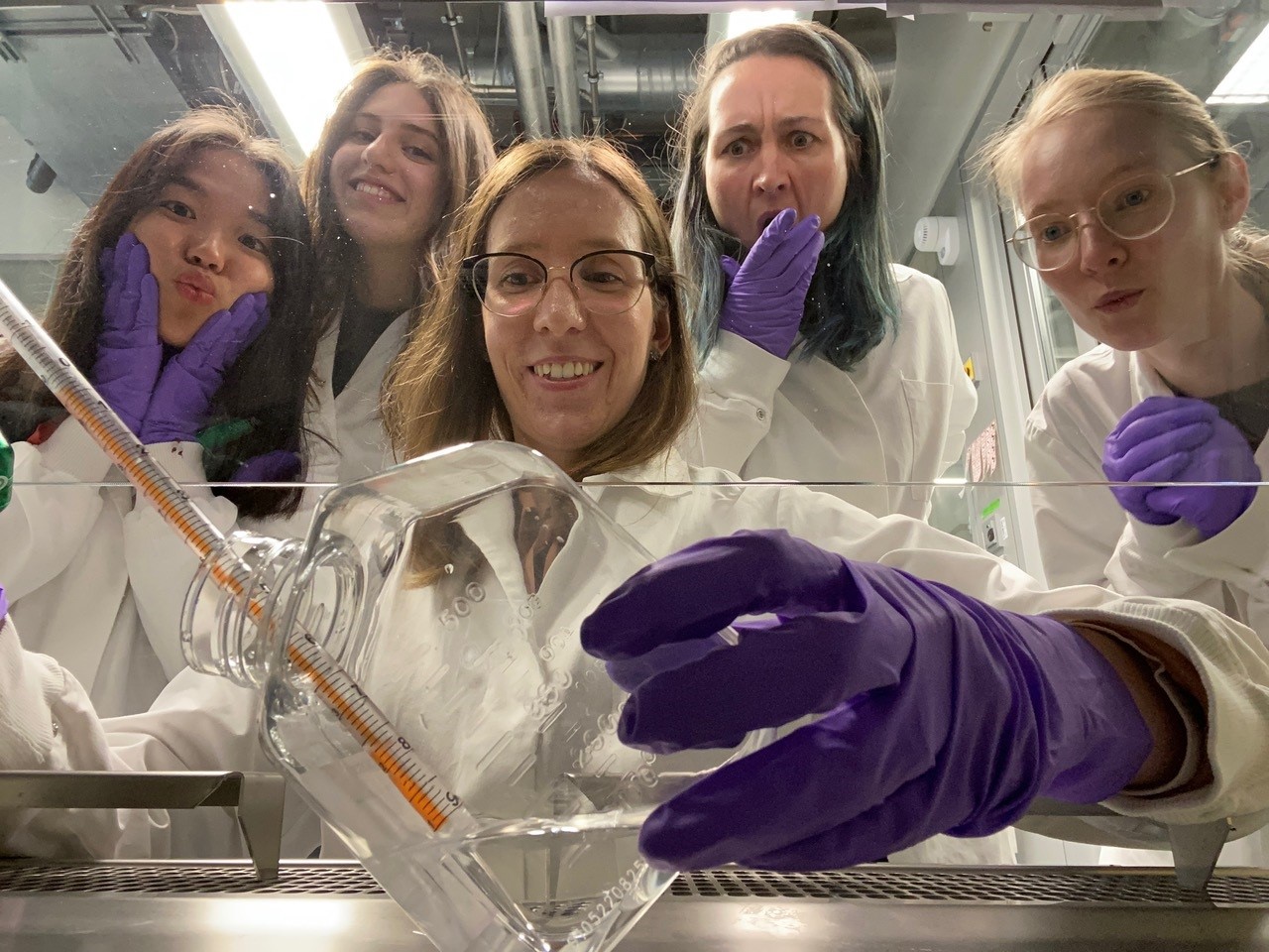
Gerri lab members
Lab roll call
Michi: lab technician. I like to help in any situations and projects. Also, I like to work on my own little project, which is focused on discovering similarities and differences in early embryonic development of different species of mammalian pre-implantation embryos.
Johanna: PhD student. I work on the role of oxygen sensing mechanisms during early human placenta development.
Ornella: master student. I am investigating the impact of specific genetic mutation on human trophoblast stem cell differentiation and trophoblast organoid self-organization.
Weiwei: bachelor student. My project is focused on testing different live imaging approaches on 2D and 3D culturing systems.
Favourite technique, and why?
Claudia: My favourite approach in studying biology is imaging. I am fond of everything that allows us to visualize biological processes. It is in this way that we can describe new phenomena and decipher how biology works. Analysis of both live and fixed samples, for example using immunofluorescence or transgenic lines, accompanied by microscopy at different resolutions can allow us to look at biological samples at different scales, from intracellular tiny structures to multicellular complex tissues.
Apart from your own research, what are you most excited about in developmental and stem cell biology?
Claudia: Biological processes require a complex coordination of genetic, molecular and physical events in space and time. Several physical principles have helped understanding developmental processes in a more unified way. I am always impressed when reading multidisciplinary papers where experimentalists and theorists work together to identify emerging principles of the living matter.
This is why I have decided to join the MPI-CBG and the Dresden campus in general, where scientists across disciplines, biology and physics, actively work together. I hope with time to develop collaborations into this direction.
How do you approach managing your group and all the different tasks required in your job?
Claudia: We are a young lab, so I believe that at this stage it is important for me to be focused on the science and to be there for the lab members. I am still active in the lab and I try to be available to answer queries and discuss issues when the team members require to do so. In addition to institute seminars and conferences, I try to organize opportunities for the team to interact with other labs working with similar and different topics (e.g. lab retreat with Huch, Veenvliet and Toth-Petroczy Labs, but also joint lab meetings with Rodenfels Lab).
In general, to manage all the tasks required for this job, I try to plan my goals monthly, weekly and daily.
I am still learning and when in doubts I ask advice to other young group leaders and mentors.
What is the best thing about where you work?
Claudia: I have experience in other institutes, bigger and smaller than the MPI-CBG. I think the MPI-CBG is great in many aspects. MPI-CBG is an internationally renowned institute and that is why many great scientists from around the world come here to give talks. Also, the MPI-CBG is big enough to have diversity in terms of science and people, but also it has the right size to allow people to interact after seminars, or in the canteen. This really helps to create a tight community, where newcomers are welcome and can easily integrate into the institute.
We have also great excellent facilities helping us in our daily routine (e.g. organoid stem cell facility) or in developing new tools (e.g. genome engineering facility or technology development studio).
Michi: I like studying the develop of the placenta, because we discover unique aspects of developmental biology. I also really like the institute with its great facilities and the great international people working here. Also, there are small things, like the smell of coffee when you enter the building. All these reasons make me happy to come to work!
Johanna: I really like the way service is provided by all the facilities, and the sense of community. People of the lab and general from the institute are always very happy to help and support me with my projects.
Ornella: In my opinion, what makes the Gerri Lab truly exceptional is the continual support and guidance you get from everyone. We all have different backgrounds and personalities, but we have a great balance that makes the lab a fun and enjoyable place to be.
The Institute’s greatest aspect is the tight-knit community, where we all come together. Thanks to the international and interdisciplinary environment, there are abundant opportunities for personal growth and learning. Being exposed to internationally renowned researchers adds immense value to my experience. Plus, I particularly enjoy the social events, where we can relax and have a good time.
Weiwei: I really like the lab environment, everyone is very helpful, caring and patient. There are seminars to follow and many other activities, also a weekly get-together.
What’s there to do outside of the lab?
Claudia: Dresden is a great city to live in! I find that there are things for every taste. There is Altstadt, where you can go to visit museums and learn more about history. There are residential areas with amazing parks, where is great to spend time with the family. There is Neustadt, which is the vibrant and young part of the city. There is also amazing nature nearby.
Michi: I think Dresden is a great city with many things to do. Most of the city rivers are folded in concrete but we have the Elbe-meadows where you can bike, run, walk, having a picnic or even skiing in winter. In summer there is an outside theatre next to the river with an amazing view on Dresdens historical Altstadt. I love going on hikes in the unique Sächsische Schweiz.
Also, Dresden is the best place to raise a kid! There are so many playgrounds, museums, sportive or cultural offers.
Johanna: Dresden is great for walks through parks, the forest and also the historic center. There are also several people of the institute that organise free time activities such as volleyball, soccer or improv theatre.
Ornella: Dresden has it all! You can go on awesome hikes or bike rides surrounded by beautiful nature. In the lively Neustadt neighborhood, you can hang out, grab a drink or beer, and have a great time with friends. If you’re into techno music, Dresden is the place to be!
Browse through other ‘Lab meeting’ posts featuring developmental and stem cell biology labs around the world.
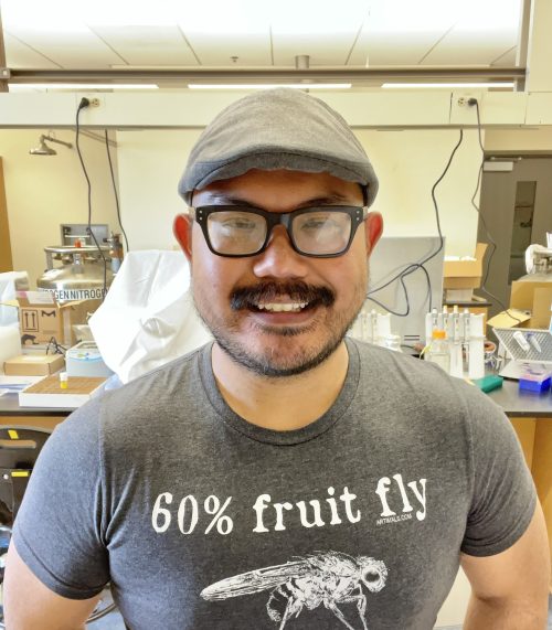
In our latest SciArt profile, we spoke to Frank Macabenta, a developmental biologist who freelances as a scientific illustrator. Franks share with us his lab’s work on quality control mechanisms regulating organogenesis in the fly embryo, and how art influenced his decision to pursue studies in developmental biology.
Can you tell us about your background and what you work on now?
I am an immigrant and first-generation scientist. My family emigrated from the Philippines to the island of Guam when I was very young, which is where I lived for most of my early life. I studied biology at the University of Guam, initially with aspirations of going to medical school; however, after trying out research in Dr. Mari Marutani’s vegetable horticulture lab at the UOG College of Agriculture and Life Sciences, I decided to change my trajectory and focus on pursuing a career in research. I wasn’t sure what I ultimately wanted to study, but I was lucky enough to be selected for a summer research opportunity at Rutgers University just before starting my senior year, where I worked in the lab of Dr. Sunita Kramer. I fell in love with the Drosophila model system and developmental biology in Sunita’s lab, where we used confocal microscopy to study heart tube formation in the embryo. I ended up applying for grad school at Rutgers, where I conducted my doctoral thesis work in Sunita’s lab. After obtaining my PhD, I decided to stick with Drosophila as a model system (while also seeking warmer climes!) and worked as a postdoc in Dr. Angelike Stathopoulos’ lab at the California Institute of Technology, where I studied the genetic programs that guide the collective migration of a small population of muscle precursors in the Drosophila embryo called the caudal visceral mesoderm (CVM). CVM cells undergo collective migration along the length of the embryo to form the longitudinal fibers of the larval midgut muscles. We not only identified a number of complex multi-tissue interactions that drive CVM collective behavior, but also learned a lot about the intersecting signaling pathways that control growth and cell death to ensure stereotyped assembly of these muscles. I am currently an Assistant Professor at the California State University, Monterey Bay, where I teach both introductory and advanced courses in general and developmental biology, as well as run a research lab where we continue to study quality control mechanisms regulating organogenesis in the fly embryo.
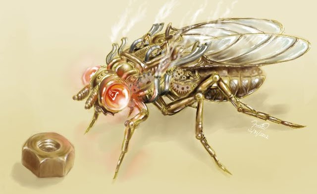
Genesee Scientific Fly Art commission, titled “Drosophila steampunkii.” Created with GIMP.
Were you always going to be a scientist?
I definitely believe I was always going to be pursuing science in some capacity — since I was a little kid, I enjoyed going out in the yard to observe and collect various insects, or making a mess in the kitchen after seeing some experiment demonstrated in a TV show (often to my mother’s chagrin!). My parents encouraged and nurtured my love for science by buying every book, even gifting me with a simple compound microscope and slide-making kit — however, in my culture, children who have an affinity for science are typically expected to pursue a degree in medicine. I rationalized the choice of studying to become an MD as a career path that would still allow me to do science, and held onto this aspiration all the way up to college. However, my first research experience at the University of Guam under the auspices of an NIH Research Initiative for Scientific Enhancement (RISE) program, combined with some conversations with my professors convinced me to fully embrace scientific research as a career goal.
And what about art – have you always enjoyed it?
Art has always been a passion of mine. Growing up as an only child with few children my age in the neighborhood, drawing was an outlet that allowed me to express my imagination. Like my love for science, my parents also indulged my love for art — I’ve dabbled in pretty much every medium while growing up. Art also influenced my decision to pursue studies in developmental biology; I was awed by the beautiful high-resolution images that one can obtain of immunostained tissues. In a way, developmental biology research combines two of my greatest passions!
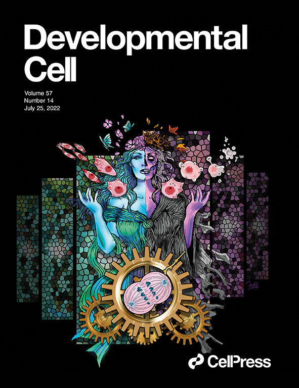
A cell cycle-coupled quality control system for migrating embryonic muscle precursor cells in Drosophila illustrated as Persephone, the dual goddess of Spring and the Underworld. The image depicts how cells either undergo continued growth and myogenesis after cell division, or programmed cell death. Created with Adobe Illustrator.
What or who are your most important artistic influences?
I would say I’m heavily influenced by detailed sci-fi/fantasy art. Growing up, I admired the works of artists like James Gurney, who combined meticulous depictions of dinosaurs with fantastical scenes for ‘Dinotopia,’ evoking a world where humans and dinosaurs coexist. As a cell biologist, I also greatly admire the work of Dr. David Goodsell — his ability to bring biomolecules and the spectacular, microscopic worlds they inhabit represent a truly elegant union of science and art!
How do you make your art?
I typically use black and white media like pencil and pen on paper for sketches and such. Some time back I bought myself a drawing tablet and started dabbling in digital art. I didn’t go through any formal training, so it’s been a lot of trial and error for me. I used to use the GNU Image Manipulation Program to make pieces, but I’ve since moved on to using Adobe Illustrator or Adobe Photoshop.
A stylized illustration of a gynandromorphic fly, depicting a friend’s discovery of a mechanism for piRNA-dependent regulation of sexually dimorphic traits in Drosophila. Created with Adobe Illustrator.
Does your art influence your science at all, or are they separate worlds?
Art definitely has a profound influence on my field of research! Throughout the grad school and postdoc phases of my career, I often illustrated figures (and occasionally cover art) for manuscripts in addition to benchwork. My lab studies how embryonic muscles undergo precise and highly-stereotyped patterning, so my group spends a lot of time collecting images of immunostained cells. Scoring defects in muscle assembly takes quite a bit of observation and pattern recognition, which are skills that both artists and developmental biologists use regularly. Trying to understand the various phenotypes we observe also takes quite a bit of creative thinking — I think a lot of people underestimate how big a role imagination plays in planning and executing studies!
What are you thinking of working on next?
I’m thinking of creating more fruit fly-related work. I love the Drosophila research community, and want to inspire my students to see the beauty in studying developmental biology through model organisms like the fruit fly!
I became interested in the evolution and development of skin appendages (such as scales, spines and feathers) during my PhD with Dr. Gareth Fraser, when I was studying the patterning of scales and teeth in the shark. I wanted to continue working in this field, and luckily the LANE(Milinkovitch-Tzika lab) advertised a postdoc position at just the right time. This lab is renowned for studying skin appendage development in really diverse non-classical models such as snakes, lizards and exotic mammals, so it was the perfect destination for me. One key finding by Michel Milinkovitch & his then post-doc Nicolas Di-Poï, was that not only hair and feathers, but also reptilian scales, develop from placodes – localised thickenings of the skin that constitute the foundations of most skin appendages (2). When I arrived in the LANE in 2020, there was already an ongoing project investigating chicken scale development, so it was easy for me to get started straight away.
What was already known about ectopic feather induction in chicken embryos?
There have been a few published articles reporting a scale-to-feather conversion in the chicken embryo (3-5). However, the results of these studies are variable. First, not all of these scale-to-feather conversions result in true, regenerative feathers, second, the coverage of these ectopic feathers was not ubiquitous, and third, the post-embryonic development of these feathers had not been examined. Also, our lab had recently developed a really nice protocol for injecting precise quantities of drugs directly into the veins of developing chicken embryos (as well as other hard-shelled eggs) (6). You can check this out in the video below. This project provided a great opportunity to try out this method.
Intravenous injection protocol for treating hard-shelled amniote eggs (Cooper et al. 2023)
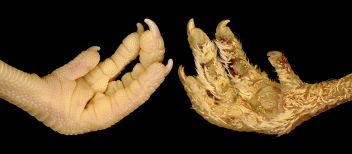
Can you summarise your key findings?
We investigated the role of the sonic hedgehog (Shh) pathway in mediating skin appendage fate in the chicken. This pathway is essential for loads of developmental events, and is really important in controlling the early development of placodes. We injected a precise dose of a Shh pathway activator directly into the blood stream of developing embryos (6), at the exact time that scales begin to appear on their footpads. We then collected the embryos at various stages after injection, and even allowed some to hatch so that we could examine the effect in adult chickens. Incredibly, every single chickens injected with the Shh activator had abundant ectopic feathers covering their feet. We tracked the development of these ectopic feathers in hatched chickens, and saw that they transitioned from juvenile down-type feathers into true contour feathers observed in adult birds. This confirmed that our experimentally-induced feathers are true, regenerative feathers.
When doing the research, did you have any particular result or eureka moment that has stuck with you?
I distinctly remember looking at the first treated sample under the microscope – it was collected after 4 days, meaning that the ectopic feather buds were still relatively undeveloped. At first glance it looked like an incredibly hairy chicken foot. I showed the sample to my boss, and he suggested that we should see what happened if we allowed the embryos to develop until hatching. A few weeks later we had our first feather-footed hatchling chickens!
And what about the flipside: any moments of frustration or despair?
Whilst attempting to harvest samples for RNAseq, the treatment suddenly began to kill all of the embryos. As there was a short power outage in our lab during the incubation of the eggs, I initially assumed that the embryos were slightly less developed than before. I then repeated the experiment with fresh eggs, and exactly the same thing happened. Finally, I contacted the farmer who provides the eggs, and he told us that they had just replaced their laying hens, meaning that the eggs were now coming from much younger chickens, which lay much smaller eggs. After adjusting the treatment to account for this change, the experiment worked perfectly.
What is next for you after this paper?
I’m working on some more really exciting skin appendage-related projects in the Milinkovitch-Tzika lab. We have an additional manuscript on chicken scale development in the pipeline, and Michel has had me heavily involved me in another project on crocodile scale patterning – they’re both really cool stories, and hopefully we will be able to share them with you soon!
References
1. R. L. Cooper, M. C. Milinkovitch, Transient agonism of the sonic hedgehog pathway triggers a permanent transition of skin appendage fate in the chicken embryo. Science Advances9, eadg9619 (2023).
2. N. Di-Poï, M. C. Milinkovitch, The anatomical placode in reptile scale morphogenesis indicates shared ancestry among skin appendages in amniotes. Science Advances2, e1600708 (2016).
3. D. Dhouailly, M. H. Hardy, P. Sengel, Formation of feathers on chick foot scales: A stage-dependent morphogenetic response to retinoic acid. Journal of embryology and experimental morphology58, 63-78 (1980).
4. R. B. Widelitz, T. X. Jiang, J. Lu, C. M. Chuong, Beta-catenin in epithelial morphogenesis: conversion of part of avian foot scales into feather buds with a mutated beta-catenin. Dev Biol219, 98-114 (2000).
5. P. Wu et al., Multiple Regulatory Modules Are Required for Scale-to-Feather Conversion. Mol Biol Evol35, 417-430 (2018).
6. R. L. Cooper, G. Santos-Duran, M. C. Milinkovitch, Protocol for the rapid intravenous in ovo injection of developing amniote embryos. STAR Protocols4, 102324 (2023).
How stem cells decide to choose between two conflicting fates, division vs. differentiation, is an unsolved mystery of stem cell biology. The overarching goal of Shariati lab is to determine the mechanisms that link cell division to cellular differentiation in pluripotent cells. We will combine emerging genome-editing technologies with single cell imaging to determine regulatory principles of cell fate decisions in pluripotent cells.
Applicants with demonstrated experience in any of the following areas are highly encouraged to apply: Stem Cell Biology, Transcription, Quantitative Single Cell imaging, CRISPR screening and Mouse Genetics. The potential start date is Fall 2023.
As your postdoc advisor, I will support you to reach the career goals that you set for yourself. In addition, UCSC has exceptional career development programs for postdoctoral fellows. Santa Cruz is a great town for postdocs as you can easily refresh your mind by strolling in beautiful redwood forests on campus or going to one of the many nearby beaches. Please email me (alish {a} ucsc.edu) a cover letter stating your scientific background and interest, your CV and names/addresses of your advisors.
Applicants from underrepresented groups in sciences are highly encouraged to apply. Informal inquires also are welcomed!
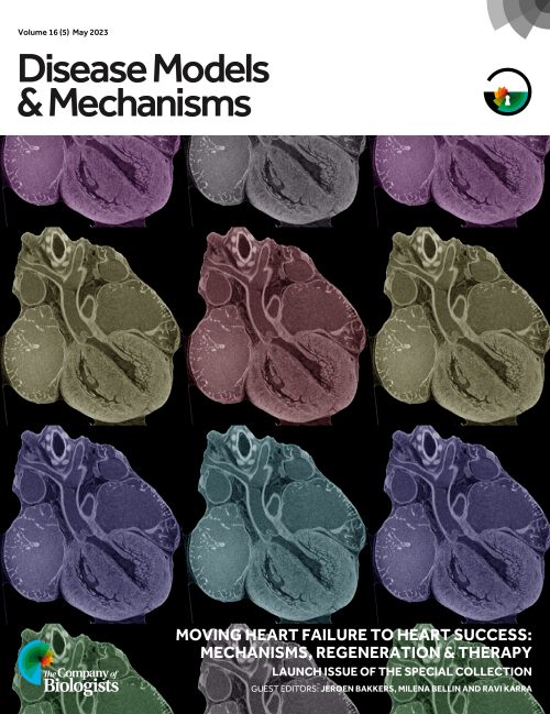
Heart failure occurs when the heart is weakened or damaged, or when there is a problem with the heart’s valves or rhythm. Strikingly, it is the leading cause of hospitalisation and deaths in men and women worldwide. Nevertheless, significant gaps remain in our understanding of heart failure, and treatment, at best, results in disease remission. Our goal in shaping this Special Issue of Disease Models & Mechanisms (DMM) was to compile original Research, Resource and Review-type articles that investigate the genetic and biological mechanisms of heart failure and identify potential therapeutic strategies, as summarised in an Editorial from Guest Editors Jeroen, Milena Bellin and Ravi Karra.
Guest Editors Jeroen Bakkers, Milena Bellin and Ravi Karra
A significant risk factor for heart failure is congenital heart defects, which are structural abnormalities affecting 8 out of every 1000 new-borns. The causes of congenital heart defects are diverse, which encourages the exploration of these disorders in the context of cardiac development. A Review from Christian Mosimann’s group summarises how defects in lateral plate mesoderm development drive congenital defects of multiple organs, including the heart, and a Research article from Didier Stainier and colleagues uses zebrafish to study the role of the epicardium, the outermost layer of the heart, in coordinating growth of the myocardium. Sally Dunwoodie and colleagues also published a study on a metabolic disease that results in congenital heart malformation.
Beyond disordered heart development, arrhythmias, which occur due to disruptions in the heart’s electrical system, represent another risk factor for heart failure. A Review by Vincent Christoffels and colleagues, published in this issue, explores the genetic causes of heart arrhythmias. Another Review from Eva Van Rooij’s group discusses the use of precise genome editing to generate models of and to potentially treat cardiomyopathies, which involve the remodelling of heart muscle and can also lead to heart failure if left untreated.
This special issue also provides an insight into the current understanding of acquired heart disease, with studies ranging from the investigation of strategies to mitigate cardiac remodelling upon injury to how the gut microbiome metabolite trimethylamine-N-oxide affects heart function.
Finally, the Special Issue includes ‘A Model for Life’ interview between leading experts Jeroen Bakkers and Didier Stainier, who discuss the advantages and disadvantages of zebrafish models of heart disease. Model organisms, both large and small, have been instrumental in advancing this field, with articles in this Special Issue using or discussing the use of mice, rats and pigs as valuable models of heart failure with strong translational potential. In addition, in vitro models, such as organoids, will provide new research opportunities to study cardiac development and related diseases, as summarised in the beautiful At a Glance poster article by Lika Drakhlis and Robert Zweigerdt and in ‘A Model for Life’ interview with Professor Christine Mummery. The Research, Resource and Review-type articles in this Special Issue cover a wealth of seemingly diverse topics. However, preventing, understanding and treating heart failure are the common threads that bind these articles. Follow the link below to read all the articles in this Special Issue for free.
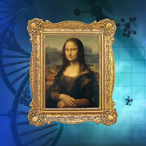
“In an attempt to fight modern-day art forgery, researchers have been developing techniques that involve using DNA to tag pieces of art”
Dr Kat Arney
In the latest episode of the Genetics Unzipped podcast, we’re taking a journey into the world of art and artefacts, extracting DNA from paintings, hair and even chewing gum, and unearthing the genetic secrets of long-dead legends like Da Vinci, Van Gogh and Beethoven.
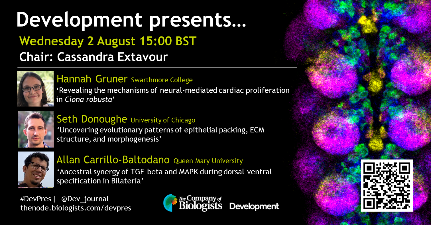
Our August webinar will be chaired by Development Editor Cassandra Extavour (Harvard University) and features three early-career researchers studying evo-devo. The webinar will be held using Zoom with a Q&A session after each talk.
Wednesday 2 August 2023 – 15:00 BST
Hannah Gruner (Swarthmore College) ‘Revealing the mechanisms of neural-mediated cardiac proliferation in Ciona robusta’
Seth Donoughe (University of Chicago) ‘Uncovering evolutionary patterns of epithelial packing, ECM structure, and morphogenesis’
Allan Carrillo-Baltodano (Queen Mary University) ‘Ancestral synergy of TGF-beta and MAPK during dorsal-ventral specification in Bilateria’
By Ioakeim (Makis) Ampartzidis, Courtney Lancaster, Danielle Liptrot, and Rosie Marshall
Happy Birthday YEN!
The Young Embryologist Network (YEN) turns 15 this year. YEN serves as a vibrant platform for knowledge exchange and collaboration, with a particular emphasis on showcasing the work of early career researchers (ECRs — postdocs and PhD students). It has been 15 years full of inspiring science and what better way to celebrate than with a debrief of this year’s fantastic conference:
Hot Off the Press: Scientific Talks
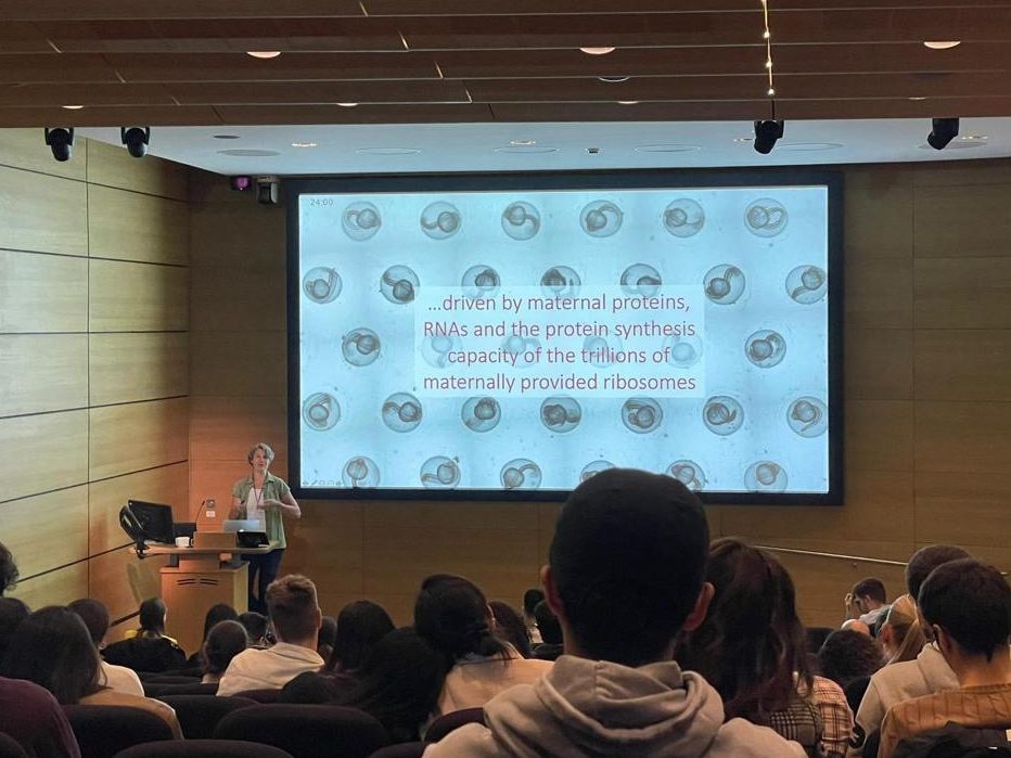
Pre- and peri-implantation development was a particular focus in the scientific talks this year, highlighting the drive to improve in vitro embryo work relating to fertility. Two of the invited speakers presented impressive work on the subject: Katsuhiko Hayashi from Osaka University kicked off the day with an impressive overview of his ground-breaking work on reconstructing oogenesis from pluripotent stem cells, which he hopes will be used to save the endangered Northern White Rhino (only two females of this species remain); Andrea Pauli from the Institute of Molecular Pathology in Austria later showed us her fascinating work on how closely related species prevent interspecies fertilisation (in the case of zebra- and medaka-fish, it’s just one protein expressed on the egg!)
Many of the selected talks also focused on these early stages of development: Chloe He showed us how deep learning and non-invasive imaging of cleavage-stage human embryos can help us to study how cell arrangement affects blastocyst quality; Johanna Gasler demonstrated the requirement of orphan nuclear receptors on early development and zygotic genome activation in murine embryos; Lessley Sepulveda-Rincon produced apoptosis-deficient primordial germ cells in mouse chimeras to understand their functional incorporation to the mouse embryo; Zukai Liu presented the specification of extra-embryonic mesenchyme in primate development, at a genome level revealing ape-specific attributes.
Andrea Pauli’s exciting talk on fertilization.
Once again, YEN showcased the wide range of fascinating organisms used in developmental biology research. Michael Emmerson revealed how zebra finches can detect sound cues to alter their growth rate in utero (including expert impressions of zebra finch heat calls)! The utility of the old favourite Drosophila was once again on show, with Gloria Jansen’s impressive talk on how paternally inherited transposable elements alter penetrance of germ cell loss. O. fusiformis, a bilaterally symmetrical organism, is another neat model on which Allan Carrillo-Baltodano studies dorso-ventral patterning. Finally, zebrafish once again lived up to its reputation as a beautiful model for imaging, with Agatha Ribeiro da Silva utilising the model to study endothelial cell migration defects in heart development.
Wrapping up the short talks was the final invited speaker, Laura Pellegrini. She is a current postdoc in Madeline Lancaster’s lab who is soon moving to King’s College London to start her own group. She showed us how choroid plexus organoids have a wide range of applications, from a drug screening system to test whether drugs can cross the blood-CSF barrier, to a model for studying viral infection of the brain. Certainly ground-breaking work and we look forward to seeing how her group progresses!
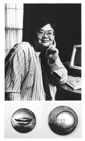
Sammy Lee Memorial Lecture
The final talk of the day was the Sammy Lee Memorial Lecture, which was established to remember Sammy and to reflect on his contributions to developmental biology and his passion for inspiring the next generation of scientists.
Dr Sammy Lee and the memorial Sammy Lee medal designed by Felicity Powell.
Sammy was a visiting professor in Cell and Developmental Biology at University College London (UCL), whose life-changing work was responsible for outstanding advancements in reproductive health and fertility. Sammy started his scientific journey with a PhD at UCL, where he was working on nerve muscle interaction. Not long afterwards, Sammy became interested in developmental biology. This led to his work on gap junctions in mammalian embryos where he discovered a link between gap junction communication and early embryonic development. He then became a clinical embryologist in 1985, helping to perform some of the UKs first egg donations, before being appointed as the scientific director of the Wellington IVF department. Sammy’s team was the first in the UK to perform gamete intrafallopian transfer (GIFT), pioneered a method of mechanical assisted hatching in the UK to improve chances of in utero implantation and produced the world’s first virus free ICSI (intra-cytoplasmic sperm injection) baby to a HIV discordant couple.
Fitting with Sammy’s character, the Sammy Lee Memorial Lecture was delivered by Henrik Kaessmann who entertained us with the wonders of sex chromosomes. He drew our attention to the small size of the Y chromosome which has a striking feature of chromosome wide gene decay. He then discussed gene dosage across different evolutionary lineages from mammals to birds.
It was Sammy’s wish to present a medal to a young developmental biologist on the merit of scientific communication and outstanding research. The 2023 Sammy Lee medal for best short talk was awarded to Clara Munger, who impressed the judges with her presentation about marmoset amnion specification. She told us about her work using marmoset embryonic stem cells to make spheroids in a microgel culture system, and how this system can be used to interrogate signalling pathways required for lineage specification in the post-implantation embryo. The runner-up went to a fantastic talk from Ashley Libby who discussed progenitor cell dynamics and gene regulatory networks during neural tube development.
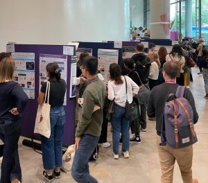
Poster Sessions
First poster session of the day.
The day was split up by two fantastic poster sessions in the morning and afternoon, with over 30 posters being presented both in person and online. The topics ranged from sea urchin larval skeletogenesis to the identification of unwanted genetic variants in stem cell populations, having implications in evolution, developmental biology and regenerative medicine. In addition, it was excellent to see projects being presented from a range of early career researchers, from master’s students to senior postdocs. The diversity in presenters and topics incited long discussions in both poster sessions, making it difficult to coax everyone back to their seats. It will certainly be exciting to see what collaborations were spawned in these sessions, and how it will influence the direction of developmental research in years to come.
With all the excellent projects being presented, it was surely a hard decision for the judges to award the poster prize and runner up. That being said, on observing the work of prize winner – Francisco Manuel Martin-Zamora – and runner up – Adiyant Lamba – it is evident their projects are both deserving of recognition. Francisco, a PhD student at Queen Mary University, presented his work on how epigenetics may underpin temporal shifts in the diversification of larval and bilaterian life cycles. Adiyant, also a PhD student based at the University of Cambridge, described the heterogeneities which influence early cell fate decisions between the inner cell mass and the trophoectoderm. The future of these two young embryologists is very bright!
Perspectives: EDI Awareness in Academia
A refreshing addition to the science-rich day was the addition of scientific perspective sessions, introduced by the committee in 2022. This year, the focus was on Equity, Diversity, and Inclusion (EDI) in science, raising awareness for the importance of embracing EDI in scientific meetings.
This year’s perspective experts Alison Forbes, Head of Inclusion at The Francis Crick Institute in London, UK, and Rafael Galupa, a social entrepreneur and group leader at the Centre for Integrative Biology in Toulouse, France, shared their valuable perspectives on the topic. They focussed on the current state of underrepresentation in science and offered practical strategies to enhance inclusivity. When asked how ECRs can realistically improve diversity, Alison suggested to “[…] start with self-education. Read some books and listen to podcasts;” while Rafael encouraged us all to “[…] find a way to contribute that is aligned with their personality and availability”. By prioritizing EDI in scientific meetings, we collectively advance knowledge and ensure that the science we all love and care about reflects the full spectrum of human perspectives and capabilities. For a more in-depth conversation on this topic, read this interview with Alison and Rafael.
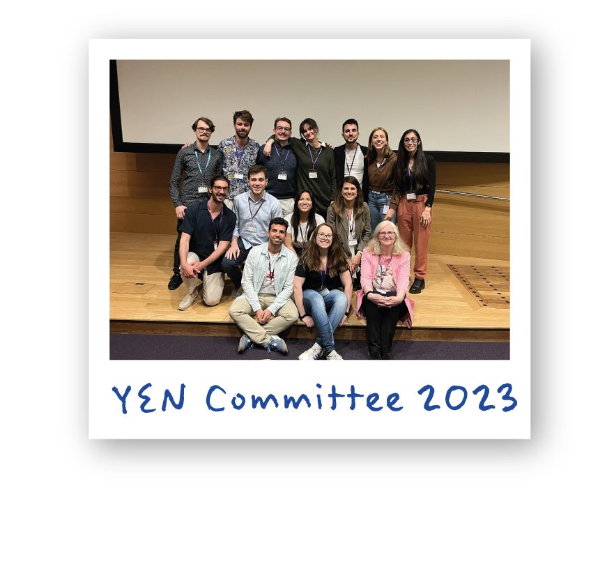
The faces behind YEN 2023
A big shout-out to the organising committee of YEN Conference 2023, for their hard work, fresh ideas, and organising skills. This year’s chairs Foteini and Jeremie along with the fantastic team of Michelle, Sergio, Jack, Ollie, Luca, Ferran, Olivia, Oliver, Matyas, Jesus, Mint, Claudia, Irina, and Christos, celebrated the 15th birthday of Young Embryology Network with a memorable Conference. The team is composed mainly from PhD students and ECRs based in London and if you like to learn more about their journey follow them on twitter@YEN_community.
 (No Ratings Yet)
 (3 votes)