Hi everyone, my name is Laura Hankins and I’ve recently joined Development as a Reviews Editor. I haven’t moved far, as I initially joined The Company of Biologists back in 2021 as their Science Communications Officer. That role gave me the opportunity to work with lots of different teams within the company and provided me with an insight into how publishing operates behind the scenes. I’m excited to build on this experience as I settle into the Development team.
Before joining The Company of Biologists, I completed a PhD at the University of Oxford via the Wellcome Trust’s four-year DPhil in Chromosome and Developmental Biology. After rotation projects with Paul Riley and Neil Brockdorff, I settled in Jordan Raff’s lab where I worked on centriole biogenesis in early Drosophila embryos. This involved plenty of microscopy! During this time, I was lucky enough to be awarded a place on the 2019 Physiology Course at Woods Hole, MA. This was a fantastic experience that exposed me to even more microscopy and various quirky organisms, as well as allowing me to meet many wonderful scientists from a range of career stages and research backgrounds. This sense of community was something that I really missed when the pandemic struck towards the end of my PhD.
I’m therefore delighted that I get to stay in touch with the scientific community in my new role. I’m looking forward to working with authors to produce front section content for the journal, and to getting to know the community better as I start to attend meetings and workshops. Please feel free to contact me via email or LinkedIn, and hopefully I’ll get the chance to meet some of you in person over the coming months!
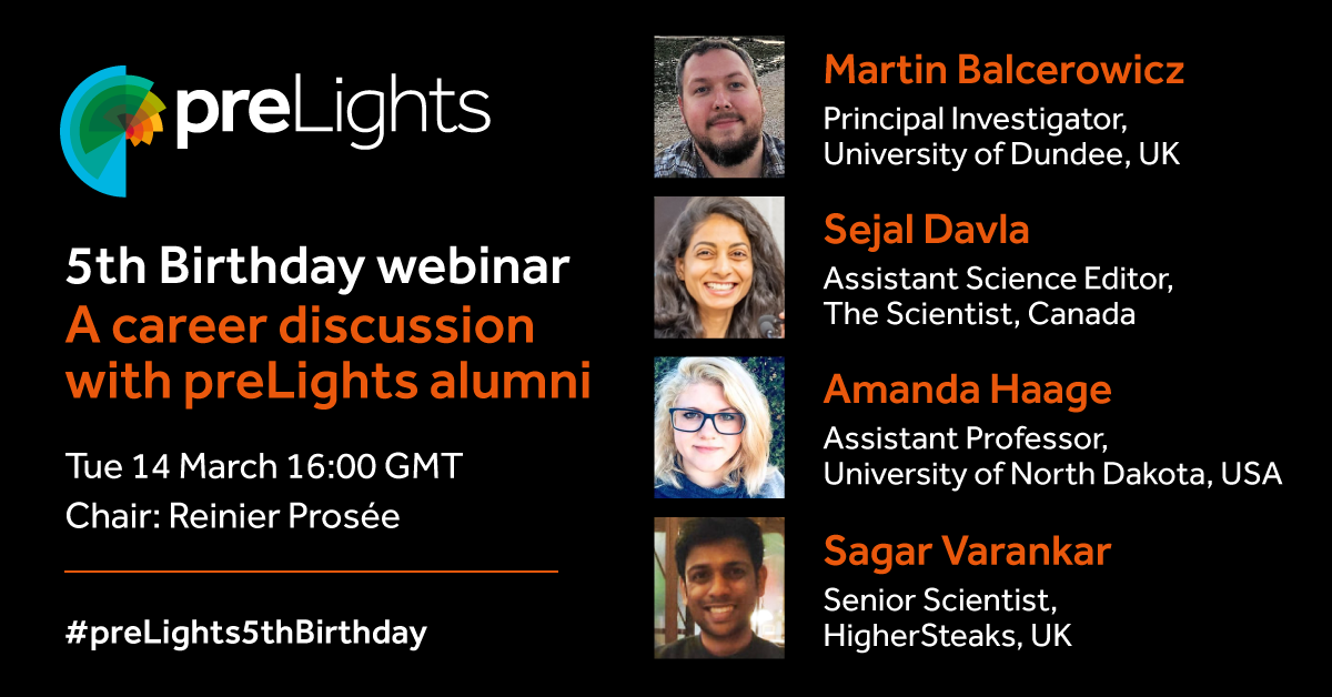
Our sister community site preLights is celebrating its 5th birthday! 🎉
To mark the occasion, a festive webinar has been organised that features talks given by preLights alumni who have all made significant contributions to preLights, and have since taken a range of career paths. In these talks, they will tell us how they have managed to identify and navigate the challenges, opportunities, and obstacles they faced as early-career researchers and how these have all led them to where they are today. Also, they will tell us a bit about their current position and future goals and ambitions.
Following a brief presentation by each speaker, there will be a Q&A session.
The webinar is free and open to all, and a link to the recording will be made available to registrants.
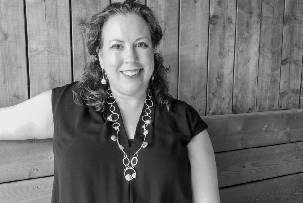
In our latest SciArt profile we hear from Robin Cassady-Cain, a professional artist based in Canada. Robin’s scientific background is in immunology and many of her artworks are inspired by the human immune system. As well as creating stunning jewellery, Robin uses her artwork as educational tools.
Where are you originally from and what do you work on now?
I was born in Barrie, Ontario, but mostly grew up in Toronto. I did my undergrad in Biochemistry at University of Waterloo, then a Masters in Immunology, and my PhD in Immunology at University of Cambridge. My last research post was at the Roslin Institute, University of Edinburgh, working with Professor Mark Stevens. I was a core-funded Research Fellow, so I worked on large animal models of bacterial enteric infection (the big 3-Campylobacter, Salmonella and various pathogenic E. coli) part of the time, and part of the time directing my own research looking at the molecular mechanisms, biophysical and structural characteristics of a bacterial toxin homologue, Lymphostatin, known to block lymphocyte activation in vitro/ex vivo. In 2018, I moved back to Canada, giving up my research career to become a professional artist.
Yes! The exact time that I decided that I wanted to be a scientist is lost to the sands of time, but it was somewhere in middle school, doing all those science fair projects. I had dreams of being a professor, it seemed so glamorous to me (how little I knew!!). The idea of being a scientist really cemented itself in high school, particularly as I had several supportive and influential science teachers. Science held an endless fascination for me, and although I could imagine doing other things, I felt that research was where my mind and heart were leading me.
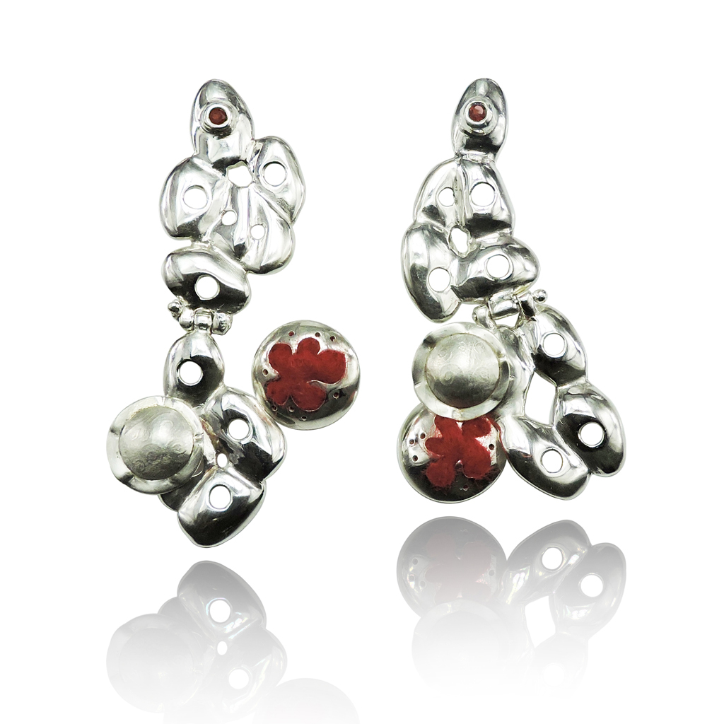
Tumbling Earrings: sterling silver, garnet and needled felt
And what about art – have you always enjoyed it?
Yes! I’ve enjoyed looking at art and making things since I was very young. My earliest memory is making clothes on my child’s Singer machine for my Barbie dolls (the only thing I thought they were good for!) Over my life I have done photography, wet and needle felting, papier mache sculpture, quilting, collage, Scottish kiltmaking and, of course, jewellery.
What or who are your most important artistic influences?
I love impressionism, and abstract art, but if you want to know about jewellery/metal arts, Wendy Ramshaw, an iconic British goldsmith had a huge influence on how I viewed jewellery and the art of making jewellery. From her early paper jewellery, her stacking rings that come with their own unique tower storage, and her whimsical story-telling pieces, I love the breadth of her work and particularly some of the geometric lines that she used.
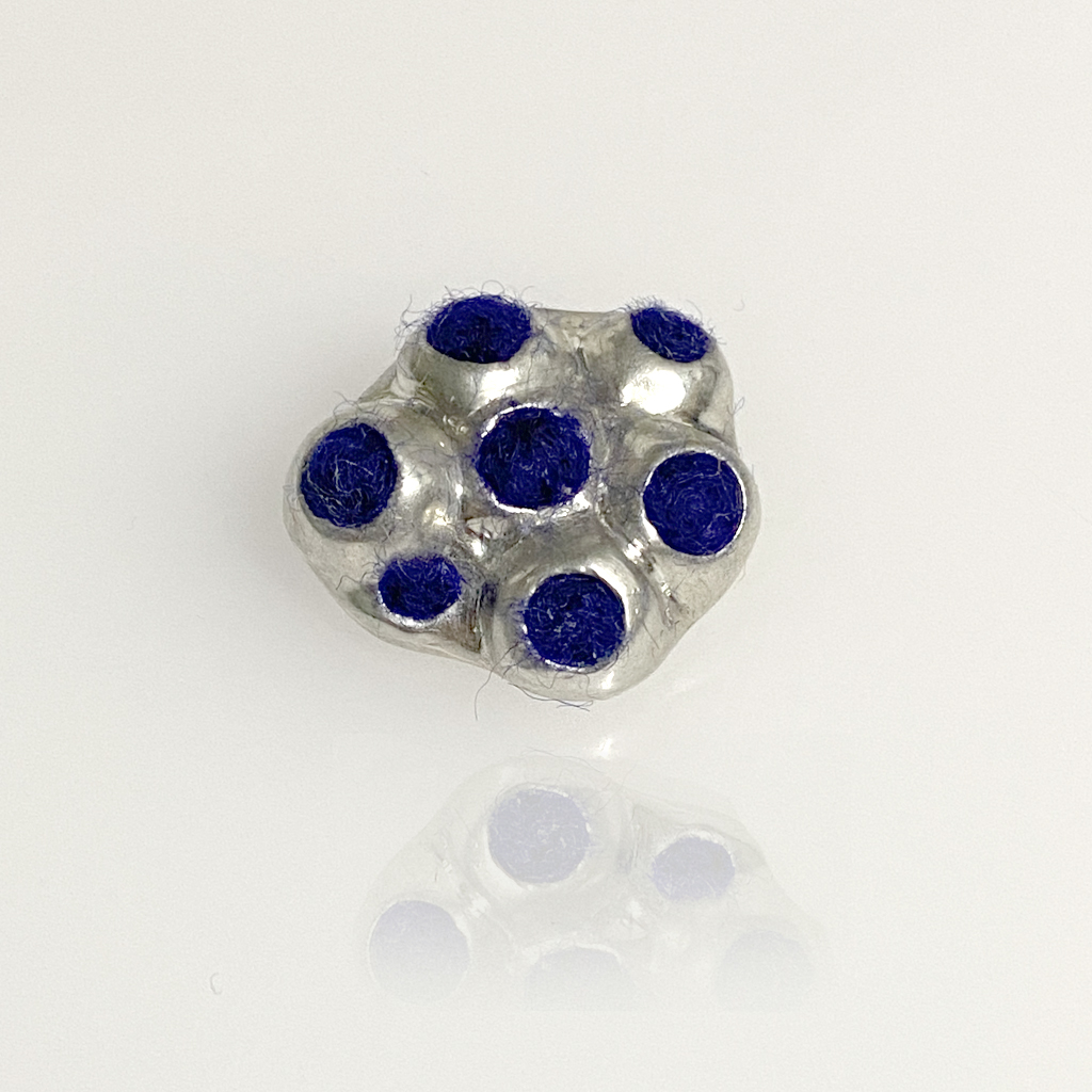
Stem cell brooch: sterling silver, needled felt
How do you make your art?
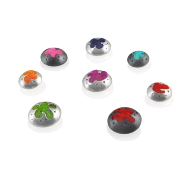
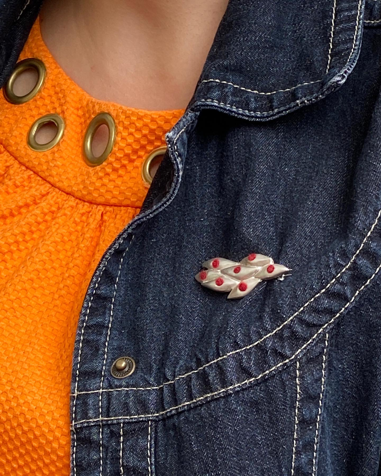
I work primarily out of a shared jewellery workshop in the west end of Toronto called Jewel Envy. Since I’ve switched to primarily working as a professional artist, I have found community there, which is invaluable. I have an ongoing obsession with immunology and cell forms and concepts clearly influence my personal art jewellery, but I do a lot of work with clients to help them realise their own concepts in jewellery form, and more collaborative work, like some of the pieces showcased here. I worked with an old friend and colleague who wanted to have some jewellery created that reflected some of the cell work that they do with stem cells, and myometrial cells. The end result were two sculptural cell forms—one representing the embryonic stem cell ball, complete with felty nuclei, taken from fluorescence microscopy images. The other is modelled after a schematic of myometrial muscle cells, again with felted nuclei. I do a lot of jewellery incorporating felt, patterned textured, and incorporating semi- and precious gemstones, using the traditional methods of metal fabrication and lost wax casting.
Does your art influence your science at all, or are they separate worlds?
My art used to influence my science, in the sense that it provoked me to think more deeply on some topics, and how things connected together. I found it almost a meditative process that would quiet the part of my mind that was always actively thinking about my science, and allowed insights to float to my conscious mind more easily. Conversely, I’ve carried through so many skills and thoughts that I had about my science through to my professional artist practice.
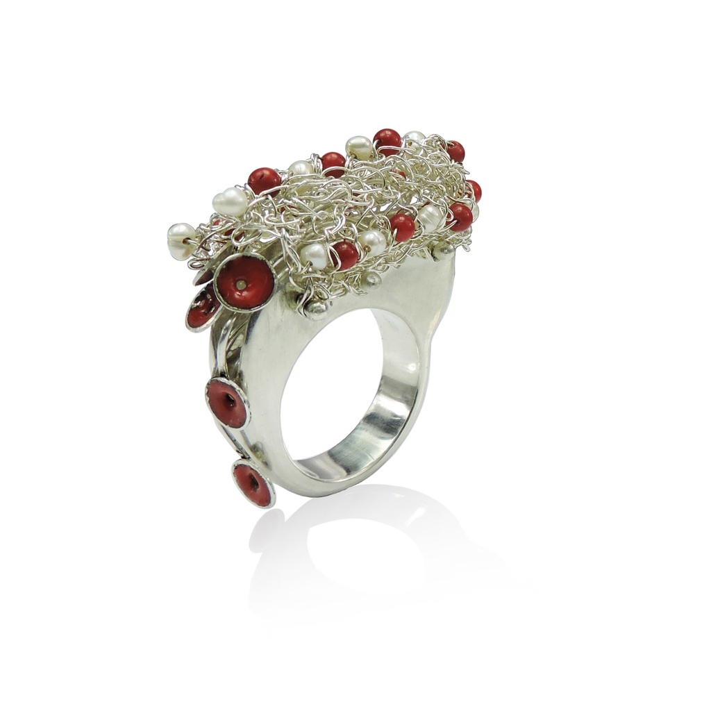
Spillage ring: sterling silver, sea bamboo, seed pearls and enamel
What are you thinking of working on next?
I continue to explore the themes of the immune system in my jewellery, and I can’t see me ever exhausting my ideas there. I love the fact that every major piece that I do is a potential conversation about immunology and/or opportunity for education. I am also embarking on a collaborative series of mixed media art pieces with another artist that is centred around the concept of herd immunity. It’s just at the very early formative stages, but I can’t wait to see where it takes us!
Thanks to Robin and all the other SciArtists we have featured so far.We’re looking for new people to feature in this series – whatever kind of art you do, from sculpture to embroidery to music to drawing, if you want to share it with the community just email thenode@biologists.com (nominations are also welcome!)
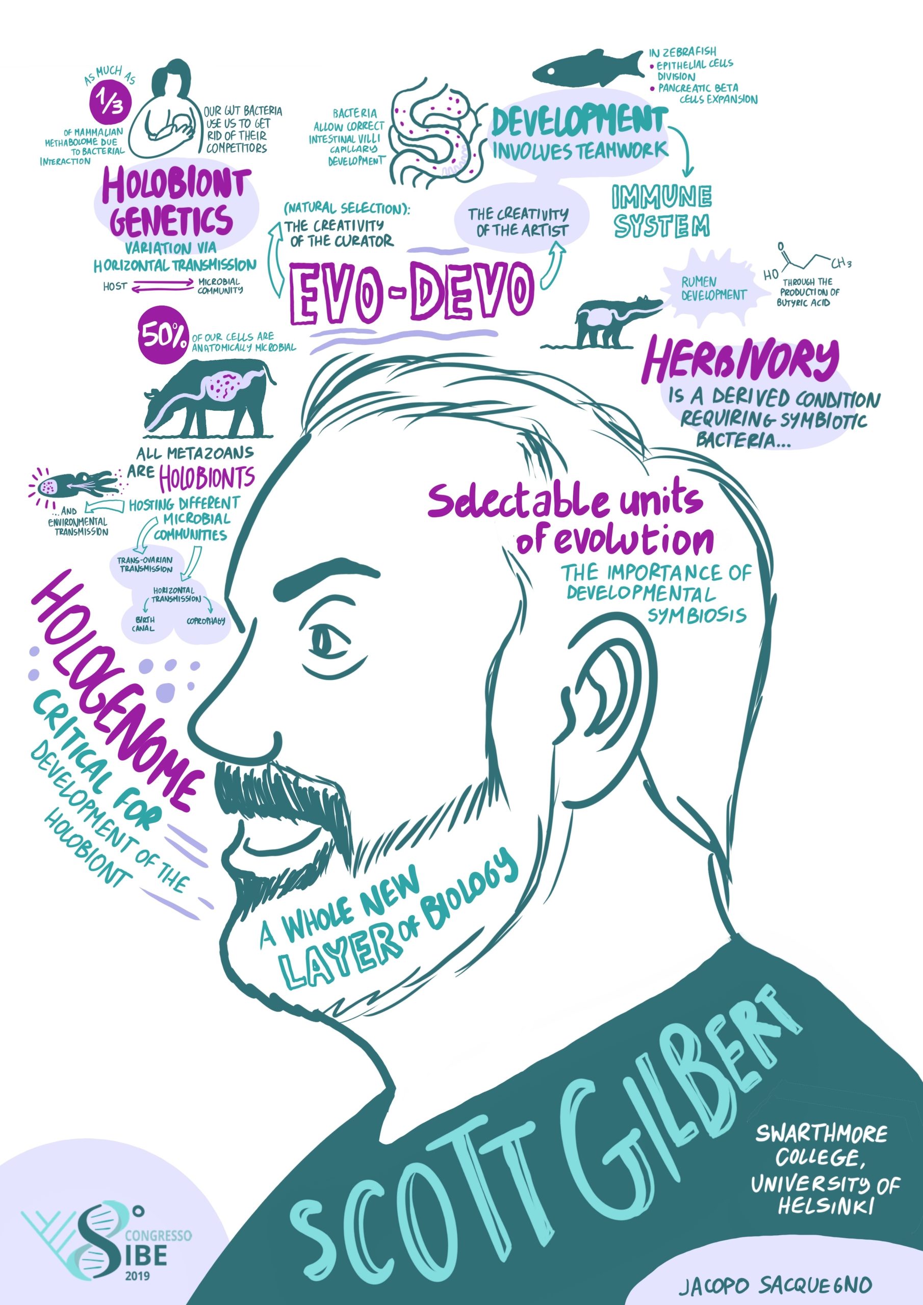
Scott Gilbert is Emeritus Professor of Biology at Swarthmore College and the University of Helsinki. He literally wrote the book on developmental biology! With the 13th edition of ‘Developmental Biology’ about to be published, we took the opportunity to find out more about the story behind the textbook, to discuss Scott’s research career and his social and political commentaries in relation to developmental biology.
Let’s start at the beginning, when did you first become interested in biology?
I think that everyone is initially interested in biology; but for some odd reason, most people aren’t able to keep that interest. Most young people love dinosaurs, turtles, and horses, but they leave those interests behind. I was always fascinated by nature. My summer days were spent in the woods and fields of northern Pennsylvania, finding salamanders, frogs, and butterflies. My scientific career peaked when I was 12 years old. I had caught a five-legged frog, and word of this somehow got to Leonard Lyons, the syndicated gossip columnist for the New York Post. So, I was mentioned in his New York Post column, alongside Josephine Baker, Leo Szilard, and Pablo Casals. I don’t expect to ever reach this pinnacle again. I was also fortunate to have met Dr Jean Cook, a Black haematologist at the Albert Einstein College of Medicine, who gave me my first research experience while in high school. He was fascinated by the ways that life could have originated in the early atmosphere of the planet. In trying to repeat the Urey-Miller experiment, I nearly fried myself, trying to splice wires together while they were still plugged into the sparking coil – a learning experience (and I did get to identify aminoacrylonitriles). In addition to teaching me about erythrocyte development and how to do column chromatography, he also taught me to appreciate and enjoy the music of Julian Bream.
You went on to study biology with religion at university, what prompted you to choose that combination?
I always thought I was going to be a biology major, but in college, I also became fascinated by the study of religion. I came to the conclusion that the ecology movement needed an alliance of science and religion, and that the two areas were actually related. I see ‘Wonder’ as the prime experience that the mind has with the world. But wonder has a short half-life and ‘decays’ into curiosity (I wonder) and awe (the wonder of the world.) Curiosity gives rise to science and philosophy; awe gives rise to reverence and the religious attitude. So, science and religion are both the ‘grandchildren’ of wonder. Moreover, science and religion have a vested interest in keeping those sources of wonder alive. I don’t care whether a person goes into the environmental movement to preserve biodiversity, or to be a proper steward of God’s creation. They’re both motivations for preserving natural wonder, which I feel is critical, among other things, for preserving science.
In 2016, I was asked to give a lecture on developmental biology to His Holiness, the Dalai Lama. After my talk, the Dalai Lama said that he enjoyed the talk, but that the questions of interest to him are: How does the reptilian brain become the mammalian brain, and how does the mammalian brain become the human brain? So, we have a religious leader who not only accepts evolution, but who wants to know the evolutionary developmental mechanisms of neocortex formation!
Do you think that it is important for scientists to comment on social and political issues that are related to their work? Have you faced any criticism for your papers and commentary?
As embryologists we have to deal with issues of sexism, racism, and abortion. If we’re working in evo-devo, we’ll come across creationists. Saying nothing about these social issues is itself a political position. If you have expertise in these fields yet don’t combat publicly stated falsehoods, then you’re basically saying that the status quo is acceptable. Most of my social commentary has been against people misusing developmental biology to say that science supports white supremacist or male supremacist arguments. I think it’s because I love developmental biology and I don’t want to see my beloved being smeared! Having a background in history of biology has helped me enormously in addressing these issues. I’ve written about how scientists in the 1800s tried to use human embryology to show that women and Black people were immature, embryonic, forms of the white male. Until very recently, the female was seen as the incomplete development of the male phenotype.
My most recent paper concerns myths that are currently being used in the abortion debate in America. Each of these myths is represented as science, but none of them has much to do with facts from developmental biology. These myths portray fertilisation as ensoulment, and depict the woman as a passive entity. I also show that different groups of biologists claim different embryonic stages to be the start of personhood (admitting that ‘personhood’ is not really a scientific concept), and that the notion that all biologists believe that personhood begins at fertilisation is ideology, not science. Some biologists say it begins at fertilisation when you get your genome. Other biologists say it begins at gastrulation, where you become an individual and can’t form twins or triplets anymore. Other biologists have claimed personhood begins when you get your electroencephalogram (EEG) pattern, because that’s when the anatomical correlates of consciousness and pain perception form (and we’re willing to say death is the loss of that pattern). Still others say personhood begin around when the fetus becomes viable outside the womb. And some people maintain that personhood begins at birth, when the first breath causes pressure differences that change endothelial gene expression and cardiac morphology, preparing the fetus for life outside the mother. I think the public should be made aware of these different possibilities. I haven’t faced criticism for my views, which probably means they are not well enough known.
Can you give us a brief overview of your research career?
I feel like the surfer that caught the perfect wave. I was at Swarthmore College with a background in developmental biology and history of biology, and that allowed me to catch and ride the waves of developmental biology and the evo-devo wave behind it. As I mentioned, I was an undergraduate at Wesleyan University in Connecticut, where I majored in biology and religion. I did research on the attachment of DNA to the nuclear envelope during sea urchin fertilisation in the laboratory of Tony Infante, and it was published in Nature New Biology. Teaching during the summers at an NSF-sponsored Pre-College Science Center run by Michael Somers convinced me that embryology was the field that most excited me. I pursued my PhD at the Johns Hopkins University in the laboratory of Dr Barbara Migeon, who is most famous for her work in X chromosome inactivation. My project concerned extending amniocentesis to look at the genes of human kidney cells, not just fibroblasts. This was before the days of DNA sequencing, and it meant doing enzyme assays in the cold room. I should mention at this point that I probably hold the record at Hopkins (and maybe elsewhere) for the number of sequential thesis advisors: four. While doing my PhD, I was also able to work on an MA in the history of biology under the direction of Donna Haraway. I did my master’s thesis on how the X chromosome was the bridge between Thomas Hunt Morgan’s embryology and his genetics.
For my first postdoc, I went to the University of Wisconsin to work with Masayasu Nomura, because I thought that E. coli ribosomes would be the best model for looking at the co-transcription of eukaryotic genes. I thought I could prove the Britten-Davidson model using E. coli ribosomes. We published some of the first gene sequences using the Maxam–Gilbert technique with restriction enzymes, polymerases and gel apparatus that we had isolated and made ourselves. Within a few months I had disproven my thesis, but we got some beautiful sequences of the regulatory region for RNA operons! I did a second two-year postdoc in developmental immunology with Robert Auerbach, while my wife was doing her residency in obstetrics and gynaecology. Here, we made monoclonal antibodies against poliovirus, and we discovered the mechanism by which antibodies neutralise the virus.
Moving on to the next step, I wanted a career where I could help raise children, have friends, and do other things besides science. But at the same time, I wanted a life that was saturated with science and research. I found this at Swarthmore College, a liberal art college in Pennsylvania. It had one of the best undergraduate biology departments in the country, and there were some phenomenal students coming into Swarthmore. My first paper from Swarthmore came after I heard a lecture on auxin transport given by our plant physiologist, Mark Jacobs. He had an assay for this transport, but he had no way to identify the proteins involved in transferring the hormone from one cell to another. I suggested using monoclonal antibodies (which I knew how to prepare). So, Mark and I published the first paper (in Science) using monoclonal antibodies to study plant development, identifying the location of the auxin transport proteins. I was able to get NSF grants because I was teaching undergraduate students how to do developmental biology. Originally, I continued my work on kidney development, and in 1990, I had the opportunity of spending a sabbatical leave in Finland with Lauri Saxén. Here, I worked on cell-cell communication and branching morphogenesis in mouse kidneys. I thoroughly enjoyed this time in Finland, and I brought back an interest in studying branched organ morphogenesis. Working with Judy Cebra-Thomas, we delineated paracrine factor involvement in the branching of amniote kidneys and lungs. Then I heard at a meeting that Brigid Hogan and Saverio Bellusci were going to work on this problem, and I knew that they could do in a year what would take us five years to do. It was time to get out! During this time, I had been writing about evolutionary developmental biology, so I decided that this would be the context for my next research project. After mooting a number of possibilities, I decided that asking how the turtle got its shell would be a really good project, and we began collaborating with several bone developmental biology laboratories in the Philadelphia area. Many undergrads got their first research experience working on turtle embryos, and grad students working on serious human bone diseases could have some relief working with our turtles. We proposed a paracrine factor hypothesis for the origin of the carapace, and we have evidence that cells migrating the trunk neural crest may actually form the plastron. Many years later, I decided that if I really wanted to get answers to these questions, I had to get into a research laboratory. So, I went half-time at Swarthmore, and received a half-time position at the University of Helsinki, working in the lab of Jukka Jernvall. We’re still working on the turtle project. I no longer have a lab, but Judy Cebra-Thomas has really interesting data that, if confirmed, will give us a great story on how the turtle gets both its carapace and its plastron. Former student Tyler Lyson has been integrating our developmental data into the paleontological record to identify the mechanisms by which turtles may have evolved their shells. The turtle has gone from being an anomaly to being a prime example of how developmental biology and palaeontology can interact to construct an account of how organisms evolved. I’m presently pursuing work on holobiont development (discussed below).
Visual summary of Scott’s lecture at the Italian Society for Evolutionary Biology meeting in 2019 by Jacopo Sacquegno (jacoposacquegno.com)
Working at a liberal arts college such as Swarthmore, you’re expected to do research because it’s the best way of teaching your undergrads. Of course, you go a bit slower because you’re teaching methods and attitudes. (I know how to do an in situ hybridization; my job is to teach the students how to do it; and they do not get it right the first time.) Also, a teacher at a liberal arts college has the ability to work with people outside one’s own department. This has allowed me to publish on the aesthetics of embryology, the roles of puns in science education, the bone that the Bible says created Eve (it wasn’t a rib!), and the use of embryos in the art of Gustav Klimt and Frida Kahlo. My students and I have collaborated to publish papers on Intelligent Design (writing this paper was their final exam), temperature-sensitive holobionts (another final exam), and feminist critiques of biology (which was one of the first papers in the field). When I gave a history of biology course, I saw that the students’ term papers on bioethics would make a fascinating book. Why should I be the sole audience for these investigations? So, with two students as co-editors, we published it. All-in-all, I’ve published nearly 200 papers, many of them concerning the history, philosophy, and social situatedness of biology.
What drew you to the field of evo-devo, and now eco-devo?
My interest in evo-devo comes from my background in the history of embryology. I took a master’s in the history of embryology, and I had directed readings with Bill Coleman on the science of Thomas Huxley, with Camille Limoges on the biology of Sir Richard Owen, and with Donna Haraway on the history of embryology, where we discussed Waddington and others. This meant that I knew the background for evolutionary theory that came out of embryology; I was exapted for evo-devo. I knew about homology and about developmental notions of adaptation. I had read EB Wilson, Frank Lilly, Fritz Müller and Aleksander Kovalevsky. I knew about these people who had a developmental approach to evolutionary biology. In our 1996 paper, John Opitz, Rudolf Raff and I wrote that we can now return to these questions that we had abandoned, that we now have the techniques to go back and look at the notions of homology, look at the notions of heterochrony, look at these notions that we abandoned largely for genetics at the turn of the last century.
So, I was similarly pre-adapted for eco-devo. Again, I knew the history. I knew about the debates between Oscar Hertwig and August Weismann and the examples of developmental plasticity and context-dependent phenotypes. I was encouraged in this area by Donna Haraway, Evelyn Fox Keller and Anne Fausto-Sterling, who were railing against genetic determinism. I found that there was another embryologist who had retired to Swarthmore, NJ (Jack) Berrill. Berrill’s developmental biology books were amazing, and he was adamant that genetics could not explain development. So, I had all these people around me who were talking about environmental regulation of development. Then, in 2001 I had a ‘eureka moment’ when I read the article by Laura Hooper and her colleagues on bacteria controlling gene expression in the mouse intestine. It made me think that this was the missing piece, the influence of microbes on development. They showed that commensal bacteria induced angiogenin-4 production in our intestinal cells, and this secreted protein induced the adjacent mesoderm to become the blood vessels of the gut. This was not only a good thing for the mouse’s life, this protein also helped the microbes by killing Listeria and other competitors. Moreover, mice that lack bacteria are asocial. Their brains were different. In making the mice social, they created the conditions for mouse reproduction, which means not only more mice, but more bacteria.
I’ve become fascinated by the development and evolution of the holobiont, the organised consortium of symbionts. We humans are not only organisms but biomes. We develop with help from other species. How do we receive bacteria and how are they structured in the body? How do they induce the maturation of the gut neurons and the auditory neurons? How do they help mature the immune system, which then patrols our bodies? Does natural selection select ‘teams’ rather than individual players? Are we losing bacteria that are crucial for our normal development? These are new questions for developmental biologists, and they relate directly to environmental concerns.
How did you come to write the first version of ‘Developmental Biology’, which was published in 1985?
The first version was written out of frustration. I had just accepted the position of teaching developmental biology at Swarthmore, and I was frustrated because I had no book that I felt I could teach from. Developmental biology had changed; there were books on ectoderm, endoderm, and mesoderm, with scant mention that genes might be involved. Conversely, there were textbook on RNA, DNA, proteins in development, with only a hint that they somehow formed mesoderm etc. I wanted a view of developmental biology that would integrate classical embryology, cell biology, and molecular biology. Apparently, many other young developmental biologists were thinking the same way and the publishers realised this. So, in 1980, Andy Sinauer, the publisher of ‘Developmental Biology’, was trying to find a person to write such a book. He wanted David Sonneborn to write one, so he went to visit David at University of Wisconsin. According to David, he told Andy that he had a great lab, a great project, and grant money, so he wasn’t going to write such a book. But he said that there was this postdoc down the hall who’s been griping that he doesn’t have a textbook to teach from, and that maybe he should talk with him. So, I got to speak with Andy Sinauer, who suggested that I send him three sample chapters that he would send out for review. The summer before I came to Swarthmore, he gave me the go-ahead to write the book. I later found out that many other people had been thinking about writing textbooks for the same reason I did; but when my textbook came out, there was lateral inhibition. My textbook was similar enough to what they wanted, so they didn’t have to write their own books.
I owe a lot to Andy Sinauer and the fact he was willing to take a chance on me as a postdoc. It was five years before the book first came out. It actually took four years to write, but we had some delays. This ended up being a good thing, because in 1984/85, John Gerhart published his Nieuwkoop center articles, and Katherine Anderson published that maternal mRNA in Drosophila is important for the dorsal ventral axis patterning. The molecularisation of the field was really starting at this time, and I was able to get that into the book.
Another example of when working with a small publishing house was an advantage came in 1997. I was in the cafeteria at my college early in the morning, reading The New York Times. I had a really nasty moment. I don’t know what I yelled, but I immediately ran up to my office, called my editor, Carol Wigg, and asked her if the book manuscript had gone out to the printers yet. She told me that it was in the outbox and would go out that morning. I asked her to grab it and bring it back to her desk. She asked what had happened, and I said they cloned a sheep! We had specifically said in the book that no adult mammal had ever been cloned from a differentiated mammalian cell. Now it had been done; Dolly had arrived. Andy Sinauer called me back a few hours later and told me that I could have one sentence – 120 characters – to replace the sentence we had written earlier. So, when that book came out, about four months later, Dolly was in it!
With the field often moving so quickly, how do you decide what to update in each edition?
I update every chapter in the book (and with my new co-author, Michael Barresi, on board, I have fewer chapters to revise). The small revisions are made piecemeal. Usually, when I read something that I find interesting, I’ll write a paragraph about it and file it away. Then I can go back to it when I’m revising the chapter. Going to meetings is absolutely crucial for revising the chapters, because at meetings, especially at the poster sessions, bars, and meals, I’ll hear about the unpublished work. For instance, I first heard about the SRY gene from Albert de la Chapelle at a meal at a conference. The research hadn’t been published yet. That’s why, as good as Zoom is, it shouldn’t replace scientific meetings. Revising is also much easier now because of the internet. I used to have to go to the college library with my list of journals. I even had a key because I often got there before the librarians! Now, I go to PubMed or Web of Science, and I type in sea urchin fertilisation, chick gastrulation etc. and I’ll find things that I never would have seen by looking in journals.
The new chapters happen when there are big changes in the field. The evo-devo chapter came about from anger and bewilderment! I was at a meeting where some theoretical biologists were claiming that you can discuss evolution mathematically, without knowing about development or even the organism. I felt that this was clearly wrong, and so I put an evolution and development chapter into the book, giving examples of how evolution actually occurs by developmental changes. The ecological developmental chapter was written after a challenge by Cor van der Weele, who criticised my textbook at a meeting, saying that all my examples of developmental plasticity were in the ‘sidelights and speculations’ sections, literally marginalising them. I replied that there was no chapter on ecological development because there was no coherent theory linking these phenomena together, and I couldn’t write a chapter of mere episodes. Cor told me to find such a link. And that year (1995), three publications came out. Firstly, Lynn Nyhart’s book ‘Biology Takes Form’, showed me that the earliest experimental developmental biology was indeed eco-devo. Then, Jessica Bolker had a wonderful article on how the model organisms that we use in developmental biology were all chosen to reduce or eliminate environmental effects, so you could compare your results to the results between labs and highlight genetic causation. And then David Epel wrote that insightful paper, ‘Beakers versus breakers: how fertilisation in the laboratory differs from fertilisation in nature’, discussing how studying sea urchin fertilisation is in the laboratory was different from studying it in the field. At this point I thought, ok, I can now write a coherent chapter on the environment and development that would fit into the textbook.
To play devil’s advocate on your comment about information being so readily available on the internet: why is your textbook still important?
Textbooks summarise, organise, and synthesise an enormous amount of research. They also are de facto ‘gateways’ that interpret what is considered accurate scientific fact. This last is a problematic function because it can prevent new ideas from becoming accepted. However, I think that it’s more important than ever to have someone or some group vetting scientific information.There is so much garbage and misinformation on the web, especially if one is looking for information about fertilisation or human development. Initially, I hated the idea of textbooks being gatekeepers; but that’s what they’re becoming.I think textbooks are becoming an important resource inthe age of the internet and social media, where everybody can be a broadcasting company.In 1994, we started a website for the textbook so that researchers could update, disagree with, and download new information onto the website. We didn’t want the book to be a gatekeeper; we wanted it to be a community resource. We tried that for four years, but it didn’t work, people were not downloading anything. (We had a Quadra 950 computer that could hold 16 people at a time, and a LaCie storage disc that could hold 1 entire gigabyte of information! Yes, it crashed.) But I think, as the internet has become a huge part of our lives, the idea of vetting information has become critically important in science. So, I think that in a crazy way, textbooks have increased in importance.
What’s new in the upcoming (13th) edition of ‘Developmental Biology’?
We have two new and important chapters. One new addition comes straight after the chapter on fertilisation and gives an overview of early development throughout the animal and plant kingdoms. So, before going into the details of sea urchin, Drosophila, chicken, mammalian development, we provide the reader an overview of cleavage and gastrulation, and how it’s done. We talk about the mechanics of cell division, convergent extension, all these things that are going on in different ways in the different phyla. The idea is that we show the forest before we show the trees. The second new chapter is on human embryology. The inclusion of this chapter allows us to discuss things that we couldn’t detail in the mammalian development section. For instance, we can now detail the research on implantation and discuss the fascinating studies on how the embryo and the uterus interact during development. It also meant that we could bring forth some of the bioethics issues. For instance, we talk about when human life begins and also about what is ‘normal.’ (We found eight definitions of what is normal in the biomedical literature.) This chapter will be provocative. It should make people question their ideas. It may not change them, but I think it will give students something to think about and something to talk to their roommates and families about.
How has writing the book changed since Michael Barresi came onboard as your co-author?
The book has benefitted enormously from Michael’s passions and expertise. He is uniquely qualified, as he is pleasantly obsessed by those areas of developmental biology that are growing in importance – developmental neurobiology, environmental disruptions, and stem cells. He has been adamant that plants be fully represented, and that human embryology be a separate chapter. Michael is also doing pioneering work in electronic pedagogy and has helped keep developmental biology teachers active during the Covid-19 pandemic. He is using such electronic pedagogy to show students that developmental biology is an international, interracial, intergender discipline, and that anyone in his class should feel comfortable going into it. He has become the point person for the e-book and for the web-based material. Michael is also teaching classes (whereas I am retired), so he gets immediate feedback from his students, which is invaluable.
What do you think are the next big questions for developmental biologists?
I obviously think that ecological developmental biology is going to be enormously important because it addresses some of the problems of climate change. Climate change is happening, and developmental biology belongs in the context of climate change: what is going to happen to turtles (who have temperature-dependent sex determination), what is going to happen to coral reefs (where the life of the coral depends on its endosymbiotic algae)? What happens to the interactions between plants and their pollinators if a flower’s sexual development is determined by photoperiod, and the pollinators’ eclosion depends on temperature? These all depend on development.
Related to that, I think that studying plant and fungal development is becoming even more important. Our foods, our building materials, perhaps even our energy sources, may be coming from modified plants and fungi. Knowledge of plant and fungal development may be critical for all life on this planet.
Thirdly, I think that brain development will always be one of the great frontiers of developmental biology. As Young Frankenstein says, “Hearts and lungs are simply tinker toys when stacked against the brain!” Liver cells have may have a dozen or so connections to other cells, while brain neurons can have 105 such connections. How are these organized?
Then of course, there is evo-devo. I think that if we are going to discuss the origins of biodiversity seriously, then we have to discuss evo-devo. Nothing about evolution makes sense except in the light of developmental biology. If one wants to talk about how variations occur, you need developmental biology. I’ve started to think about evo-devo in terms of it being the holobiont that is evolving and developing. Then the question becomes: What happens when an organism acquires a new symbiont, does that give it a different phenotype? (We know that some of our agricultural pests used to be rather benign critters until they found a different symbiont, for example the red turpentine beetle, it wasn’t a pest until it found a different fungus.) Our genome has and uses several genes from retroviruses, and some beetles seem to have acquired genes for cellulose digestion from symbiotic fungi. Acquiring symbionts was critical for the evolution of herbivory, and the bacteria helps the bovine rumen to develop! So, looking at the evolution of the holobiont in an evo-devo way is going to be really interesting, and very important.
Finally, is there anything that the Node community might be surprised to find out about you?
I have played piano in a klezmer band and have given concerts of “Schlock Rock of the 60s.” My teaching philosophy comes largely from what Leonard Bernstein said when he was asked why he conducted so much of Mahler’s music. Bernstein replied that since he loves Mahler and loves his audience, he wanted his audience to love Mahler as much as he did. It’s easy to transfer that to developmental biology.
Muhammed Simsek, Ertuğrul Özbudak and colleagues have discovered that oscillations in the ppErk gradient, driven by the Her1-Her7 oscillator, is sufficient for sequential segmentation during zebrafish somitogenesis. Muhammed, Angad, Didar and Ertuğrul share the story behind their research, which was recently published in Nature.
How did you get started on this project?
E.M.Ö.: We are broadly interested in the mechanisms governing spatiotemporal control of somite segmentation. Sequential formation of those embryonic tissues is a landmark example of developmental pattern formation. How this process is controlled in space and time has long been debated. In this project we aimed to tackle this long-standing question. To put it in a framework, maybe I should give a brief description of the field’s status quo first.
Yes, please…
E.M.Ö.: Among its alternatives, the clock and wavefront (CW) model dominated the field and became the textbook model as some evidence suited this model the best. The CW model was initially proposed approximately 50 years ago1. Seminal works by the Olivier Pourquie’s lab, who was my second postdoctoral mentor, identified critical molecular players controlling somite segmentation. Although the dynamics of discovered segmentation clock genes2 and signalling gradients3 were different than how they were envisioned in the original CW model, Olivier noticed that his discoveries could be better explained by an updated version of the CW model than other competitive conceptual models in the field. Therefore, he updated the CW model to its current form in the textbooks4. According to this later version of the CW model (let’s call it CWL, L for later), a molecular clock controls the period of segmentation while posteroanterior FGF/ppERK and/or Wnt/β-Catenin gradients determine the positions of segment boundaries. According to the CWL model: (1) the clock and the gradient act independently, (2) how cells integrate their information is unknown, (3) the gradient passively moves over cells posteriorly by tail elongation, is static in the tailbud frame, and provides positional information at a concentration-threshold.
Where did the CW model fall short of explaining things?
E.M.Ö.: While I was completing my first postdoctoral study in the late Julian Lewis’s lab, Kageyama lab published a seminal paper showing that ppERK gradient is not static but rather its amplitude (peak levels) and spatial range oscillates in the mouse PSM by carefully sorting static ppERK immunostaining data5. This clearly violated the static gradient and smoothly regressing wavefront of the CWL model. It was also counterintuitive that an oscillatory gradient could reliably encode positional information at concentration-thresholds. We (Julian and Ertuğrul) were disturbed with the implications of new findings; other colleagues in the field might have shared the same feelings. Additional data, specifically from Aulehla Lab, came out later that also did not seem to fit to the CW model. Although these results shook our trusts in the CWL model, a better model did not emerge.
One option to save the CWL model was to attribute the main wavefront function to the Wnt/ β-catenin gradient instead of the FGF/ppERK one. There is positive feedback between the FGF/ppERK and Wnt/β-Catenin gradients in the posterior PSM and perturbing the activities of each one changes the somite lengths. Wnt/ β-catenin gradient has not been shown to oscillate yet. Thus, it is theoretically possible that Wnt/β-Catenin gradient (if it is not oscillating) directly encodes the positional information while the FGF/ppERK gradient affects somite lengths indirectly through the Wnt/β-Catenin gradient. Therefore, it was critical for us to first find out which gradient directly instructs positional information. This was the first project Muhammed undertook after joining my lab for postdoctoral training.
M.F.S.: I had joined the lab with exposure to cell culture and microscopy. So, I was working on developing a 3-D explant culture for near-objective imaging of zebrafish tails without yolk6. One day I accidentally noticed some explants had stopped their axis elongation but kept making smaller and smaller somites. Decoupling axis elongation from gradient dynamics, we had a perfect tool to test what “the positional information” was for somites. We published those results from both explants and whole embryos in our 2018 study7, which set the foundation of our recently published paper8. In the 2018 paper, we showed that FGF/ppERK gradient directly instructs positional information for somites while Wnt/β-Catenin indirectly influences somite boundaries by its coupling with the FGF/ppERK gradient. To our surprise, we also discovered this instruction however was not at a fixed concentration threshold of the gradient and was not cell-autonomous. Instead, cells compare their ppERK levels with their neighbours and boundaries are instructed when the neighbour comparison passes a critical ratio (the spatial fold-change, SFC). This is mathematically equivalent to local gradient slope divided by local ERK activity.
E.M.Ö.: After this work, several new questions emerged: (1) How could ERK activity universally encode positional information if its dynamics are not conserved among the vertebrates? (2) If it was conserved, that is if ERK activity had also oscillated in zebrafish like mice, how can this oscillatory gradient reliably encode positional information? (3) Why is this ratiometric (SFC) signal encoding utilized instead of a simple concentration-threshold (i.e., what’s the advantage of the SFC over concentration threshold)? (4)How the clock and ERK activity gradient are integrated? We reported our answers to these critical questions in the recently published paper.
Can you summarise your findings?
E.M.Ö.: In this work, we first showed that ppERK gradient is not static but rather oscillating in zebrafish as well. This points to a conserved ppERK dynamics among vertebrates. We then showed that ppERK oscillations are clock-dependent and that the clock, by periodically repressing ppERK levels, projects its oscillations on the gradient. Building upon this knowledge, we were able to create boundaries in clock mutant fish (these fish lack clock genes and hence no proper somite boundaries form) by artificially repressing ppERK levels in a periodic manner using pharmacological drugs. These results also broke a long-standing dogma in the field regarding the role of traveling waves of the segmentation clock, showing that they are dispensable for the somite formation. Crucially, it resolved the hierarchy of the somitogenesis network. Unlike the CW model proposed, the clock actually works upstream of FGF signalling. Our results further showed that as long as ERK activity is periodically repressed somite boundaries can be formed.
When doing the research, did you have any particular result or eureka moment that has stuck with you?
A.S.C.: For an aspiring young scientist like me, it was the use of systems approach to resolve this decades old problem of pattern formation. Taking part in this project, I closely witnessed the power of systems approach in teasing out the working principles of nature. Especially, when Ertuğrul and Muhammed came up with an experiment to test if the clock’s only role in boundary formation is to periodically repress ERK activity. At first, I was not able to believe the results but once repeated, it was an eureka moment for me. While reading the stories of discoveries, I was always amazed by the feeling that there were secrets of nature known only to the researchers. Working on this project gave a me a taste of how that felt.
M.F.S.: We first simulated this pulsatile drug inhibition idea to see if imitating the clock’s action with drugs was really feasible. Affirmative outcome was a big motivation for experimentally searching for the optimal treatment regimen. Those days were joyful that I was seeing chevron shape somite boundaries even in clouds and kept spamming lab’s chat group with pictures of drug-induced somites.
D.S.: As a graduate student in biology with a background in physics, I am trained to use math to derive answers in physics and my curiosity brought me into biology where most complex molecular mechanisms take place. While working on this project, it was amazing to see how the predicted dynamics was emerging bit by bit from every experimental data. I was blown-away seeing how mathematical modelling can predict the function of biological signalling pathways in developing embryo.
And what about the flipside: any moments of frustration or despair?
M.F.S.: The dynamics we were quantifying were quite fast that not having a live ERK activity reporter to capture it was kind of frustrating. I think Angad did a perfect job at implementing kinase translocation reporters developed for ERK signalling to generate a zebrafish line. It was satisfying to see the cytoplasmic localization of the live reporter was perfectly capturing underlying ppERK gradient.
A.S.C.: Live imaging of double reporter (the segmentation clock and ERK activity) was quite challenging. Segmentation and tracking of single cells of presomitic mesoderm (PSM) was turning out to be an impossible task, given that cells are quite dynamic, motile and have relatively large nuclei. Muhammed and I had to manually verify each software-tracked cell.
Where will this story take the lab?
E.M.Ö.: One big question remained in this work was how the clock molecularly and mechanistically regulate the ERK activity. It is quite surprising that the clock, known as a bHLH family transcriptional repressor, can lower ppERK levels quite speedily. We are currently working on this aspect of the problem. Another direction is discovering the decoding mechanism that cells use to understand SFC dynamics and execute the boundary-making decision.
What is next for you after this paper?
E.M.Ö.: Muhammed is looking for a place to establish his own lab where he will work on similar problems. Others in the lab will continue working on non-overlapping problems.
M.F.S.: I want to understand the design principles behind how position is sensed in embryos and why sequential segmentation is so widespread in animal body plans. Somitogenesis has been and will continue to be my main sandbox to play with those ideas.
A.S.C.: This work has furthered my interest in science and especially about those moments when you are one of the lucky few whom nature reveals how it works. I feel more very passionate to finish my own projects which also deals with similar fundamental questions.
D.S.: This project has been a fascinating experience for me to see the conference of biology and mathematical modelling which motivates me to further understand how embryos form spatiotemporal patterns by encoding and interpreting biological signals in real-time.
REFERENCES
1. Cooke, J. & Zeeman, E. C. A clock and wavefront model for control of the number of repeated structures during animal morphogenesis. J Theor Biol58, 455–476 (1976).
2. Palmeirim, I., Henrique, D., Ish-Horowicz, D. & Pourquié, O. Avian hairy gene expression identifies a molecular clock linked to vertebrate segmentation and somitogenesis. Cell91, 639–648 (1997).
3. Dubrulle, J., McGrew, M. J. & Pourquié, O. FGF signaling controls somite boundary position and regulates segmentation clock control of spatiotemporal Hox gene activation. Cell106, 219–232 (2001).
4. Hubaud, A. & Pourquié, O. Signalling dynamics in vertebrate segmentation. Nat Rev Mol Cell Biol15, 709–721 (2014).
5. Niwa, Y. et al. The Initiation and Propagation of Hes7 Oscillation Are Cooperatively Regulated by Fgf and Notch Signaling in the Somite Segmentation Clock. Dev Cell13, 298–304 (2007).
6. Simsek, M. F. & Özbudak, E. M. A 3-D Tail Explant Culture to Study Vertebrate Segmentation in Zebrafish. Journal of Visualized Experiments2021, e61981 (2021).
7. Simsek, M. F. & Özbudak, E. M. Spatial Fold Change of FGF Signaling Encodes Positional Information for Segmental Determination in Zebrafish. Cell Rep24, 66-78.e8 (2018).
8. Simsek, M. F. et al. Periodic inhibition of Erk activity drives sequential somite segmentation. Nature613, 153–159 (2023).
It’s very difficult to know about all rare conditions, but it’s not difficult to know about rare conditions as a kind of collective, and we need to have some better awareness about how healthcare professionals can support their patients when they do present with one.
Natalie Frankish, Genetics Alliance UK
In the latest episode of the Genetics Unzipped podcast, we’re off on a journey to the world of rare genetic disorders, exploring the diagnostic odyssey that patients go on in search of answers, research into variants of unknown significance and new approaches for treating the rare disease Aicardi-Goutières Syndrome (AGS).
If you enjoy the show, please do rate and review on Apple podcasts and help to spread the word on social media. And you can always send feedback and suggestions for future episodes and guests to podcast@geneticsunzipped.com Follow us on Twitter – @geneticsunzip
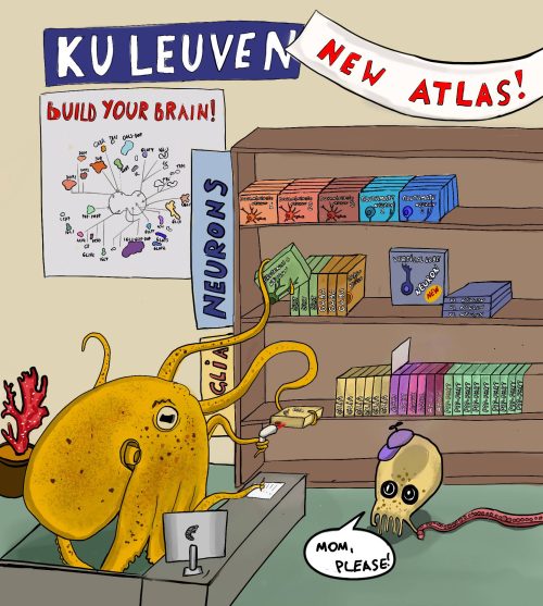
Ruth Styfhals and Dr. Eve Seuntjens at the KU Leuven, Belgium, recently published a cell type atlas of a developing octopus brain in Nature Communications. The team behind the paper was diverse, bringing together the different expertise needed to pull off this challenging project. The authors used both single cell and single nuclei RNA sequencing to identify the different cell types within dissected brains of recently hatched Octopus vulgaris (common octopus). They identified the location of several of these cell types within the brain and compared their molecular profile with brain cell types in other species.
How did you get started on this project?
In general, our lab is interested in understanding the molecular and cellular mechanisms of complex brain development. By making genetically modified mice to model human neurodevelopmental disorders we tried to identify the mechanism behind these disorders. Our focus was on mutations in Protocadherin genes. The latter were known to be important in vertebrates for cell sorting and neuronal wiring. When the first octopus genome was published in 2015, it revealed massive expansions in genes encoding Protocadherins (Albertin et al., 2015). Our lab got intrigued – could these molecules represent an evolutionary convergent mechanism to build and wire up the complex octopus brain as well? We chose to work on Octopus vulgaris, which is easy to obtain in large quantities as eggs. Before being able to study this question, we first needed to set up a system to keep and hatch the Octopus eggs in the lab (Deryckere, Styfhals, Vidal, Almansa, & Seuntjens, 2020), and we described brain development using modern technologies such as light sheet imaging (Deryckere, Styfhals, Elagoz, Maes, & Seuntjens, 2021). After identifying the neurogenic niche during embryonic development, one very important missing piece of information was molecular knowledge about cell type diversity in the brain. What was the end point of embryonic brain development, and how many cell types were present in this alien brain? Our angle was a developmental one, and we therefore focused on the brain of freshly hatched ‘paralarvae’, which is the swimming intermediate stage that grows over the course of about 5 weeks into a juvenile that settles and adopts the benthic lifestyle of adult Octopus vulgaris.
Credit artwork: Grygoriy Zolotarov, second author of the study
What was already known about the cell types in the octopus brain?
In 1971, a detailed overview of the anatomy of the adult nervous system of Octopus vulgaris was published (Young, 1971). Therefore, we had a good idea of the different brain lobes, their connections and their function in the adult. In addition, nuclear sizes and the morphology of different cell types were described. Nothing was really known about the number of cell types, what molecular markers these cell types had and how to link molecular types to “morphotypes” present in the brain. Right after hatching, the brain only has about 200,000 cells – therefore it still needs to multiply 1000-fold to reach the cell numbers present in the adult nervous system, which is around two hundred million. Therefore, we were not sure whether we could really compare this hatchling brain with the adult one. Molecular knowledge at the embryonic or larval stage was very limited to studies on selected transcription factor gene expression, often not really at cellular resolution. We also knew that certain neurotransmitters should be present, based on the adult work. We could not even guess how many clusters to expect, and did not have any marker genes to annotate clusters.
Can you summarize your findings?
Our results revealed that the octopus hatchling brain already contains a stunning diversity of cell types. These cell types are often organized according to molecular profile to appear in specific locations showing that this brain is already highly organized. We found that most of the cells are neurons, but there are also distinct glial cell types, and some seem to be spatially confined. We tried to distinguish ancestral cell types from novel cell types by using comparisons to mouse and Drosophila brains, and found that cells of octopus vertical lobe (the brain structure necessary for memory and learning) are transcriptionally similar to cell of the fly mushroom body, indicating functional convergence. We also found that novel Octopus-specific genes, like Protocadherins, are used to delineate specific cell types that might represent evolutionary novel cell types. Working with an unusual species brought additional challenges. A first key step was getting sufficient high-quality samples, by performing micro-dissection, optimizing isolation of cells and having expert help with nuclei isolation. A second key step was to ameliorate, in a significant manner, the gene model annotation of the genome, even when this genome already had a chromosome-level assembly. Using long-read Iso-seq and FLAM-seq data, we could extend 3’ ends in a data-driven manner, increase mapping statistics and more than double the amount of data. A third important step was the spatial mapping using hybridization chain reaction, a very powerful method for revealing gene expression in situ in non-model species. This enabled us to create an initial map of the cellular diversity.
When doing the research, did you have any particular result or eureka moment that has stuck with you?
When comparing octopus brain cell types to mouse and fly brain cell types, we initially didn’t really expect to find anything useful, because of the immense evolutionary distance (the ancestor of octopus and mouse lived about 600 million years ago). It was striking to see that glial cells in all three species were alike, as were neuronal cells important for memory and learning in fly and octopus. This was most amazing, to see evolutionary conservation -or convergence- on a cellular level!
And what about the flipside: any moments of frustration or despair?
Starting up an entirely new non-model, marine aquatic animal culture in a lab with background mainly in mouse development was challenging and took its time. Many grant reviewers were not convinced we were able to pull this off, leading to most grants being rejected. This meant we needed to be very creative with our minimal resources, and we were dependent on help from more fortunate collaborators who did see the innovation and the potential of the idea. Firstly, Stein Aerts, who co-founded FlyCellAtlas, chipped in some of his resources to perform a bold dual single-cell and single-nuclei experiment. Stein is a long-time collaborator and his no-nonsense attitude kept us focused on the goal: to get an initial octopus brain cell atlas. Secondly, Nikolaus Rajewsky developed an interest into octopus brain RNA profiles, and attracted the hyper-dedicated and talented master student Grygoriy Zolotarov to work on this project. We teamed up and were able to massively ameliorate annotation and gene models which more than doubled the amount of usable data. Thirdly, we did not start from the void. Previous collaborations with Gregory Maes, at that time IOF manager at the genomics core facility of KU Leuven, had yielded isoseq long read transcriptome data. Last but not least, our long-standing collaborator Eduardo Almansa made sure we had access to egg clutches and provided them to us at no charge. We wanted to give a shout out to these key people and their generosity; without them this story would not have existed.
What is next for you/the lab after this paper?
Ruth (first author) is finishing her PhD and is currently looking forward to working on neural development in even more unknown, weirder organisms, which have a less complex brain than that of the octopus.
Where will this story take the lab?
This project for sure has opened up a number of future research lines. Having a molecular view on cell types, the next challenge is to link these types to the morphotypes found by JZ Young and others. Another challenge is to understand how this diversity is generated during development: is there a spatial and temporal logic to these cell types? Do neurons and glia have a common stem cell or not? What transcription factors and signaling molecules determine cell fate and migration? How does this brain grow beyond hatching? Are larval cell types retained or replaced? How are these cell types wired up? And how do they lead to (innate) behaviors one can observe in the paralarval phase? There are still many unknowns, but with this molecular profiling of cell types, we can now better formulate hypotheses that might bring new insights into the function of this enigmatic big brain.
by Ruth Styfhals and Dr. Eve Seuntjens (eve.seuntjens@kuleuven.be)
References
Albertin, C. B., Simakov, O., Mitros, T., Yan Wang, Z., Pungor, J. R., Edsinger-gonzales, E., … Rokhsar, D. S. (2015). The octopus genome and the evolution of cephalopod neural and morphological novelties. Nature, 524, 220–225. https://doi.org/10.1038/nature14668
Deryckere, A., Styfhals, R., Elagoz, A. M., Maes, G. E., & Seuntjens, E. (2021). Identification of neural progenitor cells and their progeny reveals long distance migration in the developing octopus brain. ELife, 1–32. Retrieved from https://doi.org/10.1101/2021.03.29.437526
Deryckere, A., Styfhals, R., Vidal, E. A. G., Almansa, E., & Seuntjens, E. (2020). A practical staging atlas to study embryonic development of Octopus vulgaris under controlled laboratory conditions. BMC Developmental Biology, 20(6), 1–18. https://doi.org/10.1101/2020.01.13.903922
Styfhals, R., Zolotarov, G., Hulselmans, G., Spanier, K. I., Poovathingal, S., Elagoz, A. M., … Seuntjens, E. (2022). Cell type diversity in a developing octopus brain. Nature Communications, 13(7392), 1–17. https://doi.org/10.1038/s41467-022-35198-1
Young, J. Z. (1971). The anatomy of the nervous system of Octopus vulgaris. London, UK: Oxford University Press.
The IBDM invites applications for group leader positions. We seek researchers who define and address fundamental questions in biology, including the development, the function, and the dynamics of complex biological systems.
Research activities at the IBDM synergistically connect developmental biology with molecular, cell, and computational biology, as well as evolution, biophysics, neurobiology, physiology, and physiopathology. The IBDM, affiliated with CNRS and AMU, uniquely fosters interdisciplinarity (Centuri) by its intimate connections with physicists, computational scientists, and mathematicians. IBDM is also engaged in other federative programs of AMU to address major challenges in Neuroscience, Cancer and Immunology, Rare Diseases, and Imaging.
The IBDM strongly benefits from its collaborative and international scientific culture, English working language, and a fantastic campus, located in the heart of the Calanques National Park.
The IBDM is committed to promoting equality, diversity and inclusivity. The selected candidates will receive a start-up package, and will benefit from outstanding core facilities, including light and electron microscopy, as well as state-of-the-art animal facilities (mouse, Drosophila, Xenopus) for functional studies. The IBDM will also provide engaged mentoring to the selected candidates to obtain a tenured position (CNRS or AMU) and to secure extramural funding (ATIP/Avenir, ERC, etc…).
Candidates should provide :
A single PDF file containing a cover letter explaining their motivation to join the IBDM,
CV,
Summary of their main research achievements (2 pages maximum),
Future research project (5 pages maximum),
Contacts of three references
Applications and queries should be sent to the search committee (ibdm-call@univ-amu.fr) before March 1st 2023. In-person interviews will be scheduled from June 2023.
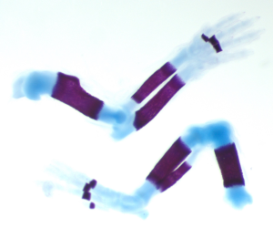
Robinow Syndrome is the best known of a set of genetic disorders that affect the growth and development of the skeletal system. Patients with these conditions have facial abnormalities, such as cleft palate, and develop short-limb dwarfism by around 18 months. Now, in a study published in Development, scientists from Nationwide Children’s Hospital in Ohio, USA, and the Van Andel Research Institute in Michigan, USA have shown the first successful correction of limb length in a mouse model of a very similar disorder known as FZD2-associated autosomal dominant Robinow Syndrome, providing hope for future therapies.
The forelimb (top) and hindlimb (bottom) of a mouse embryo, stained to reveal the bones (purple) and cartilage (blue). Image credit: Sanika Vaidya.
Although autosomal dominant Robinow Syndrome disorders are extremely rare (affecting around 50 families worldwide), they’re associated with genetic variations (mutations) in a group of genes that can be inherited from one parent or arise spontaneously, meaning diagnosis is not always trivial. Professor Rolf Stottmann who led the study said, “we began the project by studying the genomes of families with structural birth differences of the brain and face who had not yet received a genetic diagnosis. We identified that one of the initial families in this cohort had a mutation in the FZD2 gene.”
FZD2 is now known to be one of the known genes linked to autosomal dominant Robinow Syndrome. Like the other genes in this group, FZD2 makes a protein involved in sending signals that cells use to organise themselves into tissues. In their study, Professor Stottmann and colleagues used CRISPR/Cas9 genome-editing technology to induce mutations in a precise region of Fzd2, reproducing the specific types of mutations found in human patients. The researchers found that mice with these mutations had facial and skeletal malformations resembling those seen in the patients, including cleft palates and limbs less than half the normal size.
The researchers predicted that these types of Fzd2 mutations would disrupt signalling and hinder skeletal growth. To rescue the missing signals, the scientists intervened by treating pregnant mice with a drug that stimulates the signalling pathway. “This drug is an attractive option because we think we know how it works and previous work had shown that it could rescue cleft palates in a mouse model,” Professor Stottmann explained. Strikingly, they found that the pups exposed to the drug had significantly longer limbs than the untreated model mice.
The success of these experiments in mice suggests the drug could also be used as a therapeutic treatment in human patients. “The idea of treating the limb bones medically rather than surgically is a really important proof of principle, which we demonstrate in this study,” said Professor Stottmann, “we are very excited to test if this could work in the context of other genes associated with autosomal dominant Robinow Syndrome.”
Liegel, R.P., Michalski, M.N., Vaidya, S., Bitterman, E., Finnerty, E., Menke, C.A., Diegel, C.R., Zhong, Z.A., Williams, B.O., Stottmann, R.W. (2023). Successful therapeutic intervention in novel mouse models of Frizzled 2-associated congenital malformations. Development, 150, dev201038. doi:10.1242/dev.201038
 (No Ratings Yet)
 (3 votes)