Bioinformatics postdoctoral position in single-cell genomics, University of Basel, Switzerland
Posted by Patrick Tschopp, on 22 October 2019
Closing Date: 15 March 2021
A fully funded bioinformatics postdoctoral position is available in the Laboratory of Regulatory Evolution (Tschopp lab) at DUW Zoology, University of Basel, Switzerland.
We study the gene regulatory mechanisms of cell fate specification in the vertebrate skeleton. Depending on anatomical location, the vertebrate skeleton develops from three distinct progenitor populations – neural crest, somitic and lateral plate mesoderm. We are interested in the gene regulatory network (GRN) dynamics that transcriptionally re-code these distinct progenitor pools towards functionally analogous skeletal cells.
As part of a larger Swiss National Science Foundation (SNSF)-funded project, we are looking for a bioinformatics postdoc to analyze developmental single-cell RNA-seq and single-cell ATAC-seq data, followed by CRISPR/Cas9 perturbations, to infer the GRN dynamics underlying this progenitor convergence towards a common skeletal cell fate. These analyses will be performed in collaboration with the group of Prof. Erik van Nimwegen, experts in computational GRN inference, at the Biozentrum Basel. The project builds on a solid foundation of confirmed preliminary data. For more information please visit http://evolution.unibas.ch/tschopp/research/
The successful candidate will hold a PhD with a strong background in one or several of the following fields: bioinformatics; single cell analyses; statistics; computational data analysis; as well as interests in developmental and molecular biology. Good communication skills in oral and written English are essential.
We offer a highly interactive and interdisciplinary research environment, state-of-the-art technology platforms, attractive employment conditions and very competitive salaries by international standards. Full funding is available for 1+2 years.
Please send your application as a single PDF with a brief statement of motivation, a current CV and contacts for at least two references to patrick.tschopp@unibas.ch . Evaluation will begin on December 1st 2019 and suitable candidates will be contacted shortly after. Earliest starting date is January 1st 2020.
 (No Ratings Yet)
 “Cnidarian Neural Development” (Fabian Rentzsch) at the University of Bergen/Sars Centre in Bergen, Norway.The group uses the sea anemone Nematostella vectensis to study molecular, cellular and evolutionary aspects of nervous system development (e.g. Richards and Rentzsch, Development
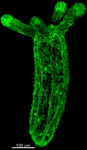
 (1 votes)