The O’Brien Lab (http://www.stemdynamics.org) is part of the Department of Molecular and Cellular Physiology at Stanford University School of Medicine. Our goal is to uncover the ongoing dialog that adult stem cells have with the tissues they support, and understand how this dialog happens on the molecular and cellular levels. We use a simple invertebrate organ, the Drosophila midgut, to pioneer new imaging and computational approaches that address basic questions in stem cell biology.
The O’Brien Lab is seeking a highly motivated, detail-oriented Life Science Research Professional 1 (LSRP1) to join our dynamic laboratory environment. The LSRP1 will perform functions and activities involved in defined research projects, which principally involve live and fixed tissue microscopy of Drosophila organs, computerized image analysis (ImageJ/Imaris), basic MATLAB coding, and construction and maintenance of sophisticated Drosophila genetic stocks. The goals of the projects are to understand cellular interactions and signaling in stem cell based Drosophila organs.
*- At the time of the interview, applicants will be asked to provide evidence of their imaging skills in the form of publications, posters, report, or other documentation.
A postdoctoral position is available in the laboratory of Dr. David Matus at Stony Brook University to investigate symbiotic and developmental processes with Selective Plane Illumination Microscopy. We have recently received funding for three years of postdoctoral support to develop protocols for extensive in vitro and in vivo imaging of cell invasion processes with a focus on the tissue and cellular entry of endosymbiotic algae as they enter their spotted salamander embryo hosts (Ambystoma maculatum). This work is funded by the Gordon and Betty Moore Foundation in collaboration with researchers from Columbia University, The American Museum of Natural History, and Gettysburg College (see Shelf Life Episode 11). The project will combine molecular biology, embryology, cell biology, and extensive light sheet imaging. Preferred candidates will have backgrounds in light sheet and/or confocal microscopy as well as an interest in advanced imaging methods and the intersections of cell and developmental biology with ecology and evolution. Opportunities will exist to develop projects and assist in advanced imaging using C. elegans and zebrafish as well. My laboratory is a part of a modern and well-equipped Department of Biochemistry and Cell Biology at Stony Brook University on Long Island, NY. For further information on our work, please see the following publications on techniques,cell invasion, and the symbiosis.
To apply, please send a letter of interest detailing your expertise, CV and names and contact information of three references to david.matus AT stonybrook.edu. You can also apply directly through the Stony Brook University Human Resources portal here or by searching for position #1702902 at the Stony Brook HR site.
It’s all about the wires. But what about the glue?
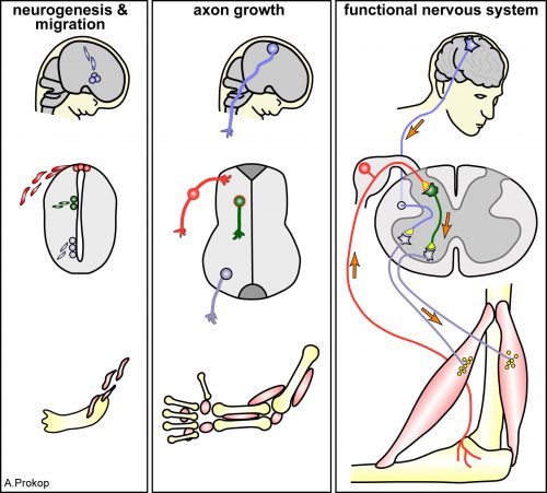
Networks make us who we are. I am not talking about social networks but about neural networks that define how we perceive the world and how we act. For a century, neuroscientists have sought to understand functions of neural networks in condition and how such networks are established in the first place still remains an active area of research. Neuronal networks consist of a large set of wires that need to be connected and organized in defined, functional ways, similarly to telecommunication wiring pervading our cities or like the motherboards of our modern computers. To work, signal carriers of such networks must be of appropriate bandwidth, with proper paths, and connected in a specific way. Likewise, in neuronal networks, cell fate must be specified, neurons must migrate to proper locations, grow and navigate their axon processes through specific paths and generate well-defined synaptic connections.
In the 1990’s, the “Decade of the Brain,” as declared by the United States Congress, developmental neuroscientists made extraordinary progress in understanding cell fate determination, axon guidance and synaptogenesis. Seminal studies identified the principal families of guidance molecules and established major paradigms, such as the role of floor plate Netrin, a secreted guidance cue, in commissural axon guidance 1,2. At the same time while some principles were established others were revisited. The non-neuronal cells of the nervous system, called glia (from the Greek γλία/ γλοία for glue), which as their name implies were long though to provide a passive substrate for neuronal growth, were realized to play active roles in neuronal physiology and function. Glia were first acknowledged to be neuron’s best friend, housekeeper, insulator, nurse 3,4and the list of glial roles expanded to include roles of glia as neural progenitors, tracts for migratory neurons and synaptic plasticity facilitators. Despite this progress, today’s classical neuroscience textbooks count only a handful of mentions of glia and their extensive roles. When I was an undergraduate, one would be lucky to attend a seminar on glial biology by one of the few scientists of the field.
I discovered the extensive glia literature when planning my post-doctoral research path. Focusing on the unknowns of neural development, I realized the enormous potential of the view that glia can actively regulate neuronal development and physiology. I was a descendant of the C. elegans research community, which I joined because of my first excitement about the C. elegans short life cycle and its recovery after freezing (allowing easy storage of strains for decades), but mainly for the ease of genetics, molecular biology and functional studies, possible at single-cell resolution. Yet, glial studies in this animal were few and far between. C. elegans axons are not myelinated by glia and glia were not shown to give rise to neurons nor to appose the neuromuscular junctions I studied during my PhD. In fact the presence of glia in C. elegans was not even clearly accepted. Then, midway during my PhD, Shai Shaham, my post-doc supervisor to-be, demonstrated that the so-called “support cells” of my favorite nematode are, in fact, glia 5, and soon after his lab demonstrated glial roles in neuronal ensheathment, dendrite shape and axon extension 6–8. Exciting! So many glial functions to be uncovered in C. elegans glia!
Yet, why would one study glial functions in C. elegans, where glial cells are limited to the 1/6 of the total neuron number, axons are non-myelinated and and microglia-like immune-specialized cells do not exist? For one, C. elegans has proven so useful for understanding the basic biology common to so many living things, that there was no reason to believe glia would be an exception! From a technical standpoint, most C. elegans neurons are born and grow independently of glia division and trophic support, thus perturbing glia does not cause neuronal death. In other systems, glial roles in neuron survival and support hamper attempts to address the active roles of glia in vivo in the nervous system. Finally, unlike most other settings, C. elegans glia can be studied in vivo, at single-cell resolution, using facile genetics, an invariant cell lineage and embryonic morphogenesis progresses in a transparent egg laid free in the environment. Moreover, live imaging of the same exact cell over and over again is possible in C. elegans. Who wouldn’t want to study glia in this setting!
The most complex circuit of the most well studied worm, and the forgotten cells
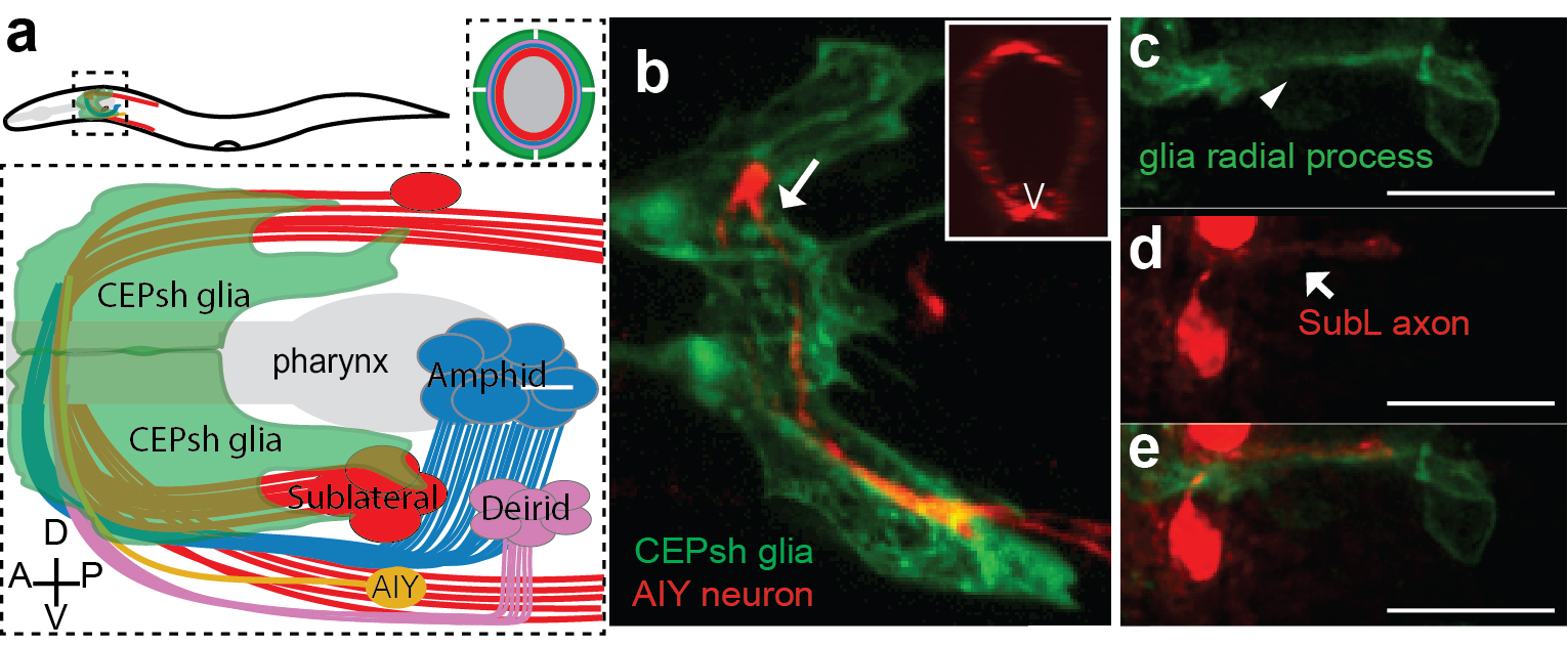
Armed with these thoughts, I joined Shai’s lab, decided to study developmental functions of glia, focusing on glial roles in the formation of the C. elegans brain-like neuropil, the nerve ring. The nerve ring (NR) consists of ~180 axons, presents the majority of synapses and interneuron connections in the animal, and is enveloped in its final form by four CEPsh glia, with astrocyte-like terminal morphology and molecular content (Figure 1; Katz & Shaham, personal communication,9). CEPsh glia express Netrin in early embryogenesis 10. Moreover, ablation of their precursors results in more profound NR defects than those of Netrin null mutants, suggesting unknown glia roles in NR assembly 7,11. Thus, I was interested to focus on glial roles in NR assembly, using biased approaches.
A few weeks into the project, writing for fellowship applications, I was struck by an important realization. While C. elegans was an important model for axon guidance cue discovery 10,12,13 and for studies of axon growth and fasciculation 12–16, how its most complex neuropil is formed during embryogenesis was subject to extensive speculation but was not yet described in detail. Only a handful of studies focused on NR structure, axon guidance or positioning 7,11,13,17, and many important questions remained unanswered. When and where does NR assembly start in the embryo? How and when do NR components enter the structure? What are the first axons to pioneer the NR and do they have functional roles in guiding later “follower axons”? Do the CEPsh glia extend early processes to define the structure or do they only enclose the structure in later embryogenesis, after axons have fasciculated, to protect them from the mechanical forces during embryonic elongation? Is NR integrity regulated only by Netrin-expressing glia (ventral CEPsh glia) or also by the non-expressing ones (dorsal CEPsh glia)? Which molecules, besides Netrin, may function from glia or from early neurons to address NR formation? Are there molecules that control specifically axon guidance, independently from neuronal migration and axon-growth initiation? I was surprised that so many questions remained open in C. elegans, a model system well appreciated for embryonic studies, tractable neuronal identities and single-cell resolution. Were the answers to these questions trivial, and not worth exploring? Or, as I have heard some neuroscientists say, is neural development mostly explained, and only the dynamic plasticity of neuronal circuits remains to be understood? Was I doomed to engage in a problem solved in other organisms, to provide the C. elegans version “merely” for the record?
I had to really understand the state of affairs in the field, and in reading the literature realized more and more how little was known. Not only in C. elegans, but at all. Indeed, my attempts to prepare introductory slides for my early scientific presentations did not prove easy. Transcriptional and morphogen signaling pathways for neuronal specification were defined in detail 18–20 and growth cone morphology distinguished pioneer and follower axons 21–23. However, how neuronal fates dictated circuit assembly initiation through a series of comprehensible axon-guidance events was, with few exceptions, not at all clear. The molecular identities of pioneer axons and the molecular mechanisms driving their functional roles remained understudied. Glia were known to form transient structures associated with axonal bundles and to express guidance cues 24. However, their functional roles in axon guidance independently of neuronal survival, growth, migration and their molecular interactions with pioneer axons appeared unresolved 25–27. I was reminded of a century-old Cajal’s quote: “What functional significance can be attributed to the neuroglia? Unfortunately, the present state of science does not allow to answer this important question but through more or less rational conjectures.” (Ramón y Cajal, 1899).
First things first: CEPsh glia and SubL pioneers initiate the nerve ring neuropil
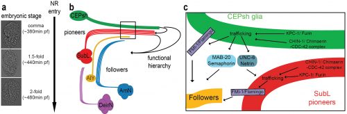
I decided to start from scratch. Instead of biasing my studies to focus on glia, I decided to observe and image NR development, and let nature be my guide. The post-embryonic NR contains two major commissures, the amphid and deirid commissures composed primarily of sensory neuron axons 17. The posterior part of the amphid commissure is also referred to as the sublateral (SubL) commissure, since its axons extend out of the NR to populate the ventral sublateral cords. Since the amphid commissure is the largest characterized bundle populating the NR we hypothesized that this may be the one growing out first. I started imaging amphid-neuron axons in the beginning of morphogenesis, initially by using recently published reporters 28. Imaging bundled membranes using the by Pdyf-7::membrane-GFP reporter, or single axons, by photoconversion of specific neurons in animals expressing Pdyf-7::kaede, revealed axon growth only after 400 min of embryogenesis (known as the comma stage). Yet, electron microscopy (EM) images acquired by Yun Lu in the lab, showed that the pharyngeal primordium was surrounded by a thick layer of axons, corresponding to the early NR, already at 440min of embryogenesis (1.5-fold embryonic stage). So many axons were present already at 440min, making us wonder whether amphid neurons were truly early NR components.
I found myself, again, going one step back, realizing that I needed a more comprehensive effort to visualize as many embryonic neurons as I could. Since C. elegans has been a valuable model system for studies of neuronal specification and diversity 29, I was not too worried about finding neuron-subtype specific markers. I started to evaluate known neuronal markers. This screen left me with a handful of useful markers, and a couple of important take-home lessons. First, markers with post-embryonic expression restricted to specific neurons are usually more broadly expressed during embryonic morphogenesis. The C. elegans research community would, therefore, benefit greatly from characterizing expression patterns also during embryonic morphogenesis. Indeed, several labs today focus on expression profiling of C. elegans embryos 30,31, a blessed endeavor. This realization, that sparse, cell-specific embryonic reporters were few and far between, also drove the development in Shai’s lab of a method for labeling individual C. elegans embryonic cells without cell-specific drivers32. Second, the willingness of the C. elegans research community to share reagents, published dozens of years ago or others unpublished, to help me with my project, proved the supportive nature of this community, something that became rare in biological research.
I was now armed with reporters for many axons populating distinct NR commissures. By performing in vivo live embryonic timelapses of process growth into the NR, I saw that axons of the sublateral bundle grow early to define the NR neuropil while other commissures, amphid and deirid, were established in an orderly fashion later, with sequential axon growth occurring even within commissures (Figure 2a-b, 9). After months of sampling and imaging live embryos, I could not sustain my excitement when we also identified two bundles of similar position and composition in EM images of comma-stage embryos, taken by Yun Lu in the lab. I kept going back to analyze those EM images; yes, there were these two bundles, and no other multiprocess-bundles were obvious at that time, navigating to the NR presumptive position!
But what about the CEPsh glia? I had begun attempts to image CEPsh glia membrane growth the minute I joined Shai’s lab, but at the time no useful information could be gleaned, since no CEPsh-specific embryonic promoters were known. I decided to dissect regulatory elements of a couple of known CEPsh-expressing promoters, but this approach was not fruitful, as deletion analysis of cis-regulatory elements resulted in derepressed expression in non-glial cells. I finally decided to use a transgenic pan-glial marker, expressed from the onset of embryonic morphogenesis, and to follow stochastic segregation of the marker in my cells with mosaic analysis. Despite the method’s low throughput nature, I was able to visualize CEPsh membranes growing in the NR early and coalescing with growing SubL axons. Those early CEPsh membranes did not present the elaborate astrocyte-like endfeet seen post-embryonically but grow thin, non-branched processes reminiscent of vertebrate radial glia.
SubL pioneers and CEPsh populated the NR early! But who actually took the lead? Were SubL and CEPsh equally important in NR initiation or does one of the cell types guide the other? The answer came from ablation experiments I performed several months later. When specific CEPsh glia are ablated by expressing an apoptotic gene and following cell killing by mosaic analysis, pioneer axons grow ectopically, while follower axons can be short or mis-guided. Cell ablation of the SubL bundle perturbed follower axon pathfinding but CEPsh membrane morphology appeared normal. Thus, the most exciting scenario I could have imagined emerged: glia are not just bystander support cells maintaining the neuropil; they are necessary for initiating NR assembly. Moreover, our later studied revealed that CEPsh also cooperate with SubL pioneers to guide later NR components (Figure 2). This was an important realization. Although glia in other systems have been reported to appose axon bundles, functional analysis of glial roles in pioneer axon guidance was lacking, or was tested in settings where defects in neuronal viability or migration complicated the analysis. 26,27,33–35.
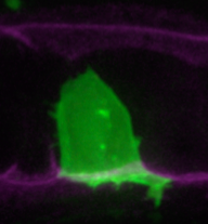
The NR is populated by neuronal commissures and CEPsh glia. Schematic (a) and imaging (b) of NR in postembryonic larval animals in lateral view. Inset: cross-sectional view. Pttx-3::mCherry, AIY and Phlh-17::membrane-GFP label AIY interneuron and CEPsh glia respectively. (c-e) In ~ 420min embryos, SubL pioneer axon bundle (SMDD neuron in red, Pttx-3::mCherry) and CEPsh glia (in green, Pmir-228::membrane-GFP) coalesce to define the NR. Figure adapted from Rapti et al, 2017 (9).
An “unsuccessful screen” & the first viable mutant with severe NR assembly defects
Defining a working model of cellular interactions for NR initiation was amazing, but not enough! Long devoted to unbiased genetics, like many C. elegans researchers, I decided to search for genes regulating NR formation through forward genetic screens. I screened tens of thousands of mutagenized worms under a compound microscope, recovering from the slides, back to bacteria-seeded plates, worms with abnormal axon trajectories. I started the screen well before my embryonic imaging uncovered the identities of early NR components. Back then I was thinking about neuropil formation, and selective fasciculation of axons. I hypothesized that fasciculation molecules, specific to neuron-subtypes, would ensure stereotypical interactions between axonal partners. I, therefore, designed screens for mutants in which two normally fasciculated axons, are properly guided but fail to adhere. My screens gave no viable mutants harboring the defects I was expecting. The reason for this remains unknown, but may suggest a different scenario of axon partnership based on the relative cell positioning of specific neurons at the time of their axon growth.
Regardless, in these screens I did find mutants with other NR axon defects. By now, I knew of the requirement of glia for NR assembly and decided to focus on mutants I found whose NR axon defects resembled those of glia-ablated animals. After cloning several previously-identified guidance genes with mild NR guidance defects, I decided to focus on a mutant with very severe NR defects, in which no mutations in the classical guidance genes were found by whole genome sequencing (WGS). And here started the painstaking tale of identifying the relevant gene. During my PhD, I mapped the first mutant of an uncharacterized gene without resorting to whole genome sequencing (WGS). It was difficult for me to understand why cloning my current mutant was so challenging, even with WGS data. The answer came when I proved in multiple ways that not one, but two mutations in two different unlinked genes were causal for the defects in the mutant. These mutations were synergistic, such that either single mutation caused only mild defects (5-15%) compared to the severe NR defect of the double mutant (>75%). To add to the complexity, one of the mutations was semi-dominant and subject to partial maternal rescue. After months of effort, I could finally announce that mutations in the Chimaerin gene chin-1, and the Furin gene kpc-1 were the cause of the mutant defects in NR axon guidance. When, in a joint group meeting, Shai, Leslie Vosshall, and Cori Bargmann commented on the low probability of recovering and identifying such a mutant, I felt that my hard work had really paid off. I was also filled with optimism that today’s ease of WGS, facilitating such complicated endeavors, will hopefully help identification of mutants that in previous decades would have been frozen and forgotten. This should allow researchers to choose their mutants of interest mainly based on interesting phenotypes and not how easy their identification is. For one, our mutant was definitely of interest; overall it showed ~80% navigation defects in any NR axon we imaged by optical means and a severe defective overall NR structure by electron microscopy of newly-hatched animals.
A wrong educated guess and the non-canonical functions of “old players”
The identities of the genes we found and previous studies in C. elegans or vertebrate nervous systems prompted me to favor cell-autonomous functions of these genes 36–38. I devoted several months to unsuccessfully prove such cell-autonomous roles of those in axon guidance. I was finally forced to consider the possibility of non-cell-autonomous roles. Rescue studies using combinations of cell-specific embryonic promoters finally revealed that the C. elegans Chimaerin and Furin act from glia to guide pioneer SubL axons and from both glia and pioneer SubL to guide follower axons. And they do so at the onset of NR assembly, by specifically affecting NR axon guidance, and not neuronal or glia survival, fate specification, migration or differentiation. To our knowledge, this is the first mutant affecting NR formation in such a specific way.
This mutant gave us an inroad in studying what turned out to be highly redundant signaling pathways for neuropil assembly initiation, a problem that has plagued genetic analysis of the process not only in C. elegans, but in vertebrates as well. Using the synergistic nature of Chimaerin and Furin mutants, we screened for mutations enhancing either single mutant and identified a network of conserved axon guidance cues redundantly regulating NR assembly. Importantly using our system of hierarchical NR assembly we could precisely define the cell-specific contributions of those factors. It turned out that knowing the site of action of these cues was crucial for modeling their actions. We demonstrated that C. elegans glia use distinct Netrin and Semaphorin signaling pathways to guide pioneer and follower axons respectively. We further showed glia and pioneer neurons together use the C. elegans Celsr/Flamingo homolog, FMI-1, to ensure proper navigation of follower axons into the neuropil. Moreover, our results support roles for Chimaerin and Furin in trafficking guidance cues, a novel combined function for this pair of proteins (Figure 2).
Model of cellular and molecular events of embryonic NR assembly (a)The NR is assembled, orderly, from the comma till after the 2-fold embryonic stage. (b) CEPsh glia and pioneer SubL axons enter the path first, later followed by “follower” axons of commissures or non-commissural paths. This follows a functional hierarchy of CEPsh glia guiding SubL pioneers and cooperating with them to guide follower axons. (c) Molecular pathways function redundantly from CEPsh glia and SubL pioneers to drive NR assembly. Figure adapted from Rapti et al, 2017 (9).
In summary, our studies uncovered the initiating events of NR formation in C. elegans, identified key pioneer roles for glia, defined the identities of the first pioneer neurons that enter this brain-like neuropil, and uncovered a molecular framework governing axon guidance events early on. Our studies uncovered a genetic strategy for dissecting the highly redundant cellular and molecular interactions driving axon guidance, and defined a new role for two old players in guidance factor trafficking. This sounds like a lot of work. And it was. But really, it is just the beginning. There are so many questions left unanswered, and the logic of assembly is still not understood for most of the neurons entering the NR. How relevant these studies are to other systems also remains to be explored, but some exciting similarities have recently emerged (see below).
New ways of thinking about old molecules and established concepts
In retrospect, our findings did not provide a short, easy-to-digest, easy-to-sell story. Instead, by tackling a multifaceted problem without shying away from the complexity we took to heart Einstein’s quip that phenomena must be explained in the simplest possible terms, but no simpler. This is an important lesson- while there is great temptation to understand biology in well-packaged sound bites, life turns out usually to be a lot more complex. Oversimplifying can have the unwanted effect of preventing fundamental understanding of a process, and can lead to proclamations of entire scientific fields being “solved”, even when this is not the case. In our case, the benefit of revisiting old problems with unbiased approaches was the discovery of new concepts, like the new roles of previously known molecules and importantly the glia roles in initiating assembly.
Remarkably, as our manuscript was nearing publication, two papers were published that questioned the precise roles of Netrin in commissural axon guidance in vertebrates 39,40. While questioning the long-standing role of the floor plate in commissural axon guidance, they revealed important roles for Netrin derived from ventricular-zone neural progenitors that correspond to radial glia. These studies help put our discoveries in a much broader context. In our paper, we suggest that embryonic CEPsh glia resemble radial glia, both morphologically and molecularly, suggesting that vertebrate radial glia may guide pioneer axons to initiate circuit assembly, in addition to their recognized roles in neuronal migration. The recent Netrin papers greatly strengthen our hypothesis, and predict that the proteins and gene interactions we described in our paper are likely to exist in vertebrates as well.
This project taught me a valuable lesson: keep an open mind; you may be surprised.
Applications are open for the Wellcome Trust funded four year PhD programme in Developmental Mechanisms at the University of Cambridge. We are looking for talented, motivated graduates or final year undergraduates, and are keen to attract outstanding applicants in the biological sciences, who are committed to doing a PhD. We are able to fund both EU and non-EU students.
Closing date: 4th January 2018
For more details about the application process and the programme please see the website:
A fundamental aspect of vertebrates is their external bilateral symmetry, which has to some extent shaped evolutionary success. Not only is beauty associated with symmetry, enhancing an individual’s chance of mating but also, symmetry in the legs will help an animal flee from a hunter (Enquist and Arak, 1994; Johnstone, 1994; Holló and Novak, 2012). However, a remarkable feature of the vertebrate body beyond this external symmetry is the asymmetric disposition and morphology of the internal organs relative to the left-right (L-R) axis. These asymmetries are essential for optimal organ packaging and function, yet it has long remained a mystery as to how this asymmetry is achieved. The initial symmetry is first broken in the vertebrate embryo at the level of the L/R organiser during early gastrulation. As a result, asymmetric information is transmitted to the lateral plate mesoderm (LPM), restricting the expression of Nodal and its downstream target Pitx2 to the left side of the embryo. The Nodal-Pitx2 pathway, which is conserved in all vertebrates, is instrumental in controlling L/R asymmetry (Raya and Izpisua Belmonte, 2006). This left-handed information is repressed on the right-hand side by the inducer of the epithelial-mesenchymal transition (EMT), Snail1. It has remained unclear whether an equivalent right-handed pathway provides instructive information to the right LPM. Laterality defects are associated with some important diseases in humans. Thus, the mechanisms underlying the establishment of L/R asymmetry are clearly of interest to developmental biologists and fully understanding these events will have significant biomedical implications.
New Findings
The mechanisms that regulate the EMT in development and disease have for long been of particular interest to our lab. When screening for genes expressed in the LPM of chick embryos, we found the Prrx1 transcription factor to be a particularly potent inducer of the EMT, and like Snail1, not only in embryos but also in cancer cells (Ocaña et al., 2012). When the expression of these genes was studied in more detail, we realised that Prrx1 is transiently expressed asymmetrically in the same temporal window as Snail1. Moreover, and as we also described previously for Snail1 (Morales et al., 2007), the expression of Prrx1 was stronger on the right than on the left side of the embryo. Likewise, prrx1a transiently appears to be asymmetrically distributed in zebrafish embryos during a similar developmental window as in the chick embryo, again with higher levels on the right-hand side. Therefore, with great expectation, we tested whether the asymmetric distribution of Prrx1 influenced organ laterality, focusing on heart positioning as this is the first clear indication of morphological L/R asymmetry in the embryo. The fastest and easiest way to tackle this question was to knock-down prrx1a expression in the zebrafish and we found that at 48 hpf, the majority of the morphant embryos developed mesocardia and the dextral looping typically observed in embryos was completely abrogated. To determine whether this effect was conserved in chicken, we performed loss of function experiments by electroporating RNAi against Prrx1 bilaterally into cardiomyocyte precursors. Prrx1 downregulation provoked a similar effect, with heart mesocardia being the main phenotype observed. Thus, it seemed that in both the fish and chicken asymmetric Prrx1 expression was required for heart laterality.
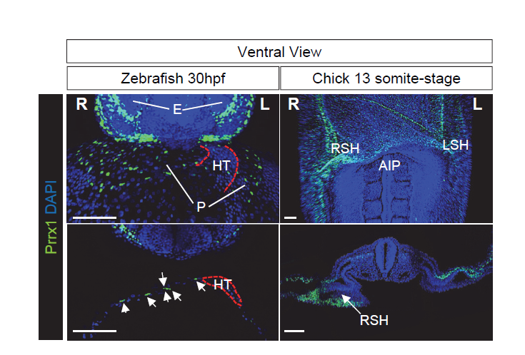
Figure 1 Prrx1 L/R asymmetric expression in zebrafish and chick embryos. Left panels, ventral view of a maximum intensity projection of whole zebrafish embryos at 30 hpf (upper) and z-plane section of confocal images immunostained to visualize Prrx1 protein expression. Note the higher number of Prrx1+ cells in the pericardium on the right hand side. Right panels, ventral view of a 13 somite-stage whole mounted chick embryo and transverse section showing the asymmetric L/R Prrx1 protein expression, higher on the right sinus horn (RSH). Nuclei were stained with DAPI (blue). Scale bars, 100 μm. AIP, Anterior Intestinal Portal; E, Eyes; HT, Heart Tube; L or R SH, Left or Right Sinus Venosus Horns; P, pericardium.
Given the conserved asymmetric expression of Prrx1, stronger on the right-hand side, and its role during heart looping, we reasoned that an instructive pathway may convey information to the right-hand side, in addition to the left pathway. Support for the existence of a right-handed program came from studies into mild Snail1 downregulation on the right-hand side, which provoked heart looping defects in the chick embryo without affecting Pitx2 expression (Patel et al., 1999). In addition, the development and position of the proepicardium, a transient specifically right-sided structure in frogs and avians, was affected by altered Snail1 expression but it was not after aberrant bilateral Pitx2 expression (Schlueter and Brand, 2009). Furthermore, zebrafish and mice carrying mutations in Pitx2 do not display heart looping defects (Campione et al., 2001; Ji et al., 2016). Together, these data suggest that heart laterality may be driven by a dominant right-handed pathway. As such, we speculated that reversing the levels of Prrx1 expression, so that higher levels were expressed on the left rather than the right-hand side, would lead to the development of embryos with reverse looping.
To test our hypothesis, we took advantage of the electroporation technique that allows unilateral manipulation of gene expression in chick. We studied the effect of Prrx1 gain-of-function in the left LPM and we found that a significant proportion of the embryos displayed reverse heart situs. These data indicate that the transiently stronger expression of Prrx1 in the right LPM was sufficient to drive heart looping. To gain further insight into the mechanism underlying this transient asymmetric expression of Prrx1 and how it affected heart looping, we started to precisely characterise the cell populations that contribute to heart development and that express this gene. This involved developing an antibody against Prrx1 and performing dual or multiple immunolabelling of cell populations associated with the heart using different markers, carrying out an exhaustive confocal analysis. As a result, we found that Prrx1 was not expressed in the primary heart tube (PHT) but that it was in fact expressed by a population of cells lateral and posterior to the cardiac venous pole.
The finding that another EMT inducer, in addition to Snail1, was strongly expressed in the right LPM, also influencing heart situs, suggestsed to us that some common features could exist between L-R pathways and the EMT program. As such, we were prompted to study whether differential L-R cell movements promoted by Prrx1 may drive heart looping. To investigate the movement and fate of the cells that express Prrx1 and that contribute to the posterior pole, new tools had to be generated and thus, we first addressed this question in the zebrafish. We generated a tbx5a-reporter transgenic line Tg(tbx5a:eGFP) as the cells expressing this gene contribute to both the PHT and the posterior pole of the heart (Ahn et al., 2002). In this tbx5a-reporter line there was complete co-localization between eGFP and Prrx1 in a subpopulation of the LPM cells, in the region of the cardiac precursors, which validated the use of these animals. When cell movements were followed in this reporter line, time lapse recordings of embryos from 28 to 48 hpf (during which time heart looping occurs) indicated an asymmetric migration of the Tbx5/Prrx1 double positive cells at the posterior pole of the heart. Significantly, there was a higher contribution of cells from the right than from the left-hand side, which correlates perfectly with the asymmetric expression of Prrx1 driving a different left/right cell contribution. In fact, prrx1a downregulation impaired this asymmetric cell migration of posterior pole cells and leads to mesocardia.
These observations raise the question as to whether these cells contribute to heart positioning and morphogenesis. Lineage tracing in zebrafish embryos demonstrated that Prrx1 expressing cells contribute to the PHT at the time of the heart looping, yet this Prrx1 expression is downregulated concomitant with their incorporation into the heart tube. Interestingly, the size of the atrium was reduced in the Prrx1a morphants, indicating that Prrx1 expression by cardiac progenitors is also required for heart morphogenesis.
The heart looping defects observed when Prrx1 was downregulated were compatible with studies describing the asymmetric contribution of cells from the right and left of the embryo to the heart after the formation of the PHT (Taber et al., 2010; Dominguez et al., 2012). Thus, we thought that differential left-right cell movements driven by Prrx1 could generate asymmetric forces and tension that would be stronger from the right. Such forces might eventually lead to the initial leftward bending of the posterior pole of the heart and the subsequent dextral torsion. In accordance with this hypothesis, cells in the tbx5a reporter could be seen migrating from the right-hand side to the posterior pole at the heart looping stage, apparently forming a structure reminiscent of a cable. Since forces in developing tissues are usually controlled by actomyosin bundles, we visualized actin stress fibres in vivo and while cardiac looping was normally accompanied by the formation of an actomyosin cable directed towards the posterior pole, this was not the case in the Prrx1a morphant embryos. Moreover, laser ablation of Prrx1/Tbx5 expressing cells on the right but not on the left side of the embryo prevented heart lateralization. These experiments demonstrate that asymmetric tension, more intense on the right-hand side, drives heart looping. Interestingly, we found that this mechanism was conserved in the chick embryo. Collectively, these data indicate that as in the fish, leftward displacement of the posterior pole and the subsequent dextral looping is driven by an actomyosin-dependent mechanism.
Having found that the asymmetric expression of Prrx1 plays a key role in heart lateralization, both in zebrafish and chick embryos, the next issue was to place this transcription factor it in the signalling pathways already known to be involved in L/R asymmetry. We knew that BMP could induce Prrx1 expression in the chicken embryo LPM (Ocaña et al., 2012), where it also induces Snail1 to repress Pitx2 expression (Raya and Izpisua Belmonte, 2006). In loss- and gain-of-function experiments performed on zebrafish and chick embryos, we confirmed that Prrx1 was activated by BMP and repressed by Nodal signalling. Hence, heart looping is driven by a BMP-mediated pathway that promotes strong Prrx1 expression, and that is repressed on the left by Nodal.
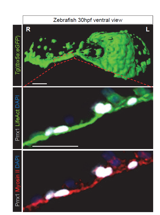
Figure 2 Prrx1 asymmetric expression generates actomyosin-mediated differential forces. Snapshot of surface rendering image from a Tg(tbx5a:eGFP) embryo at 30 hpf showing the presence of a “cable-like” structure on the right-hand side during heart looping stage (upper panel). High power confocal images of cells located in positions similar to the area boxed in a Tg(actb2:myl12.1-mCherry) reporter zebrafish embryo to visualise myosin II (red). The embryo was injected at 1 cell-stage with LifeAct:GFP mRNA to visualise F-actin (green) and subjected to immunohistochemistry for Prrx1 expression (white). Cell nuclei were stained with DAPI (blue). Scale bars, 50 μm.
A surprising evo-devo twist
Having shown the implication of Prrx1 in heart looping in both the fish and chick embryo, we turned our attention to the mouse embryo. At first, it was disappointing that our model did not seem to fit well, as there was no heart looping defect evident in Prrx1 mutant mice (Bergwerff et al., 2000). However, we knew that Snail1 mutants develop heart looping defects (Murray and Gridley, 2006) and thus, we compared Prrx1 and Snail1 expression side-by-side in mouse and chick embryos. In the territories relevant to heart looping, we found an interchange of expression patterns between these two EMT inducers (Prrx1 and Snail1). While chick embryos displayed asymmetric Prrx1 expression in the ventral posterior pole of the heart, mice displayed asymmetric Snail1 expression along with an accumulation of F-actin fibres in this territory, but no Prrx1 expression. As such, these results offered a convincing explanation for the heart laterality defects observed in Snail1 mutant mice and the lack of such defects in the Prrx1 mutants. Thus, in terms of heart laterality, it is Snail1 in the mouse that seems to carry out the role of Prrx1 in the fish and chick. While this observation may appear surprising, we had previously observed other interchanges in the lab, such as those between Snail1 and Snail2 expression at other sites in the chick and mouse embryos (Locascio et al., 2002).
General conclusions
We have identified a prominent right-handed pathway that is driven by BMP signalling and that is in turn repressed on the left-hand side by Nodal. This right-hand signalling induces asymmetric L/R activation of different EMT transcription factors, Prrx1 in fish and chick, and Snail1 in the mouse embryo, which provoke asymmetric cell movements and forces that are stronger on the right-hand side of the embryo. In fact, these asymmetric forces induce the leftward displacement of the posterior pole and dextral looping of the heart in an actomyosin-dependent manner. We are very excited as to how these studies have progressed, as they have allowed us to unravel a basic mechanism that has been conserved in vertebrates and that controls heart looping. Furthermore, this mechanism could help us better understand the congenital heart diseases that are related to heart laterality in humans.
The goal of this project is to engineer therapeutically active islet-like aggregates for future cell therapy phase 1 trials in Type 1 Diabetes (T1D)
Job description
The laboratory technicians will be responsible for the maintenance of the human pluripotent stem cell (hPSC) lab. The work will include expansion of human embryonic and induced pluripotent stem cells into characterized (e.g. marker analysis and karyotyping) hPSC line batches, development of new methods and protocols for hPSC maintenance and differentiation, transfections of hPSCs and basic characterization of hPSCs (undifferentiated and differentiated) by immunohistochemistry, qPCR and FACS. The laboratory technicians will secure the quality and reproducibility of the hPSCs to be used by scientists in the group. The candidates will work together with a dedicated team of scientists who together will tackle bottle-necks towards implementing the first phase 1 clinical trial in T1D. The working hours are 37 hours per week. The positions are time limited to the end of 2020 with a possibility of extension.
Qualifications
Highly motivated and ambitious candidates are encouraged to apply. The positions require solid experience with cell culture, including transfection (traditional and virus-based methods) and gene expression is necessary. Experience in hPSC culture, molecular biology and immunohistochemistry is required. Knowledge of cell biology, developmental biology and morphology is an advantage. The work is independent and demands flexibility and accuracy. Further, you must have good interpersonal skills and good command of English.
Terms of salary, work, and employment
The employments are planned to start as soon as possible upon agreement with the chosen candidate. The place of work is at DanStem, University of Copenhagen, Blegdamsvej 3B, Copenhagen. The positions are time limited to the end of 2020 with a possibility of extension.
Terms of appointment and salary are in accordance with the agreement between the Danish Government and HK-STAT (Danish Technician Association). The position will be at the level of salary group 5 with the possibility to negotiate due to qualifications and experiences.
Application
An application for any of the positions should be submitted electronically by clicking “Apply online” below. The application must include the following documents/attachments:
Motivated letter of application
Curriculum vitae incl. education, experience, previous employments, language skills and other relevant skills
Certified copy of diplomas/degree certificate(s)
Certified copy of transcript of records
Letter of recommendation
In all cases, ability to perform the job will be the primary consideration, and thus we encourage all – regardless of their personal background and status – to apply.
Application deadline: 15 November 2017
For further information, please contact Professor Henrik Semb by e-mail semb@sund.ku.dk
Send your application with your CV as well as names and contact details of referees electronically by clicking the ´Apply online´ below. We only accept electronic applications.
During spermatogenesis, progenitor cells must undergo tightly regulated changes to produce functional gametes. However, the genetic control of this process in humans has eluded researchers. This week we feature a paper published in the latest issue of Development that describes the changing genetic expression of cell during spermatogenesis. The co-first authors Sabrina Jan and Tinke Vormer and PIs Sjoerd Repping and Ans van Pelt of The University of Amsterdam told us more.
Sjoerd Repping, Tinke Vormer, Sabrina Jan and Ans MM van Pelt
Sjoerd and Ans, can you each give us your scientific biographies and the main questions your lab is trying to answer?
SR & AvP We work at the Center for Reproductive Medicine of the Academic Medical Center of the University of Amsterdam. Our center focusses on providing top-clinical care to patients suffering from infertility and on understanding the basic pathophysiological mechanisms underlying infertility. Our research laboratory has four active lines of research: 1- Spermatogenesis and spermatogonial stem cells, 2- Preimplantation embryo development, 3- Genomic stability of reproductive cells, and 4- Placentation and gestational diseases. Besides our laboratory research, our department is very active in healthcare evaluation research where we try to increase effectiveness and safety of medically assisted reproduction.
Sjoerd was trained as a clinical embryologist, received his PhD on genetics of male infertility at the University of Amsterdam and the Whitehead Institute in Cambridge and is currently professor of Human Reproductive Biology, head of the Center for Reproductive Medicine and Director of the Amsterdam Reproduction & Development Research Institute.
Ans was trained as a stem cell biologist. She received her PhD on the role of vitamin A on spermatogenesis and spermatogonial stem cells at the Medical School of the Utrecht University in collaboration with the Hubrecht Institute, both in the Netherlands. She is currently associate professor at the Center of Reproductive Medicine, head of the Reproductive Biology Laboratory and leader of the research line Spermatogenesis and spermatogonial stem cells.
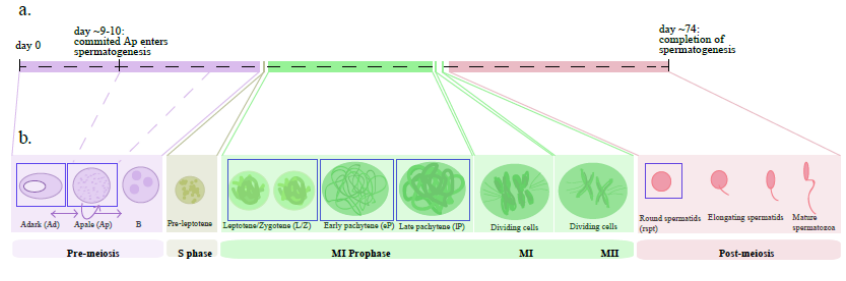
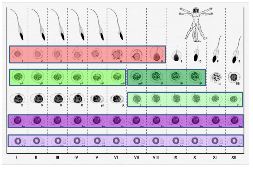
Timeline of spermatogenesis, from Figure 1, Jan, Vormer et al, 2017
Sabrina and Tinke, how did you both end up working on this project?
SJ I started working on this project as part of my PhD training. I have always been very interested in the field of reproductive medicine. In 2008 I completed a masters degree in reproductive and developmental biology. In 2011 I was looking to further academic training and as soon as I read this project’s research proposal I was immediately interested. For me, spermatogenesis is a unique process from which we can learn so much not only about gamete formation and fertility but also about the molecular control of processes such as stem cell biology, mitosis, meiosis and cytodifferentiation.
TV Before joining the Centre for Reproductive Medicine, I performed my PhD studies at the Netherlands Cancer Institute in Amsterdam, where I studied the molecular changes that occur when a healthy cell transforms into a cancer cell. As such, the step to studying the molecular changes during spermatogenesis is not as big as people sometimes think. During my undergraduate studies, I performed a literature study about molecular changes during spermatogenesis and I was absolutely amazed by this process. After finishing my PhD studies, I saw an advertisement for a postdoctoral position in the Centre for Reproductive Medicine, and immediately applied.
The stages of the seminiferous epithelium in man, from Figure 1, Jan, Vormer et al, 2017
What was known about the molecular control of spermatogenesis in humans prior to your paper?
AvP The molecular control of human spermatogenesis was largely unknown. Only recently a few groups in the world have investigated the transcriptome and epigenetic patterns of the entire human testis or some isolated testicular cell types, but not to the extent as we have now been able to do.
Can you give us the key results of the paper in a paragraph?
AvP & SJ In this paper we investigated the transcriptome of male germ cells using laser capture microdissection to isolate testicular cells based on morphology and localization in the testis. By doing so, we were able to identify the RNA profiles of many more subpopulations of germ cells than with any other method described before. This resulted in a far more detailed understanding of the molecular regulation of human spermatogenesis.
TV We identified the onset of major gene expression changes during human spermatogenesis and discovered the specific timing of transcriptional changes of individual genes. Also, we found that Adark (quiescent), Apale (actively dividing) spermatogonia display similar transcriptomic patterns and that the transcriptome of precursor cells already contains genes necessary for cellular differentiation later in development.
Can you briefly describe the technique you developed and how it could be used to answer other biological questions?
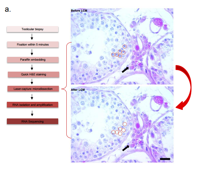
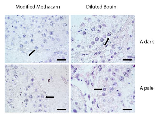
AvP & TV The technique is based on single cell laser capture microscopy out of fixed and paraffin embedded tissue. During this project, we learned that the fixative is of crucial importance. Not only for the recognition of various cell subtypes in the tissue based on their nuclear structure, but also to maintain RNA integrity to allow for in-depth transcriptome analyses of the isolated cells. In our case with testis tissue, we ended up with an alcohol-based fixative instead of the classically used Bouin’s fixative. This alcohol-based fixative maintains nuclear structure and RNA integrity and can be applied to any other tissue to allow for single cell capture. By using laser capture microscopy, we were able to isolate pools of single human germ cells from these alcohol-fixed specimens. This enabled us to analyse the transcriptome of human germ cells during the different stages of spermatogenesis. The technique can be used to study other tissues and developmental processes of interest.
Technique protocol to obtain samples for sequencing, from Supplementary Figure 2, Jan, Vormer et al, 2017
How will your technique help future research into male infertility and possible treatments?
AvP We can now use the results of the current paper as a reference for further transcriptome analyses of germ cells from patients with a maturation arrest in spermatogenesis. In doing so, we will gather information on the molecular aberrations underlying male infertility, which will give us detailed insights for establishing future treatment strategies.
When doing the research, did you have any particular result or eureka moment that has stuck with you?
SJ A moment that will always stick with me while working on this study is the moment I saw the first sequencing results. Many had doubted if this project would be feasible considering the small amounts of RNA we were attempting to experiment with, and it indeed took us years to be able to do so. It is astonishing how much one can do with extremely small amounts of RNA (picograms) (and will power!); we were able to generate whole transcriptomic profiles. This gives new meaning to the phrase “nothing is impossible”.
TV When setting up the technique, we had to find the correct fixative, microscope settings and a reliable RNA amplification method to be able to analyse our cells using next generation sequencing. Once this all worked out and we got our first sequencing runs: great!
The morphologies of the the germ cell subtypes in testes tissue using two different fixatives from Figure 2, Jan, Vormer et al, 2017
And what about the flipside: any moments of frustration or despair?
SJ There were certainly moments of frustration but certainly not despair. We had a period during the project in which the tubes used for laser capture microdissection were malfunctioning resulting in loss of our carefully collected germ cells. Capturing spermatogonia (germ cells that require careful microscopic evaluation) takes a long time – on a good day we captured 50 germ cells in 3-4 hours. So, you can imagine that loss of such precious collections causes a lot of frustration. Thankfully, it was a batch problem and we could easily solve this problem with the tube suppliers.
TV The isolation of single cells under the microscope is a concentrated job. Especially because we had to set up the technique, this involved quite some patience. Once an experiment failed after carefully isolating all the individual cells, this could be a very frustrating moment… Luckily, Sabrina and I could support each other!
What are your career plans following this work?
SJ At this moment I am in training to become a molecular clinical geneticist at the University Medical Center Groningen in The Netherlands. This project was inspirational in my current career path. Working on spermatogenesis, I was able to work on RNA expression but was also able to gain knowledge on the role genetics plays in the pathophysiology of spermatogenesis. This sparked my interest in genetics. Since RNA expression is a growing field in genetics, in my current work I get to merge these two very interesting worlds together.
TV I am currently working as a medical science liaison for a pharmaceutical company. The research for this paper was performed in close collaboration with physicians that treat infertile patients. I found this very inspiring and hope that our work will contribute to the development of future treatment strategies. In my new role, I am close to the development and application of new therapeutics in the clinic and have regular contact with physicians, which I find very rewarding.
What’s planned next for the Repping and van Pelt labs?
SR & AP One of the most exciting things we are working on is bringing autotransplantation of spermatogonial stem cells (SSCs) into clinical practise for sterile childhood cancer survivors. Often children with cancer become sterile due to the chemotherapy they receive to treat their cancer. We have pioneered the development of an in vitro culture system of human SSCs and are currently in the progress of finalizing crucial in vitro and animal safety studies before we embark on the first clinical trial in humans. Furthermore and in line with the current study, we aim to study the molecular aberrations in men suffering from idiopathic infertility. The ultimate goal there would be to treat these men with a therapy, perhaps in combination with SSC autotransplantation, that will allow these men to produce sperm again and become a genetic parent.
Lastly, what do each of you like to do when you’re not in the lab?
SR I enjoy taking long distance runs (just ran my 6th marathon), reading and spending time with my family and friends.
AvP I enjoy hiking, cycling and spending time with my family and friends.
SJ I enjoy going out dancing with my friends, swimming and various other activities such as walking in the various different beautiful dunes in The Netherlands and last but certainly not least, spending time with my family.
TV I am a big fan of flamenco dancing! I have been doing that for quite a few years, and I recently started with a flamenco-singing class for dancers. It is a lot of fun, I really enjoy the musicality and temperament of this music. Of course I also enjoy spending time with family and friends.
Sabrina Z. Jan, Tinke L. Vormer, Aldo Jongejan, Michael D. Röling, Sherman J. Silber, Dirk G. de Rooij, Geert Hamer, Sjoerd Repping, Ans M. M. van Pelt. 2017. Unraveling transcriptome dynamics in human spermatogenesis.Development. Volume 144, Issue 20, p3659-3673.
This is #30 in our interview series. Browse the archive here.
The CPH Bioscience PhD programme is designed for international talents to come to Denmark and start their research careers at one of the NNF Research Centers.
The Copenhagen Bioscience PhD programme recruits up to 16 motivated international students annually to launch their careers in the vibrant scientific environment of the Novo Nordisk Foundation Research Centers in Copenhagen. For enrollment in September 2018, applications is now open until December 2017.
Selection is based on academic achievements, research experience, academic references and interviews. A mandatory interview visit for up to 40 shortlisted applicants comprises panel interviews, one-on-one meetings with potential supervisors, and tours of the four Novo Nordisk Foundation Research Centers, and will take place in Copenhagen in March 2018. The Novo Nordisk Foundation will pay for travel and accommodation for selected applicants in association with the interview visit.
The University of Manchester, 2018/19 BBSRC DTP PhD Project
Understanding tubulin regulation during neuronal development, ageing and degeneration
Axons are slender, up-to-a-meter long, cable-like extensions of neurons which form the nerves and nerve tracts that wire our bodies and brain. These delicate cellular structures have to be maintained for an organism’s life time and are often the first to be affected in ageing, injury and neurodegeneration. To understand such conditions and identify ways to improve axon maintenance and regeneration, we study the regulation of microtubules (MTs) which form parallel bundles running all along axons to form their structural backbones and life-sustaining transport highways.
Axons are the cables that wire the nervous system (LINK)
On this project, you will study how the polymerisation of MTs within these bundles is regulated to drive axon growth and prevent senescence during ageing. The key question is how tubulins (i.e. the building blocks of MTs) are made available and continuously supplied in the narrow axons, up to a meter away from the cell body. This fascinating topic is most relevant to axon biology and pathology but surprisingly little understood. Your pioneering work will be based on our recently published mechanistic model of axonal tubulin supply and MT polymerisation, deduced from our own data and general knowledge in the field [Ref.1]. You will use cutting edge methodologies to study (1) contributions made by axonal transport and local tubulin translation, (2) roles of the chaperone machinery of tubulin assembly, and (3) mechanisms of tubulin storage and gene expression regulation.
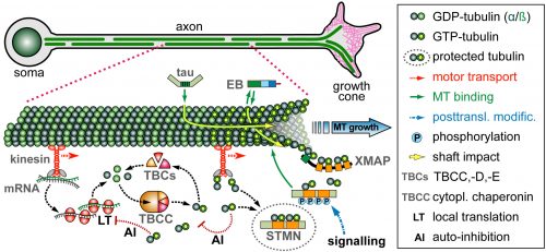
Proposed model of tubulin provision: Tubulins in axons require long distance transport, assembly via chaperones, STMN-mediated storage protecting them from auto-inhibition of their own biosynthesis (LINK)
For your studies, you will use neurons of the fruit fly Drosophila, which provide uniquely powerful genetic and cell biological means in order to efficiently generate new understanding that can then be applied to higher animals [Ref.2; LINK]. You will be able to capitalise on expertises of the supervisory team: the host group (Andreas Prokop) has long-standing experience with MT regulation and the Drosophila neuron model, the first co-supervisor’s group (Thomas Waigh) with high resolution imaging and quantitative approaches [Ref.3; Ref.4], and the second co-supervisor’s group (Mark Ashe) with RNA visualisation and processing [Ref.5].
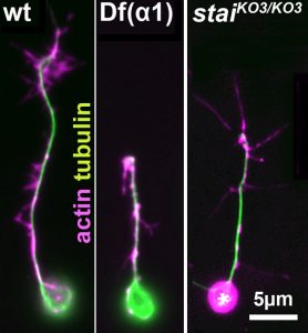
Cultured Drosophila neurons carrying tubulin mutations (middle) or STMN mutations (right) grow shorter than normal neurons (left)
Your transferable and experimental skill training will include genetics, cell biology, molecular biology, imaging techniques (live, high resolution, axonal transport, spatial detection of RNA and translational activity), quantitative analyses and modelling, as well as expertise in the important research areas of cytoskeleton and neurobiology. Finally, Andreas Prokop is an expert in science communication [Ref.6] providing further training opportunities important for your future career.
Deadline for applications is Fri 2nd of Nov. 2017, 5pm
Interviews are held on Tue/Wed, 9th/10th Jan. 2018
Offers will be confirmed in mid-Feb. 2018
References
[1] Voelzmann, A., Hahn, I., Pearce, S., Sánchez-Soriano, N. P., Prokop, A. (2016). A conceptual view at microtubule plus end dynamics in neuronal axons. Brain Res Bulletin126, 226-37
[2] Prokop, A., Beaven, R., Qu, Y., Sánchez-Soriano, N. (2013). Using fly genetics to dissect the cytoskeletal machinery of neurons during axonal growth and maintenance. J. Cell Sci.126, 2331-41
[3] Kenwright, D.A., Harrison, A.W., Waigh, T.A., Woodman, P.G., Allan, V.J. (2012) First-passage-probability of active transport in live cells, Physical Review E, 86, 031910
[4] Georgiades, P., Allan, V.J., Dickinson, M., Waigh, T.A. (2016) Reduction of coherent artefacts in super-resolution fluorescence localisation microscopy, Journal of Microscopy, 264, 3, 375-383
[5] Sfakianos, A. P., Whitmarsh, A. J., Ashe, M. P. (2016). Ribonucleoprotein bodies are phased in. Biochemical Society Transactions44, 1411-1416
[6] Illingworth, S., Prokop, A. (2017). Science communication in the field of fundamental biomedical research
 (No Ratings Yet)
 (5 votes)
 (1 votes)