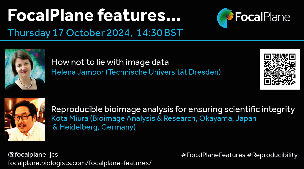
The next webinar in the FocalPlane features… series focuses on the important topic of reproducibility when acquiring, analysing and presenting imaging data. We are delighted to have talks from Helena Jambor and Kota Miura, who are both part of the QUAREP-LiMi (Quality Assessment and Reproducibility for Instruments and Images in Light Microscopy) community group, which aims to improve both quality assessment and quality control in microscopy. Helena’s talk will cover the community checklists for publishing images, which were developed by QUAREP-LiMi and published at the end of 2023, as she tells us ‘How not to lie with image data’. Kota’s talk will focus on bioimage analysis and the importance of reproducible analysis in ensuring scientific integrity.
At the speakers’ discretion, the webinar will be recorded for viewing on demand. To see the other webinars scheduled in our series, and to catch up on previous talks, please visit: thenode.biologists.com/devpres
When I began my PhD in 2020, I imagined my daily work would revolve around experiments, scientific writing, giving talks, and mentoring students. Little did I know that I’d soon be part of something quite different—a science documentary. I had the opportunity to collaborate with a team of scientists from the British Society for Developmental Biology (BSDB)and The Company of Biologists to create a film to promote UK developmental biology. As someone with no filmmaking experience, it was an exciting and daunting challenge. It was sure to be a steep learning curve full of science and fun.
We started by mapping our vision for the documentary. In film language, this included creating a film brief and storyboards. The storyboards laid out how each section of the documentary would unfold. We all pitched in with topic ideas, witling it down to four which would form our film vignettes: morphogenesis, cell migration, human brain development, and eye development. While it was hard to imagine at first how these ideas would translate into a polished documentary, we trusted the process. A crucial step in this was selecting a film production company. We wanted a collaborative team that could provide creative input whilst ensuring scientific accuracy. Fortunately, we teamed up with Cambridge Filmworks, who made the entire experience memorable.
My initial role in the documentary was to be an extra brain in the planning process. Especially since we were aiming to target a broad audience, including younger students. When the idea arose for me to help present the documentary, I couldn’t resist the opportunity. Despite my lack of on-camera experience, I’m passionate about science communication and developmental biology. The chance to work alongside brilliant scientists and collaborate with TV presenter and author Alice Roberts, who agreed to introduce and close the film, made the opportunity even more exciting.
Once we had our team of scientists and presenters on board, we focused on writing the script and drafting interview questions. The scientific team took the lead on script writing to give the documentary structure and direction. The script was then refined between our team and Cambridge Filmworks over numerous meetings and edits. This collaboration ensured that we included the relevant scientific information in a way that would be understood and be engaging to our audience. We also formulated questions which the scientists would answer throughout the documentary. The interview questions were designed to guide the scientists’ responses, keeping the flow natural and engaging. In most cases, multiple responses were filmed so that there were alternative options during the film editing process. We also had to ensure that the answers would be understood by a broad audience. With our scientific planning team and the filming team overlooking the filming, mistakes and jargon could be identified and corrected in the retakes.
Filming was an entirely new experience for me. On the first day, I learned what B-roll was and found myself awkwardly trying to walk naturally for the camera—it felt a bit robotic at first. As time progressed, I became much more confident and relaxed. I also watched Alice Roberts in action and took away some tips and tricks. During the latter stages of filming, I was recording voice over and performing solo pieces to camera, so I was thankful for the days leading up to this to hone the skills of presenting.
The main aim of the documentary is to showcase the wonder, importance, and applications of developmental biology. With that in mind, we invited a team of scientists who fitted into the selected vignettes. Helen Weavers kick starts the morphogenesis vignette by discussing her work on wound repair in fruit flies (Drosophila). Shankar Srinivas and Emily Noel follow with insights into how a small cluster of cells transforms into the complex, functioning heart. Tom Bennett rounds off the vignette by discussing plant development, offering a fascinating comparison between plant and animal development.
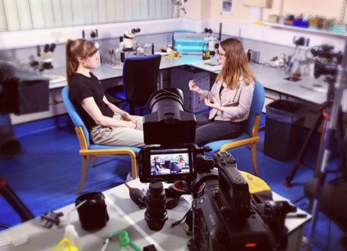
Behind the scenes of Courtney, this post’s author (left), speaking to Helen Weavers (right).
Next, the documentary explores neural crest cells, chosen for their remarkable migration abilities and their capacity to differentiate into a wide range of cell types. Karen Liu talks about the origin and migration of neural crest cells. We then have Elena Scarpa who brings a mechanical angle to the topic and highlights neural crest cell derived cancers.
In our vignette on human brain development, Katie Long explains how the brain folds during embryonic development and the differences between identical twins’ brains. My identical twin, Chloe Lancaster, joined the documentary to add a real-life twinning element. Laura Pellegrini discusses brain evolution and expresses her fascination for understanding the uniqueness of human brain development. She also introduces the audience to organoids and highlights their valuable contribution to understanding human development.
Finally, we dive into eye development with Pete Coffey and Rodrigo Young. Pete talks about his lab’s contribution to the treatment of eye diseases. His team has managed to surgically replace cells in the back of the eye with lab grown cells which enabled a patient to regain sight. He was taking a patch of cells to a patient on the day of filming which he was very excited about! Rodrigo delves into eye development with a key element being how two eyes of the same size and shape develop independently from each other.
What surprised me most during the filming process was how naturally the scientists adapted to the camera, despite not being accustomed to it. It turns out that all those conference presentations prepared them well for documentary interviews.
With the filming complete, it was time for Cambridge Filmworks to put the pieces together. Without being intimately involved in the editing, I can only say that this was some sort of self-organisation with constant feedback between the film editor and our team. We were also lucky enough to be invited to the Cambridge Studio to view the movie and provide feedback. It was surreal to see all the filming in a documentary for the first time.
I have been incredibly lucky to be involved in such a fun and exciting project with an incredible team of scientists and film creators. Brainstorming ideas and then seeing them in action during filming and editing was extremely rewarding, but the most valuable part of this experience has been all the people I met along the way. I interacted with scientists from all over the UK and picked their brains about different topics, science and career related. I might not have crossed paths with such inspiring people if I had not stepped out of the lab to embark on something completely different from my day-to-day scientific life. We also had a lot of fun along the way, laughing through retakes and mishaps.
I hope you enjoy watching the documentary as much as we enjoyed creating it. Please share it with your friends, family, and colleagues—let’s inspire the next generation of developmental biologists!
Film team: Adam Giles, Rich Millen, Barrie White, Douglas Murchie, Zheko Georgiev, Nigel Kinnings, Bronwyn Rand
Scientific planning team: Paul Martin, Shankar Srinivas, Katherine Brown, Rodrigo Young, Jeremy Green, Courtney Lancaster with help and funding from the BSDB and The Company of Biologists
Interviewees: Helen Weavers, Shankar Srinivas, Emily Noel, Tom Bennett, Karen Liu, Elena Scarpa, Katie Long, Laura Pellegrini, Pete Coffey, Rodrigo Young
Presenters + more: Alice Roberts, Courtney Lancaster, Chloe Lancaster
At preLights, the preprint highlighting service run by early-career researchers, we recently launched ‘spotLights’: a podcast series where we delve deeper into exciting preprints, featuring interviews with the scientists behind the research. These conversations allow you to hear the stories, challenges, and motivations that go into producing impactful scientific work. All episodes so far can be found here.
It goes beyond the accompanying preLight post that describes the key findings. You’ll get the chance to hear from two of the preprint authors, Dr Nafisa Jadavji and Dr Chris Smith, who share the background, process, and motivations behind their study. Don’t miss out on this insightful discussion! Tune in now and get the inside scoop directly from the experts.
Note: spotLights episodes 1 and 2 both revolve around preprinted work that describes the cephalic furrow as a “crumble zone” between head and trunk tissues (see related preLight post). Episode 1 features Bruno Vellutini, the first author of the preprint “Patterned embryonic invagination evolved in response to mechanical instability.”Episode 2 highlights the work led by Bipasha Dey, Verena Kaul, and Girish Kale, resulting in the preprint “Divergent evolutionary strategies preempt tissue collision in fly gastrulation.”
Rocío Hernández-Martínez, Sonja Nowotschin, Luke T.G. Harland, Ying-Yi Kuo, Bart Theeuwes, Berthold Göttgens, Elizabeth Lacy, Anna-Katerina Hadjantonakis, Kathryn V. Anderson
Daniel J. Stadtmauer, Silvia Basanta Martínez, Jamie D. Maziarz, Alison G. Cole, Gülay Dagdas, Gilbecca Rae Smith, Frank van Breukelen, Mihaela Pavličev, Günter P. Wagner
Stanley M. Kanai, Chloe R. Garcia, MaCalia R. Augustus, Shujan A. Sharafeldeen, Elliott P. Brooks, Juliana Sucharov, Ezra S. Lencer, James T. Nichols, David E. Clouthier
Siqi Gao, Triloshan Thillaikumaran, Martin H. Dominguez, William Giang, Kevin Hayes, Xiaowen Chen, Jesse Pace, Jenna Bockman, Danielle Jathan, Derek Sung, Sweta Narayan, Maxwell Frankfurter, Patricia Mericko-Ishizuka, Jisheng Yang, Marco Castro, Michael Potente, Mark L. Kahn
Yann Cormerais, Samuel C. Lapp, Krystle C. Kalafut, Madi Y. Cissé, Jong Shin, Benjamin Stefadu, Jean Personnaz, Sandra Schrotter, Angelica D’Amore, Emma R. Martin, Catherine L. Salussolia, Mustafa Sahin, Suchithra Menon, Vanessa Byles, Brendan D. Manning
Meryem B. Baghdadi, Ronja M. Houtekamer, Louisiane Perrin, Abilasha Rao-Bhatia, Myles Whelen, Linda Decker, Martin Bergert, Carlos Pérez-Gonzàlez, Réda Bouras, Giacomo Gropplero, Adrian KH Loe, Amin Afkhami-Poostchi, Xin Chen, Xi Huang, Stephanie Descroix, Jeffrey L. Wrana, Alba Diz-Muñoz, Martijn Gloerich, Arshad Ayyaz, Danijela Matic Vignjevic, Tae-Hee Kim
Nicha Tokavanich, Byron Chan, Katelyn Strauss, Christian D. Castro Andrade, Yuki Arai, Mizuki Nagata, Marc Foretz, Daniel J. Brooks, Noriaki Ono, Wanida Ono, Marc N. Wein
Kaitlin P. McCreery, Aki Stubb, Rebecca Stephens, Nadezda A. Fursova, Andrew Cook, Kai Kruse, Anja Michelbach, Leah C. Biggs, Adib Keikhosravi, Sonja Nykänen, Christel Hydén-Granskog, Jizhong Zou, Jan-Wilm Lackmann, Carien M. Niessen, Sanna Vuoristo, Yekaterina A. Miroshnikova, Sara A. Wickström
Eva Kreysing, Helene Gautier, Robert J Humphrey, Katrin A Mooslehner, Leila A Muresan, Daniel Haarhoff, Sudipta Mukherjee, Xiaohui X Zhao, Alex K Winkel, Andrea Dimitracopoulos, Eva K Pillai, Ragnhildur Thora Karadottir, Kristian Franze
Min Shi, Brittney Crouse, Nambirajan Sundaram, Naomi Pode Shakked, Lioba Ester, Weitao Zhang, Vinothini Janakiram, Raphael Kopan, Michael A. Helmrath, Joseph V. Bonventre, Kyle W. McCracken
Hannah R. Moran, Obed O. Nyarko, Rebecca O’Rourke, Ryenne-Christine K. Ching, Fréderike W. Riemslagh, Brisa Peña, Alexa Burger, Carmen C. Sucharov, Christian Mosimann
Alexandra de la Porte, Julia Schröder, Moritz Thomas, Johanna Geuder, Michael Sterr, Xavier Pastor, Leslie E. Sanderson, Tahsin Stefan Barakat, Wolfgang Enard, Carsten Marr, Christian Schröter, Micha Drukker
Gal Finer, Mohammad D. Khan, Yalu Zhou, Gaurav Gadhvi, George S. Yacu, Joo-Seop Park, R. Ariel Gomez, Maria Luisa Sequeira-Lopez, Susan E. Quaggin, Deborah R. Winter
Robert Mitchell-Gee, Robert Hoff, Kumar Vishal, Daniel Hancock, Sam McKitrick, Cristina Newnes-Querejeta, TyAnna L. Lovato, Richard M. Cripps, Michael V. Taylor
Christopher D Todd, Jannat Ijaz, Fereshteh Torabi, Oleksandr Dovgusha, Stephen Bevan, Olivia Cracknell, Tim Lohoff, Stephen Clark, Ricard Argelaguet, Juliette Pearce, Ioannis Kafetzopoulos, Alice Santambrogio, Jennifer Nichols, Ferdinand von Meyenn, Ufuk Guenesdogan, Stefan Schoenfelder, Wolf Reik
Megan Johnstone, Ashley Leck, Taylor Lange, Katherine Wilcher, Miranda S. Shephard, Aditi Paranjpe, Sophia Schutte, Susanne Wells, Ferdinand Kappes, Nathan Salomonis, Lisa M. Privette Vinnedge
Ramachandran Prakasam, Julianna Determan, Mishka Narasimhan, Renata Shen, Maamoon Saleh, Gareth Chapman, Komal Kaushik, Paul Gontarz, Kesavan Meganathan, Bilal Hakim, Bo Zhang, James E Huettner, Kristen L Kroll
Eirik Ryvoll Åsheim, Paul Vincent Debes, Andrew House, Petra Liljeström, Annukka Ruokolainen, Morgane Frapin, Iikki Donner, Ehsan Pashay Ahi, Jaakko Erkinaro, Jukka-Pekka Verta, Craig R Primmer
Juliane Glaser, Giulia Cova, Beatrix Fauler, Cesar A. Prada-Medina, Virginie Stanislas, Mai H.Q. Phan, Robert Schöpflin, Yasmin Aktas, Martin Franke, Guillaume Andrey, Christina Paliou, Verena Laupert, Wing-Lee Chan, Lars Wittler, Thorsten Mielke, Stefan Mundlos
Essi Wallen, Karita Ramo, Jussi Vehvilainen, Joonas Sokka, Marko Lehtonen, Timo Otonkoski, Ras Trokovic, Pauliina Auvinen, Olli Karkkainen, Nina Kaminen-Ahola
Ahmed Abdelbaki, Afshan McCarthy, Anita Karsa, Leila Muresan, Kay Elder, Athanasios Papathanasiou, Phil Snell, Leila Christie, Martin Wilding, Benjamin J. Steventon, Kathy K. Niakan
Keng Ioi Vong, Yanina D. Alvarez, Geoffroy Noel, Scott T. Barton, Changuk Chung, Robyn Howarth, Naomi Meave, Qingquan Zhang, Fiza Jiwani, Chelsea Barrows, Arzoo Patel, Jiang Xiong Wang, Neil Chi, Stephen F. Kingsmore, Melanie D. White, Xiaoxu Yang, Joseph G. Gleeson
Tayo E Adekeye, Emily M Teets, Emily A Tomak, Sadie L Waterman, Kailee A Sprague, Angelina White, Maddison L Coffin, Sabrina M Varga, Teresa E Easterbrooks, Sarah J Shepherd, Jared D Austin, Dmitrii Krivorotko, Troy E Hupper, Joshua B Kelley, Sharon L Amacher, Jared C Talbot
Juan Zhang, Lamisa Ataei, Liang Wu, Kirti Mittal, Linh Huynh, Shahil Sarajideen, Abdul Mazid, David P. Cook, Daniel Trcka, Kevin Tse, Jeffrey L. Wrana, Michael M. Hoffman, Miguel A. Esteban, Miguel Ramalho-Santos
Kiara C. Eldred, Sierra J. Edgerton, Isabel Ortuño-Lizarán, Juliette Wohlschlegel, Stephanie M. Sherman, Sidnee Petter, Gracious Wyatt-Draher, Dawn Hoffer, Ian Glass, Anna La Torre, Thomas A. Reh
Shubha Murthy, Denise A. Seabold, Lalit K. Gautam, Adrian M. Caceres, Rosemary Sease, Ben A. Calvert, Shana Busch, Aaron Neely, Crystal N. Marconett, Amy L. Ryan
Jessica K. Cinkornpumin, Sin Young Kwon, Anna-Maria Prandstetter, Theresa Maxian, Jacinthe Sirois, James Goldberg, Joy Zhang, Deepak Saini, Purbasa Dasgupta, Mariyan J. Jeyarajah, Stephen Renaud, Soumen Paul, Sandra Haider, William A Pastor
Pierre Osteil, Sarah Withey, Nicole Santucci, Nader Aryamanesh, Chi Nam Ignatius Pang, Nazmus Salehin, Jane Sun, Annie Qin, Jiayi Su, Hilary Knowles, Zhaoxiang Cai, Xiucheng Bella Li, Ernst J Wolvetang, Patrick P.L. Tam
Rajesh Ranjan, Binbin Ma, Ryan J. Gleason, Yijun Liao, Yingshan Bi, Brendon E. M. Davis, Guanghui Yang, Maggie Clark, Vikrant Mahajan, Madison Condon, Nichole A. Broderick, Xin Chen
Karolina Świtońska-Kurkowska, Jakub Kubiś, Joanna Delimata-Raczek, Bart Krist, Magda Surdyka, Żaneta Kalinowska-Pośka, Piotr Piasecki, Luiza Handschuh, Jan Podkowiński, Magdalena Rakoczy, Anna Samelak-Czajka, Michael Hayden, Nicholas S Caron, Maciej Figiel
Kutay Karatepe, Bruna Mafra de Faria, Jian Zhang, Xinyue Chen, Hugo Pinto, Dmitry Fyodorov, Esen Sefik, Michael Willcockson, Richard Flavell, Arthur Skoultchi, Shangqin Guo
James W. Swann, Ruiyuan Zhang, Evgenia V. Verovskaya, Fernando J. Calero-Nieto, Xiaonan Wang, Melissa A. Proven, Peter T. Shyu, X. Edward Guo, Berthold Göttgens, Emmanuelle Passegué
Federica Bruno, Christiana Georgiou, Deirdre Cunningham, Samantha Atkinson, Lucy Bett, Marine Secchi, Flora Birch, Sara Gonzalez Anton, jean langhorne, Cristina Lo Lo Celso
Silvia Vicenzi, Fangyuan Gao, Parker Côté, Joshua D. Hartman, Lara C. Avsharian, Ashni A. Vora, R. Grant Rowe, Hojun Li, Dorota Skowronska-Krawczyk, Leslie A. Crews
Eleanor Meader, Morgan T. Walcheck, Mindy R. Leder, Ran Jing, Paul J. Wrighton, Wade W. Sugden, Mohamad A. Najia, Isaac M. Oderberg, Vivian M. Taylor, Zachary C. LeBlanc, Eleanor D. Quenzer, Sung-Eun Lim, George Q. Daley, Wolfram Goessling, Trista E. North
Alicia Hurtado, Víctor López-Soriano, Miguel Lao, M. Ángeles Celis-Barroso, Pilar Lazúen, Alejandro Chacón de Castro, Yolanda Ramírez-Casas, Miguel Alaminos, J. Martin Collinson, Miguel Burgos, Rafael Jiménez, F. David Carmona, Francisco J. Barrionuevo
J. Luis Leal, Eva Hodková, Anja Billhardt, D. Magnus Eklund, Gustaf Granath, Pilar Herrera Egoavil, Jun Chen, Pascal Milesi, Jarkko Salojärvi, Martin Lascoux
Yaělle Dubois, Sophie Favier, Nathan Martin-Fornier, Mohyeddine Omrane, David Stroebel, Eric Perez, Sandrine Barbaux, Ahmed Ziyyat, Nicolas Rodriguez, Christine Gourier
Joost J.A.P.M. Wijnakker, Gijs J. F. van Son, Daniel Krueger, Willine van de Wetering, Carmen Lopez-Iglesias, Robin Schreurs, Fenna van Rijt, Sangho Lim, Lin Lin, Peter J. Peters, Ralph R. Isberg, Claudia Yanda, Wim de Lau, Hans Clevers
David M. Gonzalez, Rafael Dariolli, Julia Moyett, Stephanie Song, Bhavana Shewale, Jacqueline Bliley, Daniel Clarke, Avi Ma’ayan, Stacey Rentschler, Adam Feinberg, Eric Sobie, Nicole C. Dubois
The 2 October 2024 Development presents… webinar was chaired by Development’s Guest Editor, Karen Sears (UCLA) and featured three talks on the topic of environment, evolution and development. Catch up on the talks below.
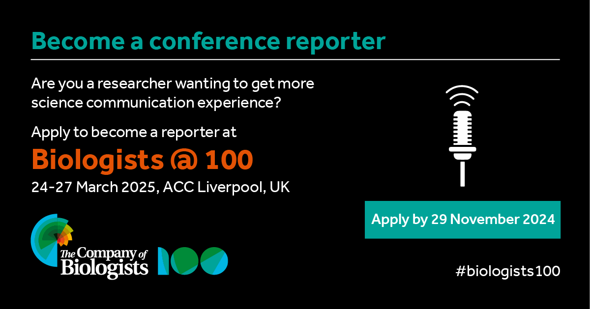
Are you keen to get more science communication experience? Is your research related to developmental and stem cell biology? The Node is looking for a reporter to attend and report from the Biologists @ 100 conference, happening 24-27 March 2025 in Liverpool, UK. This conference is a celebration of the 100th birthday of The Company of Biologists, bringing together different communities, including cell, developmental, experimental and disease biologists, and will incorporate the Spring Meetings of the British Society for Cell Biology (BSCB) and the British Society for Developmental Biology (BSDB). Registration for the conference is open and the abstract deadline is 13 December.
The selected reporter will have their conference registration fee waived (but will have to cover their own transport and accommodation costs*). They will be expected to attend the full conference. While the reporter will mainly focus on reporting from the ‘Cell and developmental biology’ scientific track, they are welcome to attend sessions from other tracks in the programme.
If you are interested, we’d love to read about your ideas of what you’d do as a reporter at the conference. If selected, we will work with you to develop your ideas and assist you at the conference.
If you have any questions about the role, don’t hesitate to email us at thenode@biologists.com.
A proposal (max 200 words) with idea(s) of what you would do as a conference reporter. We are open to suggestions of different formats of reporting. Please include a rough output you expect to produce from the reporting, and explain how the content will appeal to the Node readers.
A paragraph (max 200 words) telling us about yourself, your research, and any relevant science communication experiences you have.
Application deadline: Friday 29 November 2024
Our sister community site FocalPlane is also looking for a conference reporter. If you want to focus on reporting on cell biology and the use of microscopy in research, head over to their website to apply.
*Note: we encourage you to apply for conference grants to cover the transport/ accommodation costs, such as grants from BSDB and DMM.
The Indian Society of Developmental Biologists is the national society that represents developmental and stem cell biologists across India. We are pleased to introduce the society and our activities to the Node readers. Reading on, you will find some of our recent initiatives and posts. Do visit the InSDB website to check out more. You can get in touch with us at info@insdb.in if you would like to contribute/be a volunteer for InSDB.
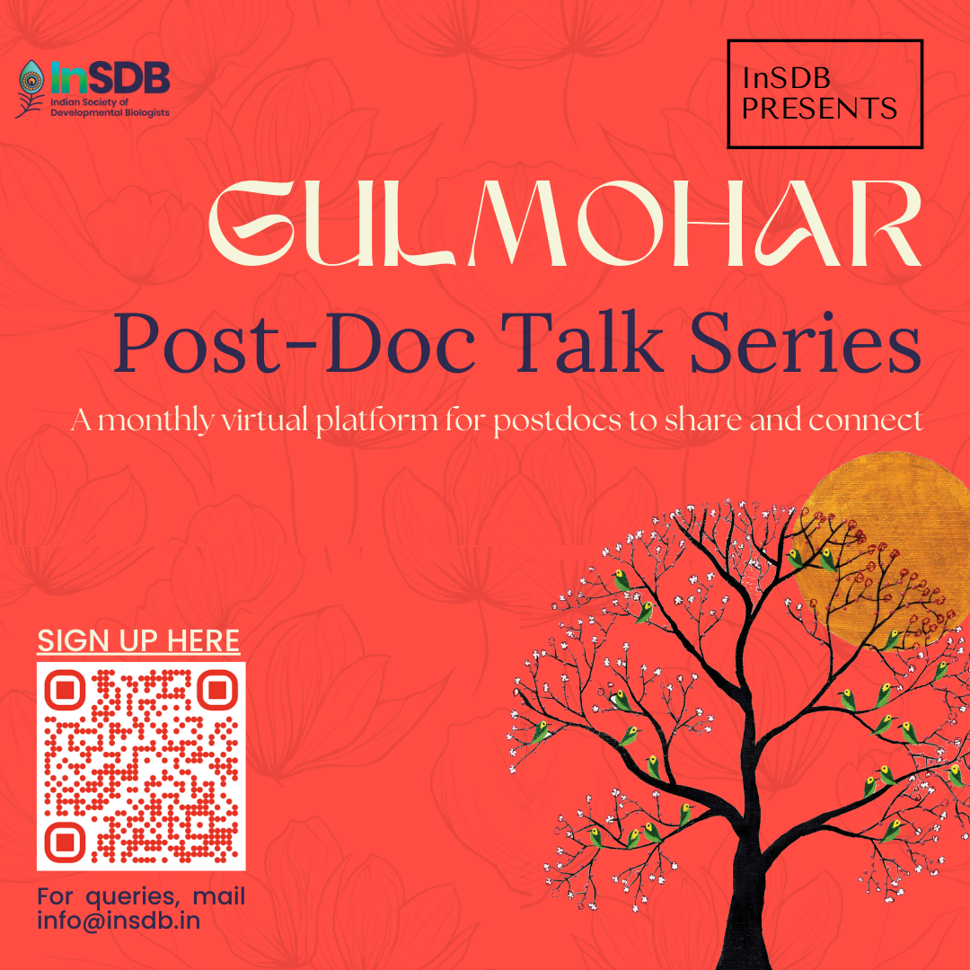
Gulmohar-Postdoc Talk Series
Gulmohar is our new, virtual, post-doc talk series through which we aim to bring together early-career scientists from around the world to share their research. This is a chance to hear from the next crop of PIs about their work, where their research is heading, and what’s next for them. Whether you are starting your research career, or already knee-deep in it, through this series, you will get to know about the different fields within the broad scope of developmental biology research. After every session, we also plan to have a post-talk informal discussion where you get to connect with the speakers and receive insight into their scientific journey and potential opportunities for your own.
We are currently accepting applications from postdocs who would like to be a speaker for the series. If you fit the bill, please sign up here, and we will get in touch with you.
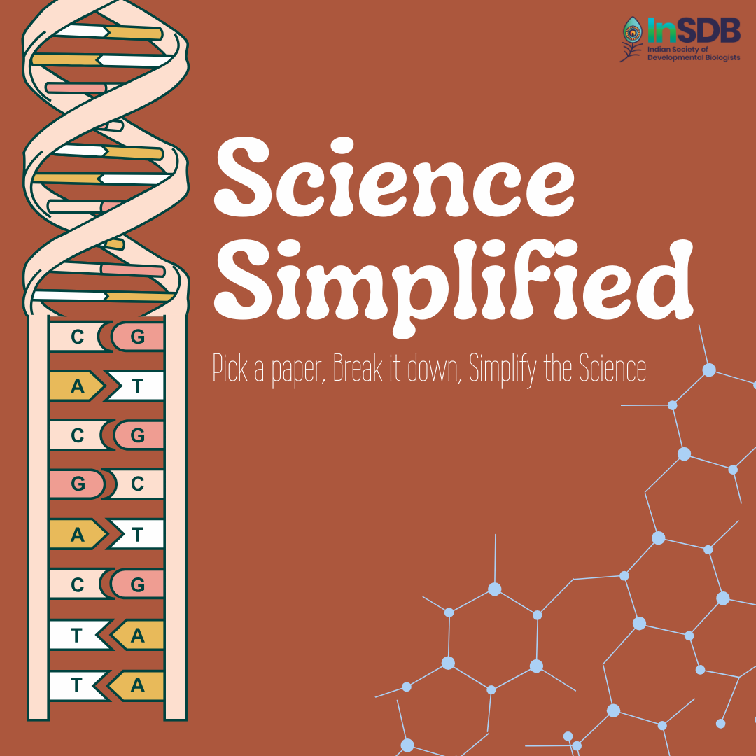
Science Simplified
Science Simplified is our series where we pick up a recently published research paper in developmental biology and distill it to the liking of a general audience. You can read the first article in this series here.
InSDB is also accepting articles from our audience! These could be stories on the latest research from the stem cell and developmental biology fields and perspectives on matters that are relevant to our audience. Anyone is welcome to pitch stories to us. We accept articles that are thoroughly researched and convey the excitement, relevance, and fascination of the subject to any curious reader. If you are interested in science writing and would like to sharpen your skills, this is your cue to get your pen and paper out! Also, do read our submission guidelines to know more. If you would like to contribute, please add your entries here.
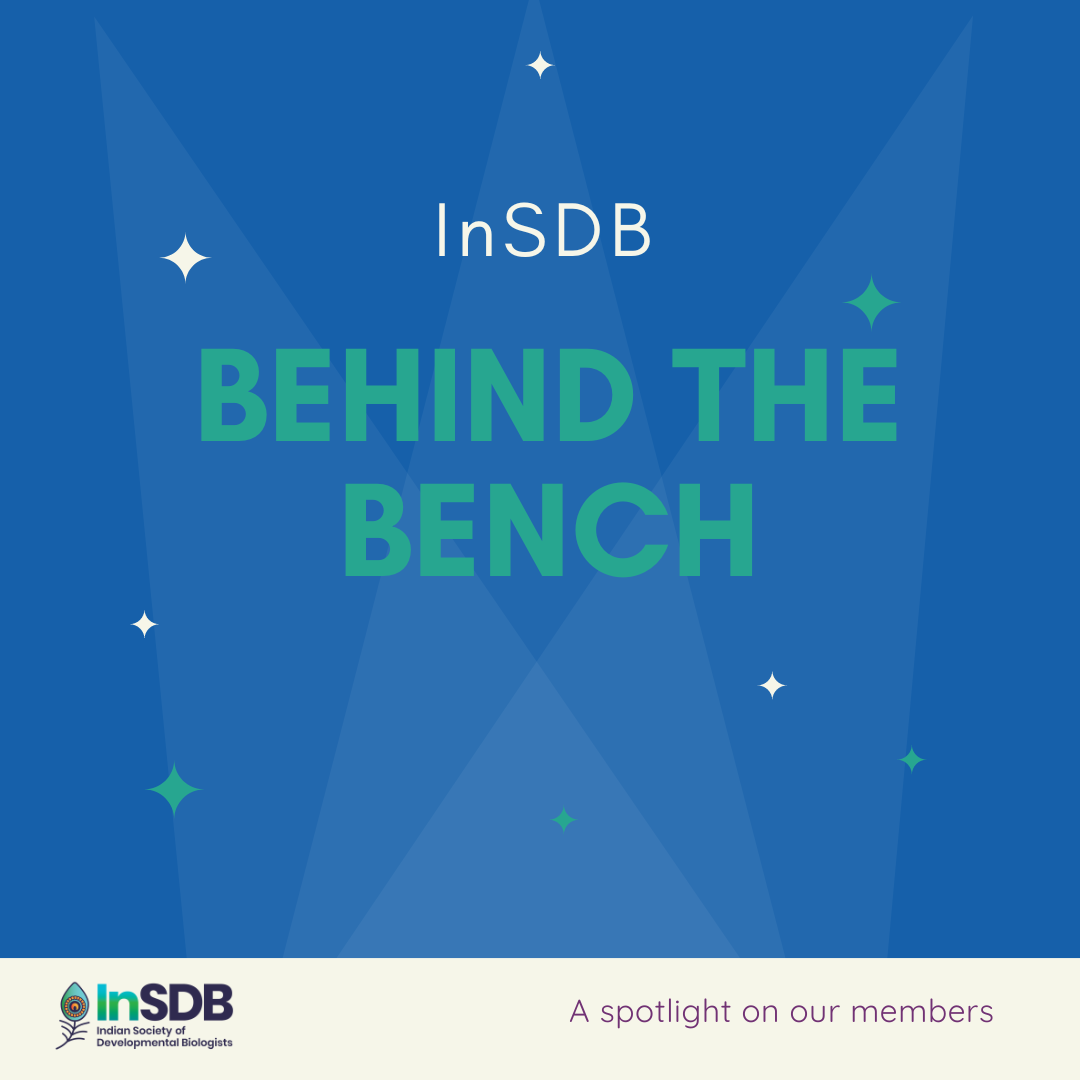
Behind The Bench
Behind the Bench is our interview series where we feature InSDB members, their stories, and the science they do. In each episode, we sit down with a member to know about their journey in science, from their early inspirations to the serendipitous moments along the way. Through this series of interviews, we aim to highlight the members who make our society what it is today. You can read these interviews here.
—
If you would like to follow our updates closely, we are on X, LinkedIn, and Instagram!
Daniel Riveline,1 Karsten Kruse,2 Mukund Thattai,3 Guntram Bauer4
Scientific communication was pioneered in the 17th century (Ref. 1). Scientists were called ‘sçavans’and had to embrace the humanities and the sciences to present their discoveries with clarity. These achievements are difficult nowadays, with the detailed technical knowledge involved in biology, physics, chemistry and mathematics research. In this context, it is challenging to present the depth of each side of an interdisciplinary scientific question, and often one field is featured more prominently, to the detriment of the other. To address this issue, we organised a Symposium in which presentations were given as tandem talks, with participants from biology and physics or from chemistry and biology. Topics ranged from cell positioning in the embryo (Eric Wieschaus and William Bialek), to liquid condensates (Anthony Hyman and Frank Jülicher), to the physics of photosynthesis (Donatas Zigmantas and Thomas Renger). We found this tandem format to be extremely successful (see Symposium online Ref. 2). Concepts from physics, biology and chemistry were explained in accessible yet informative language, and the audience could see how interdisciplinary engagements developed. Importantly, the speakers highlighted active points of debate and articulated unresolved issues, leaving space for new breakthroughs and scientific directions. As a metaphor for collaboration, a musical duo on the violin and cello illustrated how each voice in a partnership could be heard. Using a Bach counterpoint as the paradigm, the musicians from the Philharmonic Orchestra of Strasbourg explained how their pieces were harmonious only with shared and alternating voices.
References:
1- A History of Scientific Journals, Aileen Fyfe, Noah Moxham, Julie McDougall-Waters, Camilla Mørk Røstvik, UCL Press London (2022) https://www.uclpress.co.uk/products/187262
1- Laboratory of Cell Physics, IGBMC, CNRS and University of Strasbourg, Strasbourg, France, riveline@unistra.fr
2- Departments of Biochemistry and Theoretical Physics, University of Geneva, 1211 Geneva, Switzerland, Karsten.Kruse@unige.ch
3- Simons Centre for the Study of Living Machines, National Centre for Biological Sciences, Tata Institute of Fundamental Research, Bengaluru, India, thattai@ncbs.res.in
4- The International Human Frontier Science Program Organization, 12 Quai Saint-Jean, 67080 Strasbourg, France, gbauer@hfsp.org
Oxford Open Doors is an annual event that offers the public a unique opportunity to explore the historic and modern buildings of Oxford, many of which are normally closed to the public. Organized by Oxford Preservation Trust, the event takes place over a weekend in September and allows visitors to experience the architectural and cultural heritage of the city first hand.
As part of this year’s Oxford Open Doors event, researchers from the Institute for Developmental and Regenerative Medicine (IDRM) invited the public into their labs, offering a rare glimpse into their cutting-edge research. Visitors were guided through research areas and imaging facilities by enthusiastic staff eager to share their work.
The IDRM is a unique flagship institution at the University of Oxford, dedicated to meeting an ambitious challenge: two thirds of all deaths world-wide are due to non-communicable diseases, many of which are cardiovascular, neurological or immune system disorders that have a developmental origin, representing an urgent unmet clinical need. The mission of the IDRM is the development of new drugs and therapeutic strategies to tackle these chronic illnesses.
At its core is a formal merger of developmental biology and regenerative medicine in the form of over 200 cardiovascular, neuroscience and immunology scientists, integrating their expertise to foster multidisciplinary collaborations.
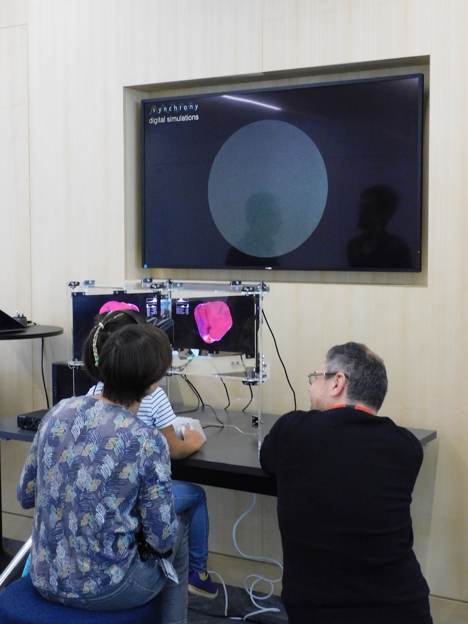
Under this research interest, we’ve developed a number of initiatives for Public Engagement that we deployed at this Open Day: the first, Synchrony, is a unique science-art project created in collaboration with computational artist Andy Lomas. Part of the public engagement efforts of the Wellcome funded Human Developmental Biology Initiative, Synchrony brings the complex world of developmental biology to life.
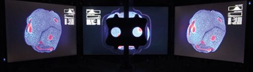
Researcher Matthew Stower talking to visitors about the HDBI research and Synchrony stereo viewer. copyright 2024 Tomoko WatanabeSynchrony stereo viewer. copyright: Andy Lomas
Synchrony allowed attendees to interact with a 3D artificial simulation of growth and pattern formation, changing the rules that govern the formation of some of the fascinating shapes that we can observe during embryonic development. This immersive experience captured visitors’ curiosity and sparked conversations about the intricate biological processes that direct embryo development and organ formation with Matthew Stower who represented the HDBI researchers. The creative process itself involved lengthy discussions between Andy and researchers Matthew Stower, Shifaan Thowfeequ, Claudio Cortes, Tomoko Watanabe and Shankar Srinivas.
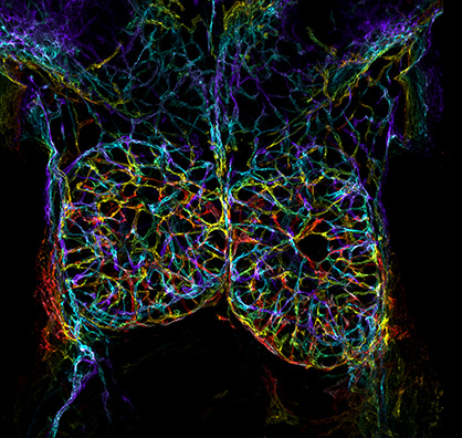
Mouse embryonic thymus. copyright David Grainger
In addition to Synchrony, visitors also engaged with scientific discovery through SyGlass, a virtual reality software for exploring, annotating and sharing 3D data. SyGlass allows users to fully immerse themselves in stunning microscopy images, rendered in interactive 3D in a VR environment. A breath-taking image of the embryonic thymus, contributed by David Grainger, enabled attendees to explore the beauty and intricacies of the 3D structure of this organ. Both children and adults delighted in slicing through the tissue sections and navigating the complexities of the thymus in real time.
copyright Tomoko Watanabe
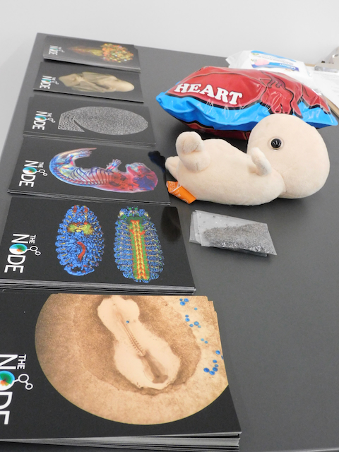
We would like to thank The Node for their support. Your extremely popular postcards gave us the perfect ‘hook’, to talk about the wonders of development and how it takes place across species. These visuals helped visitors connect the beauty and complexity of developmental biology with the cutting-edge research happening within IDRM, while at the same time sparking very interesting conversations about research and ethics.
 (No Ratings Yet)
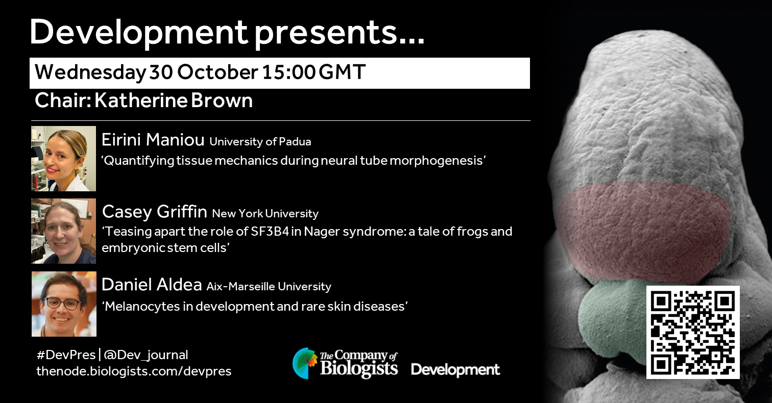
 (3 votes)