In this SciArt profile, we meet Dhananjay Chaturvedi, a developmental biologist who creates drawings inspired from nature and from his research into skeletal muscle homeostasis and repair in Drosophila.
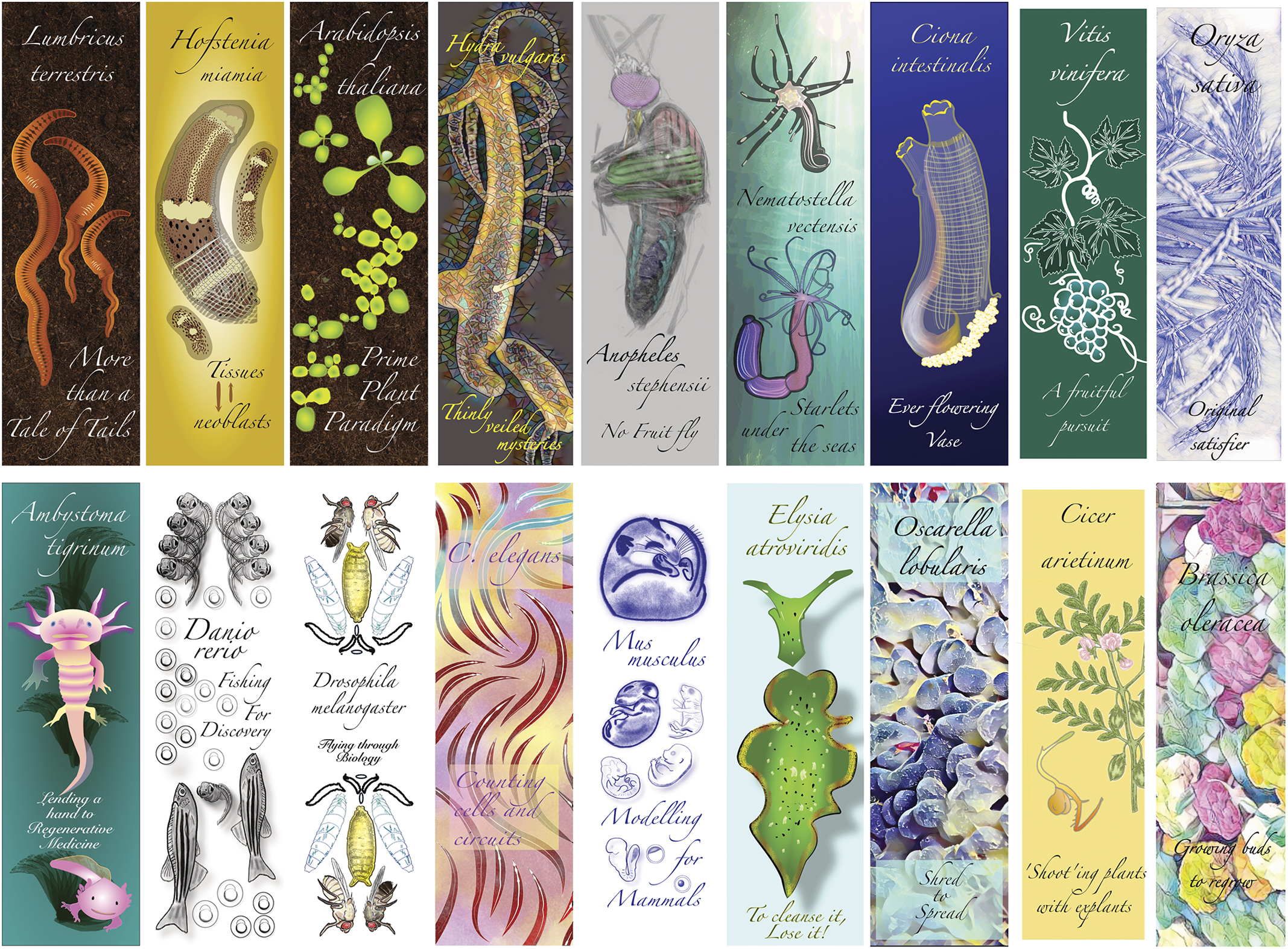
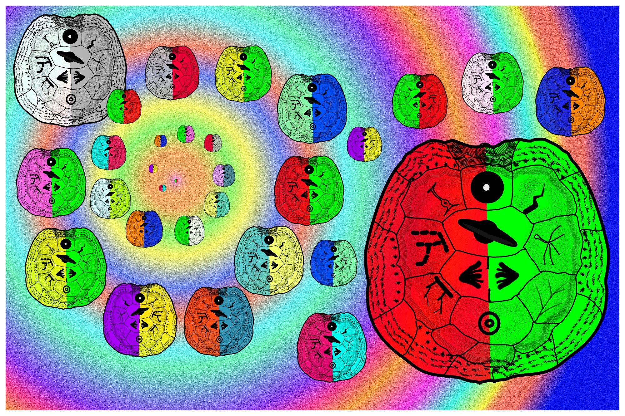
Illustrations of some of the organisms featured at the #CMMDR2024 meeting, which were distributed to the meeting participants as bookmarks. (Meeting review on Development)
Can you tell us about your background and what you work on now?
I started my lab two years ago at the Centre for Cellular & Molecular Biology (CCMB), Hyderabad, India. My group looks at multiple aspects of adult skeletal muscle homeostasis and repair. We are currently relying on Drosophila to reveal in-vivo principles that we will test later in vertebrate systems. Our new findings find their roots in work I did as a Campus fellow at the National Center of Biological Sciences, Bangalore, in the lab of Prof K VijayRaghavan, from where some brilliant findings have come across fields. My foray into Drosophila started during my PhD in the lab of Dr Michael Buszczak at UT Southwestern Medical Center in Dallas, Texas. I saw their true value and potential to assess most biological problems inside a living organism with rigour. There, I investigated the role of chromatin modifiers in germline stem cells. My master’s was in the lab of Prof Shubha Tole at Tata Institute of Fundamental Research in Mumbai, India, where I investigated eye development in the Lhx2 mutant mouse, among other things.
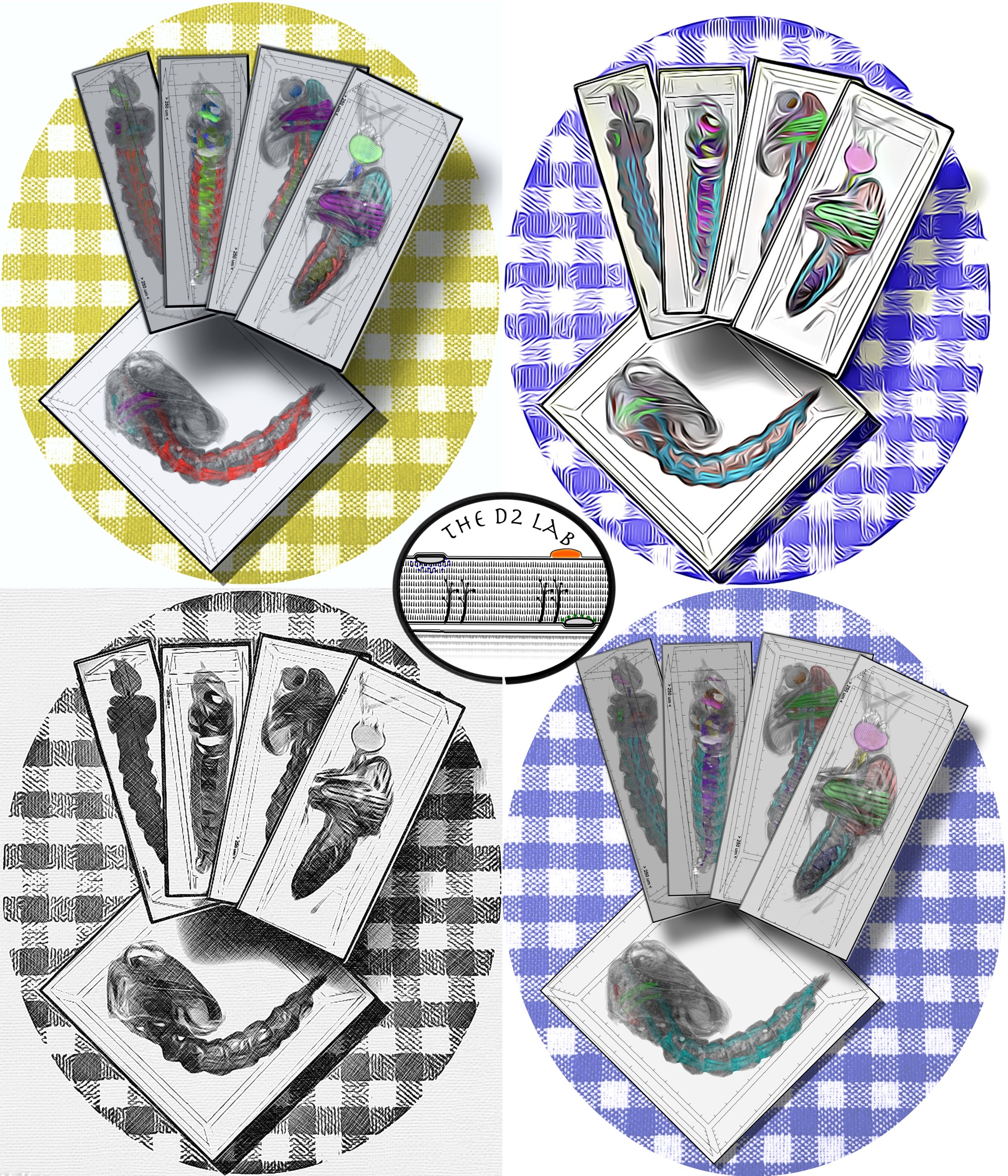
Developmental stages of Mosquitoes recorded through MicroCT scans, rendered as cards on a table.
Were you always going to be a scientist?
While it may sound pretentious now, yes. I have always wanted to do research and teach. While my classmates prepared for more secure professions, I was fascinated by what I saw on the Discovery Channel and National Geographic as a child. Having seen another scientist in the family be perfectly happy with what they do, I geared my studies towards research in Biology. Experiment-driven research has had more lows than highs, but highs are incomparable to any other experience. Also, the lows kept me grounded, honing the sense of asking the right questions and doing the right experiments. To be honest, the practicalities of growing in research, like the complete uncertainty of a career until you’ve made it, have made me question my teenage choices a couple of times so far. Having said that, I am very happy with where I am right now, only looking to discover new things in nature.
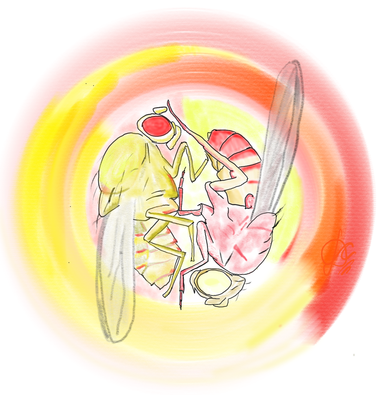
Drosophila Yin Yang
And what about art – have you always enjoyed it?
Drawing, among other ways, has been a means of self-expression since childhood. Most of my friends who like art are far more skilled than I am. I have found, however, that others appreciate my finished drawings a lot more than I expected. A combination of my own words and mental images helps me arrive at these.
Star dust, fragile compartments, forms and functions
What or who are your most important artistic influences?
I cannot think of specific artists because my exposure and training are between limited and absent. Striking images that I have seen in nature and in my research would be the biggest influences. Order and chaos in natural patterns with the added dimensions of light and colour capture my imagination. Further, superimposing, juxtaposing or inverting these with images from entirely different contexts tickles my mind.
The Big to the big bust in many iterations
How do you make your art?
Lately, I start with a specific audience and message in mind. For instance, bookmarks that were shared at #CMMDR2024 are intended for the thousands of school students who come through CCMB’s Open Day. I remember what images excited me as a child, and I channelled them into those pictures. These were meant to draw students to nature, and science by extension.
Among other things, I’ve made posters and logos for public viewing. These need to be artsy enough to stand out while directly communicating intention with some detail. This is especially true for the schematics I make for presentations and papers. Often, what is published does not communicate my exact sentiments, so I have to make my own as accurately as possible. More recently, people have been making requests for specific occasions or venues, and I do what I can.
When I get time for myself, which is very little these days, I try to visualise the jumble of thoughts and make them as appealing as possible. These might seem “stimulated”, as a cousin once commented.
I rely on software for the simple reason that it allows me to correct mistakes and rework drawings quickly. Further, there are tools that allow one to model portions of images from photographs, helping me arrive at my vision far quicker than my skills with other media would allow. I started using these, in fact, when I started making schematics for presentations and papers.
Does your science influence your art at all, or are they separate worlds?
The art I have admired is evocative, often portraying the human experience or aspirations. To me, it is a way for people to express what they see and feel. My drawings can only channel what occupies my mind the most, which is wonder for nature. So, I cannot see science and art as two separate worlds; rather, one is the manifestation of the other.
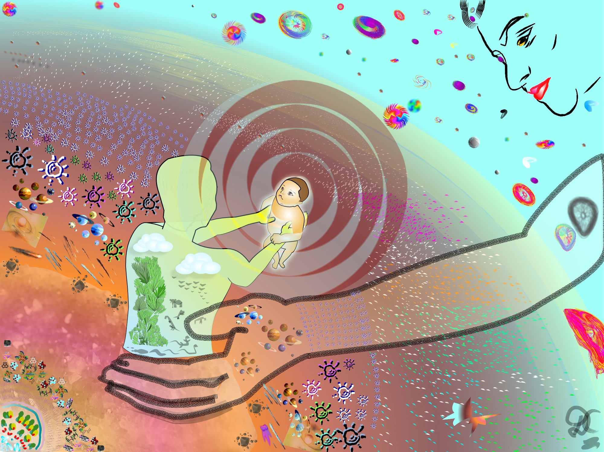
My daughter, the centre of creation
What are you thinking of working on next?
I want to draw something that conveys the oneness of the pursuit of truths of nature and society, that they are inseparable. Though, this may be hard to appreciate from siloed views. The vision has not crystallised yet. It may take a while before it does. Several simpler drawings may appear before this idea begins to materialise.
The Mary Lyon Centre at MRC Harwell invites UK-based scientists to nominate ideas and designs for our Rare Disease GEMM call to get free, novel, genetically altered mice generated and validated by our team of experts.
Apply now! The deadline for application is the 15th of September 2024. You can also contact our team for more information at gemm@har.mrc.ac.uk.
Joseph A. Bisson, Miriam Gordillo, Ritu Kumar, Neranjan de Silva, Ellen Yang, Kelly M. Banks, Zhong-Dong Shi, Kihyun Lee, Dapeng Yang, Wendy K. Chung, Danwei Huangfu, Todd Evans
Elizabeth Abraham, Brett Volmert, Thomas Roule, Ling Huang, Jingting Yu, April E. Williams, Henry M. Cohen, Aidan Douglas, Emily Megill, Alex Morris, Eleonora Stronati, Raquel Fueyo, Mikel Zubillaga, John W. Elrod, Naiara Akizu, Aitor Aguirre, Conchi Estaras
Joseph Hanna, Yacine Touahri, Alissa Pak, Lauren Belfiore, Edwin van Oosten, Luke Ajay David, Sisu Han, Yaroslav Ilnytskyy, Igor Kovalchuk, Deborah M Kurrasch, Satoshi Okawa, Antonio del Sol, Robert A Screaton, Isabelle Aubert, Carol Schuurmans
Robin P. Journot, Mathilde Huyghe, Alexandre Barthelemy, Hugo Couto-Moreira, Jakub Sumbal, Marisa M. Faraldo, Maxime Dubail, Charles Fouillade, Silvia Fre
Mireia Pampols-Perez, Carina Fürst, Oscar Sánchez-Carranza, Elena Cano, Sandra Raimundo, Eric L. Lindberg, Martin Taube, Arnd Heuser, Anje Sporbert, Norbert Hübner, Holger Gerhardt, Gary R. Lewin, Annette Hammes
Noemie Lavoie, Anaelle Scribe, Francois JM Chartier, Karim Ghani, Alexandra Jette, Sara L Banerjee, Manuel Caruso, Melanie Laurin, Andrew Freywald, Sabine Elowe, Patrick Laprise, Nicolas Bisson
Eric Surette, Joan Donahue, Stephanie Robinson, Deirdre McKenna, Crisvely Soto Martinez, Brendan Fitzgerald, Rolf O. Karlstrom, Nicolas Cumplido, Sarah K. McMenamin
Olga Lanzetta, Marchesa Bilio, Johannes Liebig, Katharina Jechow, Foo Wei Ten, Rosa Ferrentino, Ilaria Aurigemma, Elizabeth Illingworth, Christian Conrad, Soeren Lukassen, Claudia Angelini, Antonio Baldini
Mohamed T. Elaswad, Mingze Gao, Victoria E. Tice, Cora G. Bright, Grace M. Thomas, Chloe Munderloh, Nicholas J. Trombley, Christya N. Haddad, Ulysses G. Johnson, Ashley N. Cichon, Jennifer A. Schisa
Ana C. Lima, Mariam Okhovat, Alexandra M. Stendahl, Jake VanCampen, Kimberly A. Nevonen, Jarod Herrera, Weiyu Li, Lana Harshman, Ran Yang, Lev M. Fedorov, Katinka A. Vigh-Conrad, Nadav Ahituv, Donald F. Conrad, Lucia Carbone
Tobias Beckröge, Bettina Jux, Hannah Seifert, Hannah Theobald, Elena De Domenico, Stefan Paulusch, Marc Beyer, Andreas Schlitzer, Elvira Mass, Waldemar Kolanus
Zoulfia Darieva, Peyman Zarrineh, Naomi Phillips, Joshua Mallen, Araceli Garcia Mora, Ian Donaldson, Laure Bridoux, Megan Douglas, Sara F Dias Henriques, Dorothea Schulte, Matthew J Birket, Nicoletta Bobola
Jeremie Subrini, Wazeer Varsally, Irina Balaguer Balsells, Maike Bensberg, Georgios Sioutas, Obah Ojarikre, Valdone Maciulyte, Björn Gylemo, Katharine Crawley, Katherine Courtis, Dirk G. de Rooij, James M. A. Turner
Yan Zhao, Andrea Fernández-Montoro, Greet Peeters, Tatjana Jatsenko, Tine De Coster, Daniel Angel-Velez, Thomas Lefevre, Thierry Voet, Olga Tšuiko, Ants Kurg, Katrien Smits, Ann Van Soom, Joris Robert Vermeesch
Nia Teerikorpi, Micaela C Lasser, Sheng Wang, Elina Kostyanovskaya, Ethel Bader, Nawei Sun, Jeanselle Dea, Tomasz J. Nowakowski, A Jeremy Willsey, Helen Willsey
Christopher A.P. Batho, Janice D. Reid, Harley R. Robinson, Henrietta Cserne Szappanos, Lynn A.C. Devilée, Sharon M. Hoyte, Rebecca L. Johnston, Rebekah Ziegman, Sarah Hassan, Lior Soday, Rebecca L. Fitzsimmons, Simon R. Foster, Dominic C. H. Ng, Edward Tate, Enzo R. Porrello, Benjamin L. Parker, Richard J. Mills, James E. Hudson
Jack Schnell, Zhen Miao, MaryAnne Achieng, Connor C. Fausto, Victoria Wang, Faith De Kuyper, Matthew E. Thornton, Brendan Grubbs, Junhyong Kim, Nils O. Lindström
Ruochen Dong, Hua Li, Xi C He, Chen Wang, Anoja Perera, Seth Malloy, Jonathon Russell, Wenting Li, Kaitlyn Petentler, Xinjian Mao, Zhe Yang, Michael Epp, Kate Hall, Allison Scott, Sarah E Smith, Mark Hembree, Yongfu Wang, Sean McKinney, Jeff Haug, Jay Unruh, Brian Slaughter, Xunlei Kang, Linheng Li
Diana Al Delbany, Manjusha S Ghosh, Nusa Krivec, Anfien Huygebaert, Marius Regin, Chi Mai Duong, Yingnan Lei, Karen Sermon, Catharina Olsen, Claudia Spits
Evangelia Skoufa, Jixing Zhong, Oliver Kahre, Kelly Hu, Georgios Tsissios, Louise Carrau, Antonio Herrera, Albert Dominguez Mantes, Alejandro Castilla-Ibeas, Hwanseok Jang, Martin Weigert, Gioele La Manno, Matthias Lutolf, Marian Ros, Can Aztekin
Vladimir Vinarsky, Stefania Pagliari, Fabiana Martino, Cristina Mazzotti, Katerina Jirakova, Zuzana Garlikova, Enrico Di Iuri, Daniel Kytyr, Patrizia Benzoni, Martina Arici, Alessia Metallo, Kira Zeevaert, Andrea Barbuti, Wolfgang Wagner, Marcella Rocchetti, Giancarlo Forte
Ruochen Dong, Hua Li, Xi C He, Chen Wang, Anoja Perera, Seth Malloy, Jonathon Russell, Wenting Li, Kaitlyn Petentler, Xinjian Mao, Zhe Yang, Michael Epp, Kate Hall, Allison Scott, Mary C McKinney, Shengping Huang, Sarah Smith, Mark Hembree, Yongfu Wang, Zulin Yu, Jeffery Haug, Jay Unruh, Brian Slaughter, Xunlei Kang, Linheng Li
T. Sing, S. Kalamajski, J.P.M.C.M. Cunha, S. Hladkou, F. Roberts, S. Gheibi, A. Soltanian, K. Yektay Farahmand, O. Ekström, A. Mamidi, P.W. Franks, A. Rosengren, H. Semb, H. Mulder, M. Fex
Erik Jacques, Pauline Garcia, Orane Mercier, Yechen Hu, Cyril Degletagne, Jade Ravent, Sidy Fall, Maira P. Almeida, Aaron R. Wheeler, Stephane Angers, Penney M. Gilbert, Fabien Le Grand
Tony Marchand, Kemi E. Akinnola, Shoichiro Takeishi, Maria Maryanovich, Sandra Pinho, Julien Saint-Vanne, Alexander Birbrair, Thierry Lamy, Karin Tarte, Paul S. Frenette, Kira Gritsman
T. Singh, S. Kalamajski, J.P.M.C.M. Cunha, S. Hladkou, F. Roberts, S. Gheibi, A. Soltanian, K. Yektay Farahmand, O. Ekström, A. Mamidi, P.W. Franks, A. Rosengren, H. Semb, H. Mulder, M. Fex
Anne C. Lietzke, Elizabeth Bealer, Kelly Crumley, Jessica King, Ava M. Stendahl, Jie Zhu, Gemma L. Pearson, Elena Levi-D’Ancona, Belle Henry-Kanarek, Emma C. Reck, Manikanta Arnipalli, Vaibhav Sidarala, Emily M. Walker, Subramaniam Pennathur, Jesper G.S. Madsen, Lonnie D. Shea, Scott A. Soleimanpour
Francine Paraiso, Huiqiong Lin, Chengxia Li, Daniel P. Woods, Tianyu Lan, German F Burguener, Connor Tumelty, Juan M Debernardi, Anna Joe, Jorge Dubcovsky
Munan Lyu, Hiroyuki Iida, Thomas Eekhout, Meeri Mäkelä, Sampo Muranen, Lingling Ye, Anne Vatén, Brecht Wybouw, Xin Wang, Bert De Rybel, Ari Pekka Mähönen
H. Mayeur, J. Leyhr, J. Mulley, N. Leurs, L. Michel, K. Sharma, R. Lagadec, J.-M. Aury, O.G. Osborne, P. Mulhair, J. Poulain, S. Mangenot, D. Mead, M. Smith, C. Corton, K. Oliver, J. Skelton, E. Betteridge, J. Dolucan, O. Dudchenko, A.D. Omer, D. Weisz, E.L. Aiden, S. McCarthy, Y. Sims, J. Torrance, A. Tracey, K. Howe, T Baril, A. Hayward, C. Martinand-Mari, S. Sanchez, T. Haitina, K. Martin, S.I. Korsching, S. Mazan, M. Debiais-Thibaud
Jelisaveta Djordjevic, Patrick Tran Van, William Toubiana, Marjorie Labédan, Zoé Dumas, Jean-Marc Aury, Corinne Cruaud, Benjamin Istace, Karine Labadie, Benjamin Noel, Darren J Parker, Tanja Schwander
Eglantine Heude, Hugo Dutel, Frida Sanchez-Garrido, Karin D. Prummel, Robert Lalonde, France Lam, Christian Mosimann, Anthony Herrel, Shahragim Tajbakhsh
Chloe A. Briney, Jesslyn C. Henriksen, Chenwei Lin, Lisa A. Jones, Leif Benner, Addison B. Rains, Roxana Gutierrez, Philip R. Gafken, Olivia S. Rissland
Martina Jabloñski, Guillermina M. Luque, Matías D. Gómez-Elías, Claudia Sanchez-Cardenas, Xinran Xu, Jose Luis de la Vega-Beltran, Gabriel Corkidi, Alejandro Linares, Victor X. Abonza Amaro, Aquetzalli Arenas-Hernandez, María Del Pilar Ramos-Godinez, Alejandro López-Saavedra, Dario Krapf, Diego Krapf, Alberto Darszon, Adan Guerrero, Mariano G. Buffone
Maya Emmons-Bell, Grace Forsyth, Abby Sundquist, Sylvie Oldeman, Angeliki Gardikioti, Roshni de Souza, Jonathan Coene, Maryam H. Kamel, Shine Ayyapan, Harrison A. Fuchs, Steven Verhelst, Joanna Smeeton, Catherine A. Musselman, Juan-Manuel Schvartzman
Daniel de la Fuente, Maria Maroto, Yulia N Cajas, Karina Cañón-Beltrán, Raul Fernandez-Gonzalez, Ana Munoz-Maceda, Juana M Sanchez-Puig, Rafael Blasco, Paula Cots, Manuel Aviles, Dimitrios Rizos, Alfonso Gutiérrez-Adán
Helen A. Brown, Ludivine Guillet, Charles A. C. Williams, Hayley Shaw, Houjiang Zhou, Diana Rios-Szwed, Rosalia Fernandez-Alonso, Liam McMulkin, Marios P. Stavridis, Greg M. Findlay
Lieke Stockmann, Hélène Kabbech, Gert-Jan Kremers, Brent van Herk, Bas Dille, Mirjam van den Hout, Wilfred F.J. van IJcken, Dick Dekkers, Jeroen A.A. Demmers, Ihor Smal, Danny Huylebroeck, Sreya Basu, Niels Galjart
Byung Ho Lee, Kana Fuji, Heike Petzold, Phil Seymour, Siham Yennek, Coline Schewin, Allison Lewis, Daniel Riveline, Tetsuya Hiraiwa, Masaki Sano, Anne Grapin-Botton
Natalie A. Trigg, John E. Schjenken, Jacinta H. Martin, David A. Skerrett-Byrne, Shannon P. Smyth, Ilana R. Bernstein, Amanda L. Anderson, Simone J. Stanger, Ewan N.A. Simpson, Archana Tomar, Raffaele Teperino, Colin C. Conine, Geoffry N. De Iuliis, Shaun D. Roman, Elizabeth G. Bromfield, Matthew D. Dun, Andrew L. Eamens, Brett Nixon
Vukasin M. Jovanovic, Narisu Narisu, Lori L. Bonnycastle, Ravi Tharakan, Kendall T. Mesch, Hannah J. Glover, Tingfen Yan, Neelam Sinha, Chaitali Sen, David Castellano, Shu Yang, Dvir Blivis, Seungmi Ryu, Daniel F. Bennett, Giovanni Rosales-Soto, Jason Inman, Pinar Ormanoglu, Christopher A. LeClair, Menghang Xia, Martin Schneider, Erick O. Hernandez-Ochoa, Michael R. Erdos, Anton Simeonov, Shuibing Chen, Ilyas Singeç, Francis S. Collins, Claudia A. Doege, Carlos A. Tristan
Benjamin Furtwängler, Nil Üresin, Sabrina Richter, Mikkel Bruhn Schuster, Despoina Barmpouri, Henrietta Holze, Anne Wenzel, Kirsten Grønbæk, Kim Theilgaard-Mönch, Fabian J. Theis, Erwin M. Schoof, Bo T Porse
Ken To, Lijiang Fei, Jan Patrick Pett, Kenny Roberts, Krzysztof Polański, Tong Li, Nadav Yayon, Peng He, Chuan Xu, James Cranley, Ruoyan Li, Kazumasa Kanemaru, Ni Huang, Stathis Megas, Laura Richardson, Rakesh Kapuge, Shani Perera, Elizabeth Tuck, Anna Wilbrey-Clark, Ilaria Mulas, Fani Memi, Batuhan Cakir, Alexander V. Predeus, David Horsfall, Simon Murray, Martin Prete, Pavel Mazin, Xiaoling He, Kerstin B. Meyer, Muzlifah Haniffa, Roger A. Barker, Omer Bayraktar, Christopher D. Buckley, Sarah A. Teichmann
Nikola Sekulovski, Amber E. Carleton, Anusha A. Rengarajan, Chien-Wei Lin, Lauren N. Juga, Allison E. Whorton, Jenna K. Schmidt, Thaddeus G. Golos, Kenichiro Taniguchi
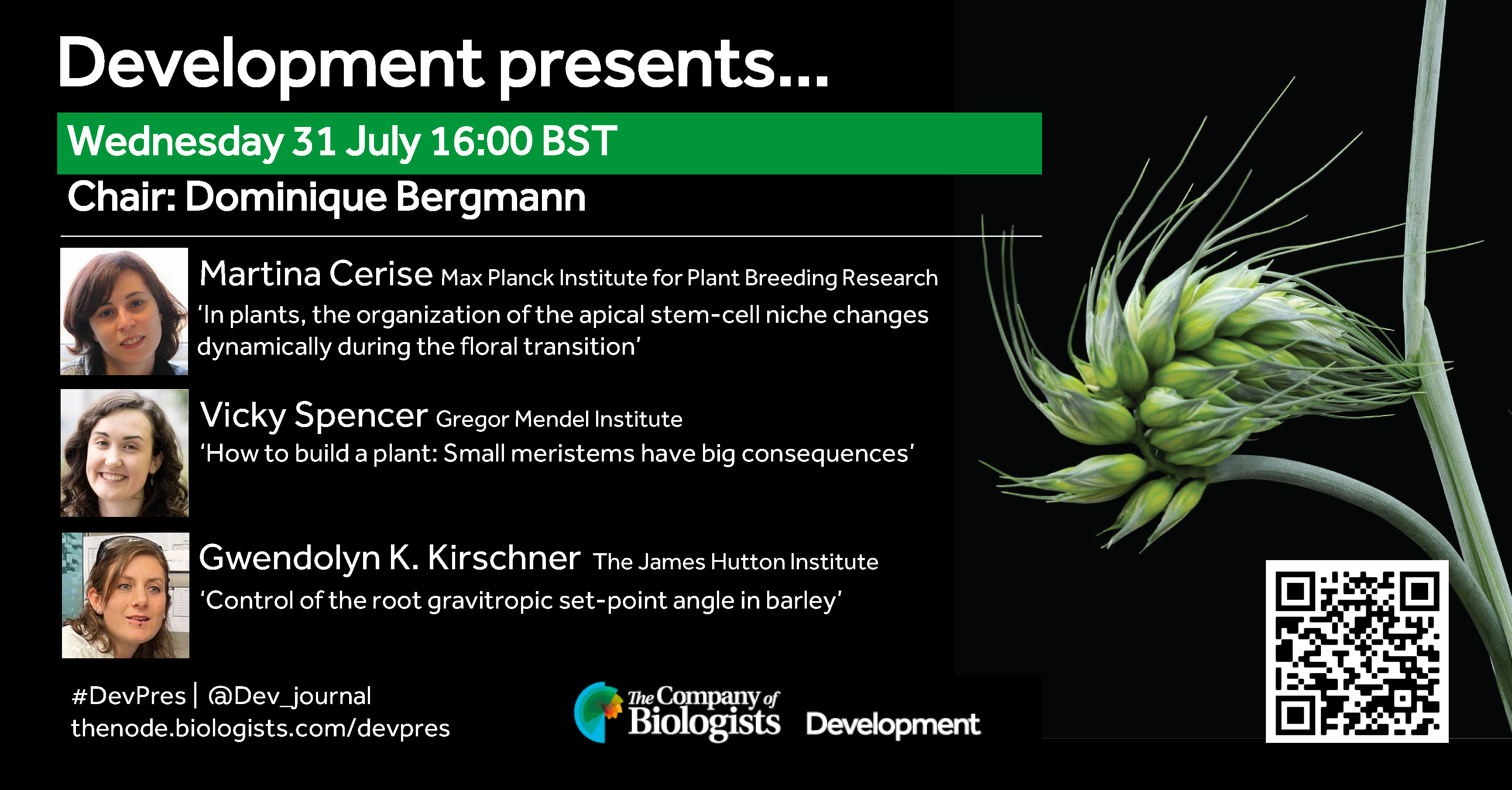
The July 31st Development presents… webinar was chaired by Development Editor Dominique Bergmann (Stanford University) and featured three talks on plant development. Catch up on the talks below.
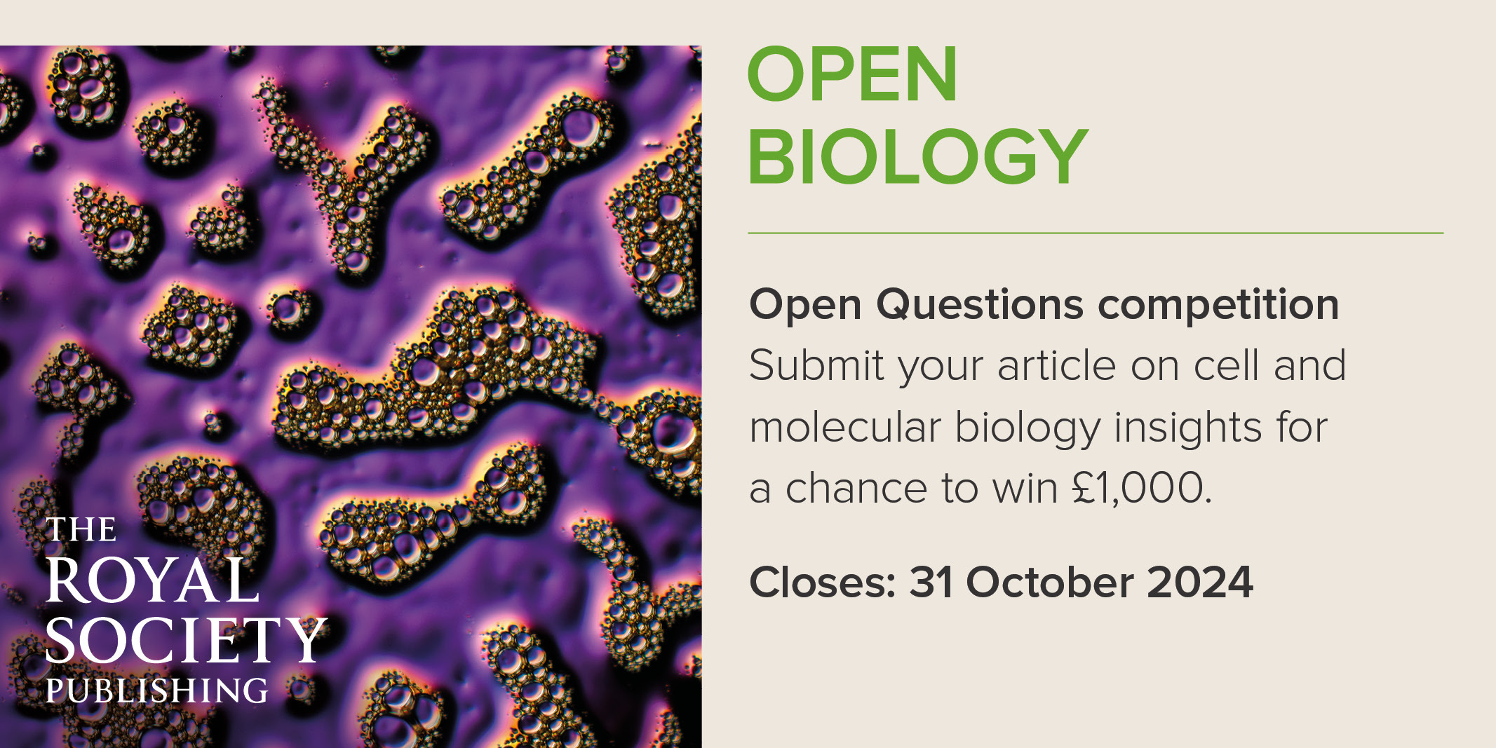
We are excited to announce the launch of Open Biology’s inaugural Open Questions competition. Submit a pressing, understudied or interesting ‘open question’ in cellular and molecular biology.
Participants have the chance to win an overall prize of £1,000 and enjoy a full Article Processing Charge (APC) waiver – submit your article before 31 October 2024.
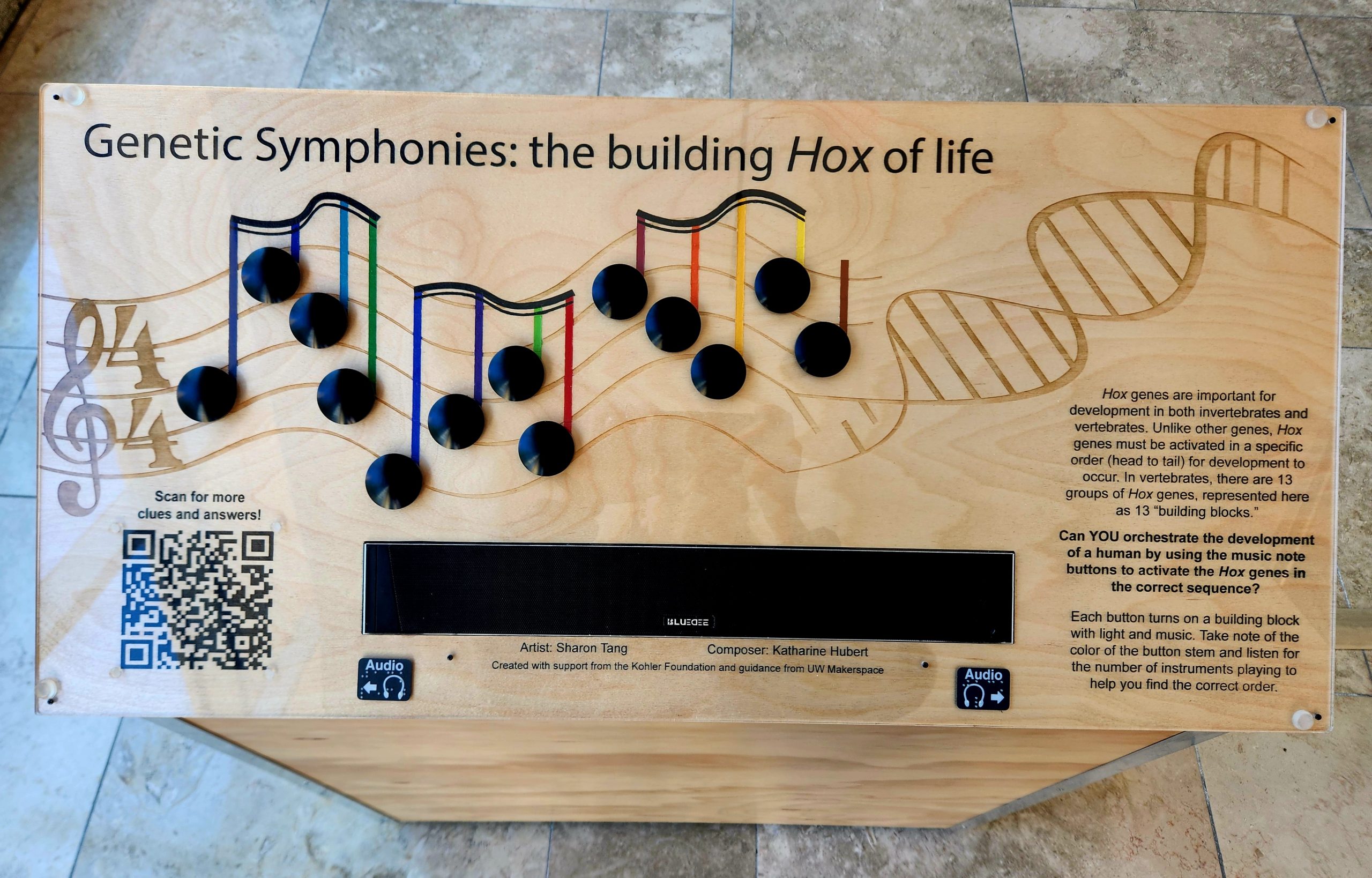
Over the past year and a half, I had the opportunity to participate in the UW-Madison Kohler Fellowship Program. In this program, an artist is paired with a scientist to create a science-art fusion project, some goals of which include fostering cross disciplinary communication and increasing the accessibility of scientific concepts through art. Though I was accepted into this program as a science fellow, my partner, Sharon Tang, and I both consider ourselves artist-scientists. Sharon is a PhD candidate in the Cell and Molecular Biology program and an avid muralist, while I’m a Genetics PhD candidate and composer/musician. Together we leveraged our scientific knowledge and artistic expertise to create “Genetic Symphonies: the building Hox of life”.
“Podium”: The control panel for “Genetic Symphonies: Building Hox of life” contains 13 black buttons arranged on a music staff that was laser cut into wood. The stem of each music note is color coded to match its corresponding Hox box. Buttons must be pressed in the correct order to activate Hox gene expression in the corresponding box.
As most developmental biologists know, Hox genes are transcription factors critical for patterning the skeletal axes. They also remain one of science’s biggest mysteries; we still have yet to determine what Hox genes actually regulate to control developmental and adult homeostatic processes. In addition to the mysterious mechanisms of action, Hox genes possess unique properties (clustered chromosomal arrangement, spatio-temporal expression pattern, regional restriction) that lend nicely to artistic manipulation and exploration.
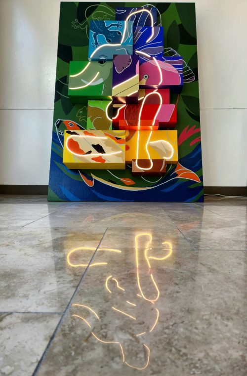
Sharon and I took advantage of these features to generate an interactive, multi-modal exhibit. In our exhibit, 13 paralogous groups of Hox genes are represented by 13 painted building blocks, affectionately referred to as “Hox boxes”. Likewise, there are 13 color-coded, randomized, buttons that control each “Hox box” on an accompanying podium. Participants must activate gene expression, via a button press, and determine the correct order of Hox gene activation (head to tail). Each correct button press produces light in the corresponding box and sounds a unique measure of music. With each successive, correct, button press participants develop their own genetic symphony and an abstract human figure via lights. By conveying development through both light and sound, we also increase the accessibility of our exhibit.
Want to know more about Genetic Symphonies: Building Hox of life? Check out Behind Building: Hox ! At this site, you can find more information about the construction, fabrication, composition, and electronics of the exhibit.
“Lights on”: When all 13 buttons are pressed in the correct order, a hidden abstract human figure illuminates from within the boxes. (3 votes) Loading...
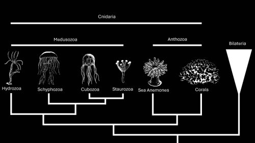
Hydractinia symbiolongicarpus is an emerging model to understand stem cell evolution
Stem cells can’t hide what they are. At least, that’s the takeaway from the newly sequenced genomes of two colonial hydroids, Hydractiniasymbiolongicarpus and Hydractinia echinata. I recently sat down with Dr. Christine Schnitzler of UF’s Whitney Laboratory for Marine Bioscience to talk about her experience assembling these genomes — a huge collaboration that started pre-pandemic, slowed down during the pandemic, and finally culminated in a paper published in Genome Research in March. Our conversation covered topics spanning from the logistics of large collaborations (including the need to find the right people), the shared molecular vocabulary that stems from genome projects, and the scientific merit of studying emerging systems.
Hydractina are colonial cnidarians, a group that consists of anthozoans (sea anemones and corals), schyphozoans (co-called “true” jellyfish), cubozoans (box jellyfish), and hydrozoans (Hydra and Hydractinia). Cnidarians have an informative phylogenetic position as sister to bilaterians. In addition to providing evolutionary insights, many cnidarian species — including Hydractinia symbiolongicarpus — are exceptionally good at regenerating. Schnitzler’s lab is studying Hydractinia symbiolongicarpus to understand how interstitial stem cells, or i-cells, impact an organism’s ability to regenerate.
“All genome projects are huge,” said Schnitzler. “I would never have been able to do this myself.”
This particular project has been 10 years in the making. In that time, sequencing technologies and analyses kept changing. The research team opted to swap the Illumina short-read platform for Pac-Bio long-read sequencing to dig deeper into two unconventional model organisms: Hydractinia echinata and Hydractinia symbiolongicarpus.
The genomes of these two cnidarians had a few surprises. First, the two genomes were quite different in size — Hydractinia echinata was 775MB and Hydractinia symbiolongicarpus was 514MB. And that’s not just based on sequence. The research team isolated single cells, stained nuclei with propidium iodide and ran them through a flow cytometer against a known standard. Second, based on the researchers’ analysis, these two species’ genomes diverged around 19 million years ago. That may seem like a long time, but if you consider that two different strains of the same species of jellyfish are estimated to have diverged 45 million years ago, then a true species divergence 19 million years ago is surprisingly recent.
“I thought that was pretty cool,” said Schnitzler.
Genomes offer a common language for biologists to understand similarities (and, importantly, differences) between species. To that end, Schnitzler’s team took a lot of time and care into making this resource available for anybody with even a cursory interest. They also developed a web portal for both Hydractinia species that allows curious biologists to dive deeper into a very, very granular level of gene evolution between two closely related genomes. They also generated a single cell RNAseq browser.
“That’s kind of fun. The whole point is to make this genome accessible and useful to the community,” said Schnitzler. “It was a priority for us to build that resource, which was not easy,” she added with a laugh.
“Collaboration is key. And having the right people is very helpful.”
At the end of our discussion, I gave Schnitzler a chance to respond to the “Questions For The Author” from PreLighter Isabella Cisneros who spotlighted the preprint last September (check out the PreLight article here). I’ve included her questions, numbered 1–3, as well as Schnitzler’s responses below.
Questions For The Authors (from PreLighter Isabella Cisneros):
1. How do you reconcile the large number of shared i-cell marker genes with the higher proportions of phylum-specific and cnidarian-specific genes in the H. symbiolongicarpus genome?
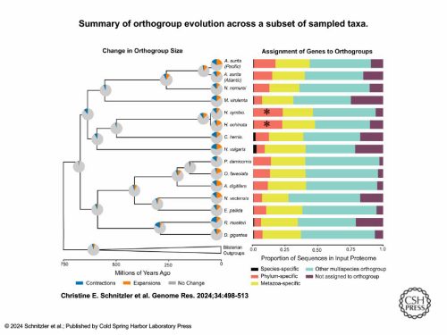
We took the entire genome and ran an orthology clustering of all predicted proteins from 49 other animals, including 16 cnidarians. We put them into bins once we got the clusters back. Are these genes in a “multi-species orthogroup?” That means they group with genes from animals outside of cnidarians, so they’re shared more widely. A lot of the other categories were cnidarian-specific.
This graph shows just the cnidarians and a few other animal outgroups. Those two [asterisked] red bars are further out than all the other cnidarians — they had the highest proportion of genes in their genome that were specific to their phylum.
If you apply that same clustering to the most highly expressed genes that are specific to just the i-cells, we found that i-cell genes are mostly shared with other animals (that was the title of the paper).
How do I reconcile that? I think it means that stem cells are exactly what they are.
They’re undifferentiated cells that have to use basic cell characteristics — cell cycle genes, genes that help with proliferation, genes that are involved in maintaining stemness — and these are universal things that all animals have developed and retained throughout evolution.
That does not mean that stem cells of Hydractinia are exactly homologous to the stem cells of other animals. Cell types can evolve very quickly, but the underlying genes that make stem cells stem cells are very highly conserved.
There are a lot of cnidarian-specific genes in their genomes. It’s just in their stem cells, they’re not using those very much. But when you think about stem cells, they’re not really unique to cnidarians. All animals have some type of a stem cell — that’s just how it works.
2. Towards the end of the preprint, you claim that it remains unclear whether other animals share the same toolkit of genes, or whether these toolkits are instead partially overlapping. While further studies will be necessary to determine this, at this stage, what do you anticipate to be the case?
It’s very hard to relate cell types and say they have a shared evolutionary origin of the cell type. If you look at stem cells in Hydractinia and then look at stem cells in planarians, they have a lot of shared characteristics. But what about all the animals in between? Where did their stem cells go? I think that’s a very interesting question.
I think getting down to this core level of genes and gene regulatory networks that are controlling these types of cells, to me, might be more interesting and more informative from an evolutionary perspective than trying to absolutely say this cell type first arose at some point and then shifted. It’s not really about the cell types. It’s more about the core genes that are involved and finding out their function. And it’s really hard. I think the only way really is to drill down on function within several animals and then try to see if they relate to each other.
It’s good to look at informative positions on the tree, as a lot of people try to do, and try to gain as much insight as you can from looking at those different places. I don’t think there’s going to be 100% overlap. But I think there will be themes that emerge — categories of genes that may be similar. You could group them by orthology, but maybe not exactly by BLAST.
It’s a question for the future. I think people are trying to tackle it, but it’s going to take more than these genome-wide approaches. We’ll have to go back to the lab and do functional testing to get at those questions.
3. Given the different evolutionary trajectories that H. symbiolongicarpus and H. echinata have followed since divergence, what kinds of studies would be better suited for each species, if any? What could be gained by using both species in a comparative framework?
Most people have dropped H. echinata as a research organism. It’s got a bigger genome that’s not as well assembled. And no one’s maintaining them in the lab, anymore. They’re just harder to keep in culture long term. There may be some ecological and other questions that might be more interesting with echinata, and it would be cool if someone picks it up now that the genome is available.
On the other hand, H. symbiolongicarpus just grows and grows and grows indefinitely. After years and years of being in culture, we can still get them to spawn on a weekly basis and get tons of embryos. It also has a smaller genome that ended up with a better assembly.
Smaller genome, easier lab culture — all of that screams, “Work on this one!”
So for us, the path forward is symbiolongicarpus. Now we can talk and really understand each other when we’re talking about a particular gene or a particular process. Now we have this resource. It’s a starting point.
One interesting thing to think about is what is unique about Hydractinia biology? I like Hydractinia because it is different. It’s colonial with a polymorphism of polyp types. We have feeding polyps, sexual polyps, defensive polyps. Because we have this diversity just within a single colony, there are a lot more questions to ask. It’s a different type of development, a different kind of asexual reproduction.
The other thing that I think is super cool is its lifecycle. It’s a hydrozoan that has lost the medusa stage. So there’s no jellyfish stage. Its next closest sister group is Podocoryna carnea, which does have a polyp and a medusa stage. That genome is being sequenced now. So when we have these two genomes — Podocoryna and Hydractinia — you can now start to understand the difference between a hydrozoan genome that produces the medusa jellyfish and the genome that doesn’t. I think some of these comparative studies with new genomes are really, really exciting. And I think with unlimited resources, we would have perfectly assembled genomes for those two groups and start doing more experiments to try to understand the unique biology of Hydractinia.
One thing our paper does not really focus on is regeneration. Hydractinia symbiolongicarpus is a model of regeneration. There’s a huge amount of knowledge we can learn by studying regeneration in this animal. There’s been some seminal ground laying papers about regeneration in this model, but doing updated studies, which we have some data, hopefully soon we can update and talk more about how this animal achieves its amazing regenerative abilities.
At the speakers’ discretion, the webinar will be recorded for viewing on demand. To see the other webinars scheduled in our series, and to catch up on previous talks, please visit: thenode.biologists.com/devpres
In this SciArt profile, we meet Friedrich Bliem, who has a background in cell biology and scientific illustration and has been creating “Art in Science” paintings for decades.
Can you tell us about your background and what you work on now?
I am Austrian and have spent a total of 24 years in the USA, UK and Australia, where I was raised. As the son of a freelance artist, I practiced the skills of drawing and painting from an early age and in my teens focussed on later enrolling in the University of Applied Arts, Vienna. Music had also been a passion and I co-founded the successful Austrian music group, Misthaufen, which is still active today.
Despite my interests in both music and art, I also had an innocent fascination for science based on the link between art and music. So, in pursuit of this interest, I chose first to enroll in a natural sciences and engineering course at a Viennese university, which was then renamed Biotechnology.
Concomittantly, I continued my art training with non-formal education, especially under the expressionist artist, Rudolf Macek.
After completing my university studies in 1981, I moved to Cambridge, England, where I continued to paint.
I had always had a passion for drawing cartoons as it allowed me to reflect on my social environment without having to say sorry. This passion motivated me to approach the Elsevier Publications office in Cambridge with some ideas and examples of my work. Soon I found a successful niche as scientific illustrator and cartoonist for various publishers.
Encouraged by the Cambridge environment, as well as my work as illustrator, I began working on scientific, especially cell biological, subject matters. This culminated in a solo exhibition in Cambridge, UK, 1984 (“Art in vivo”). As such, I might be said to be a fore-runner of today´s so-called SciArt movement.
However, in 1988 I decided to devote my time to science, before again returning to art. This period lasted 25 years!
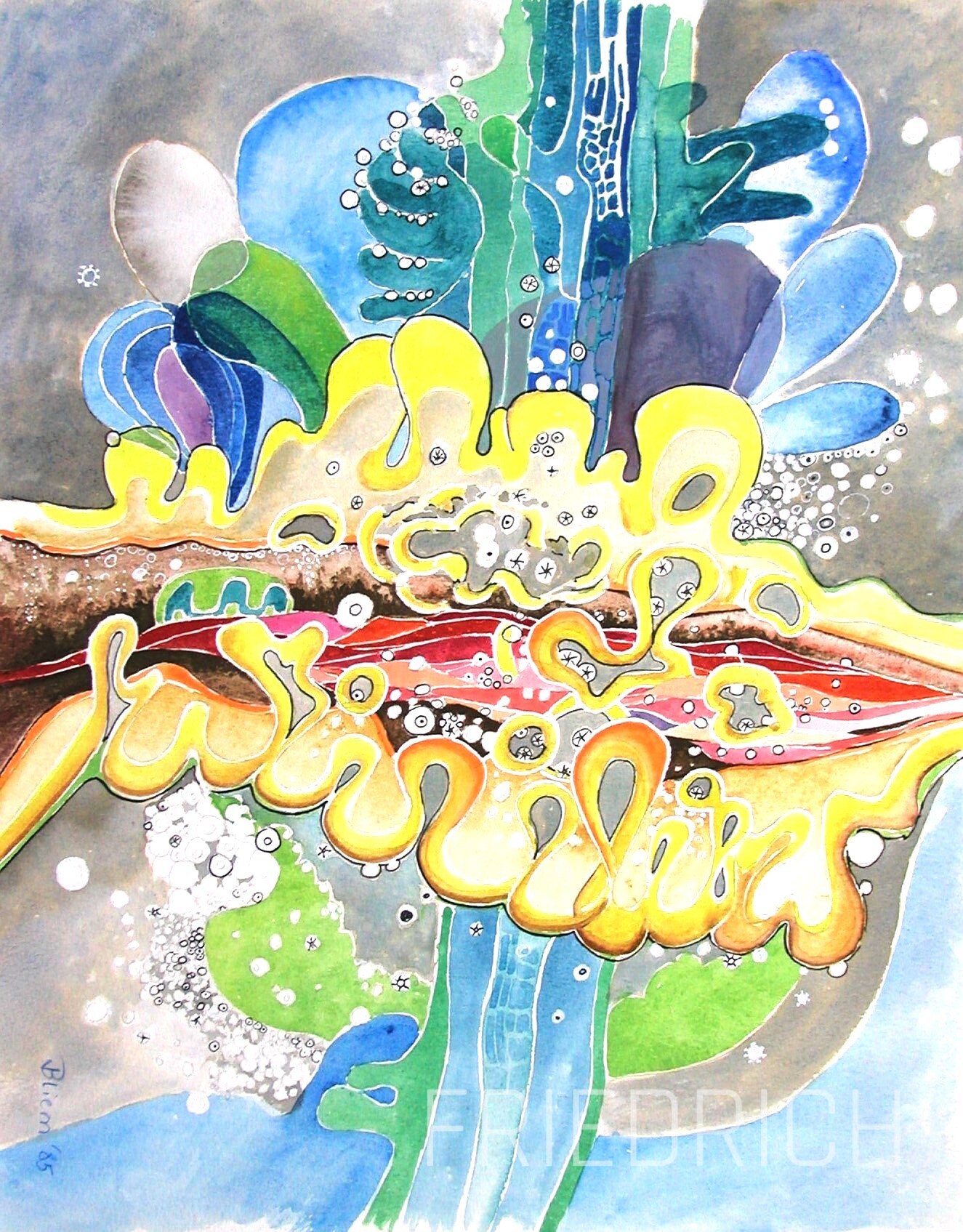
Cells Tissues Organs Watercolour, 30×45 cm 1987
Were you always going to be a scientist?
No, not really. Like so many youngsters I had a chemistry set and developed a fascination for chemistry, but the continuous smell of turps and oil in our house always drew me back to drawing and painting. Music was simply another form of expression. The intensive engagement in music led me to question the connection between music and art. I was convinced there was a physical connection, but also realised that I simply didn´t have the theoretical knowledge to proceed. This was the sole reason for choosing a study programme with a broad curriculum both in biology and engineering over art school.
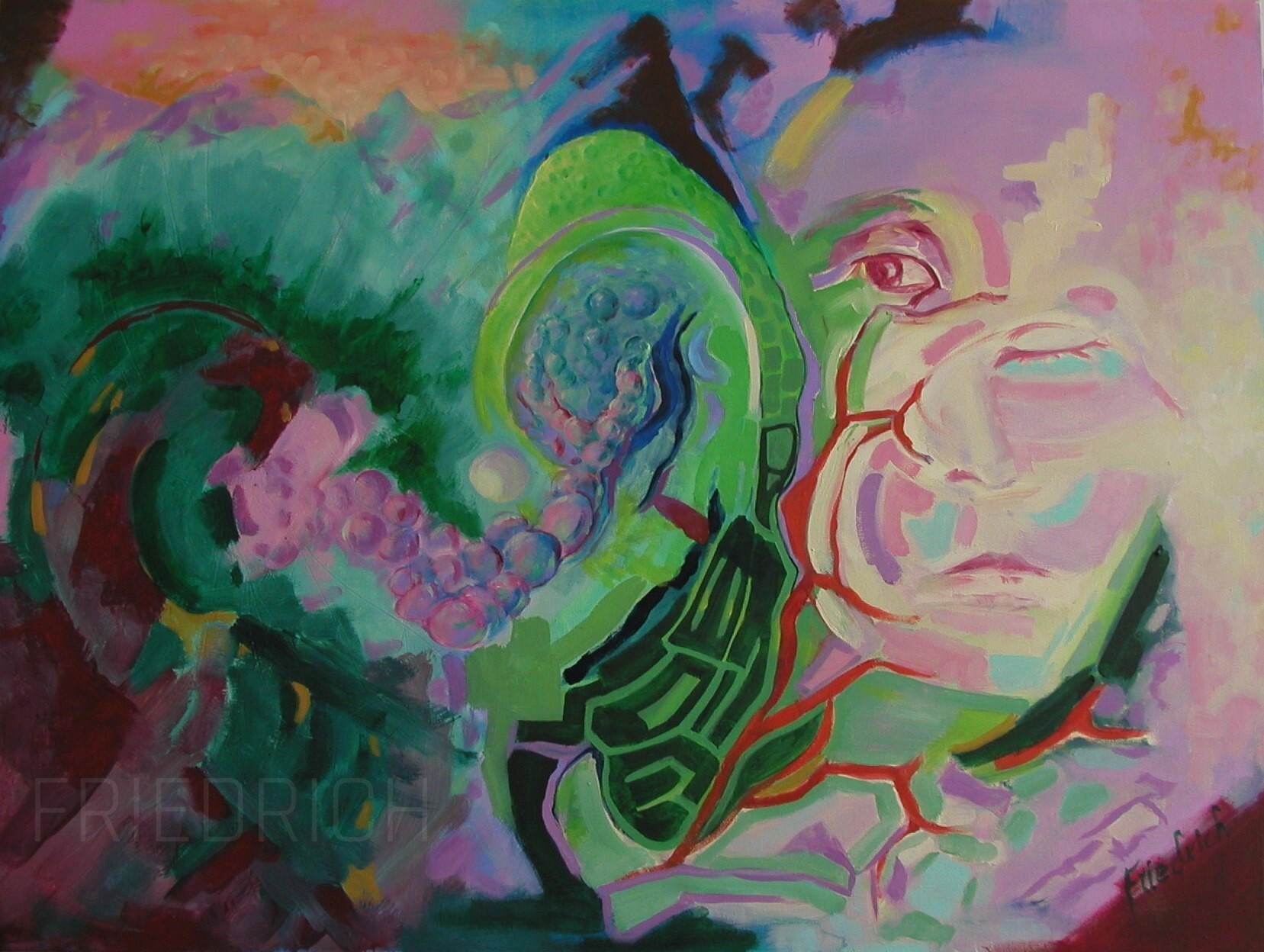
Evolution Oils on panel, 50×70 cm 2023
And what about art – have you always enjoyed it?
Well, yes and no. I wouldn´t speak of joy. My relationship with Art was set in a much deeper fashion as just to be enjoyed. It was an intuitive means of expression that came easily, be it fine art or music, which found some success, even in school days, and of course in which I reveled. But it also stemmed from an admiration for my father and our complex relationship.
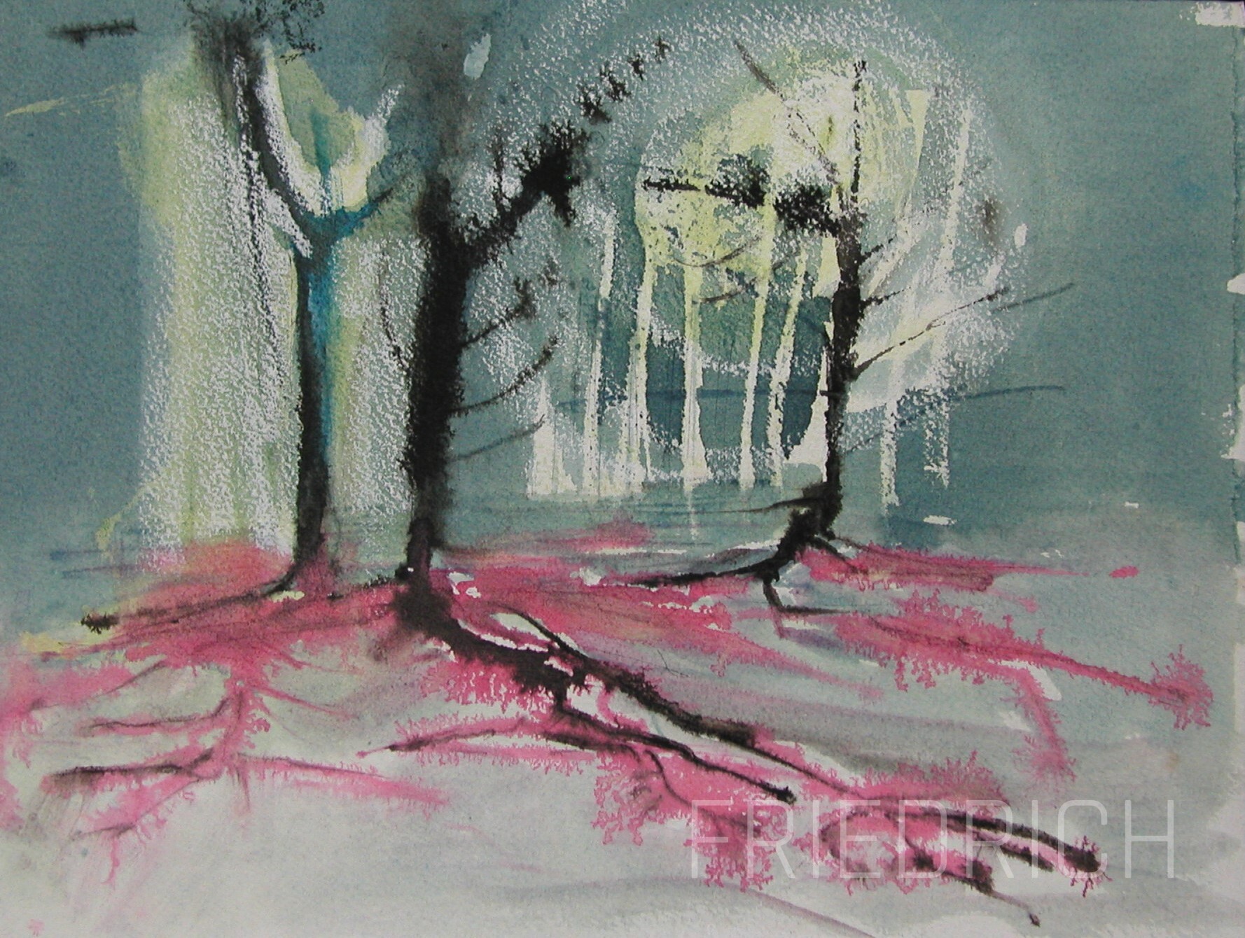
Fungal scene Watercolour, 30×40 cm 1988
What or who are your most important artistic influences?
Of course my father, whose work was largely impressionistic. And Rudolf Macek, who was clearly an expressionist of the 1950´s and 60´s. And from the public figures first and foremost they were and are Franz Marc, Wassily Kandinsky, August Renoir; painters of the romantic period, such as Ferdinand Waldmüller, and of course the old masters such as Jan Vermeer or Salomon van Ruysdael.
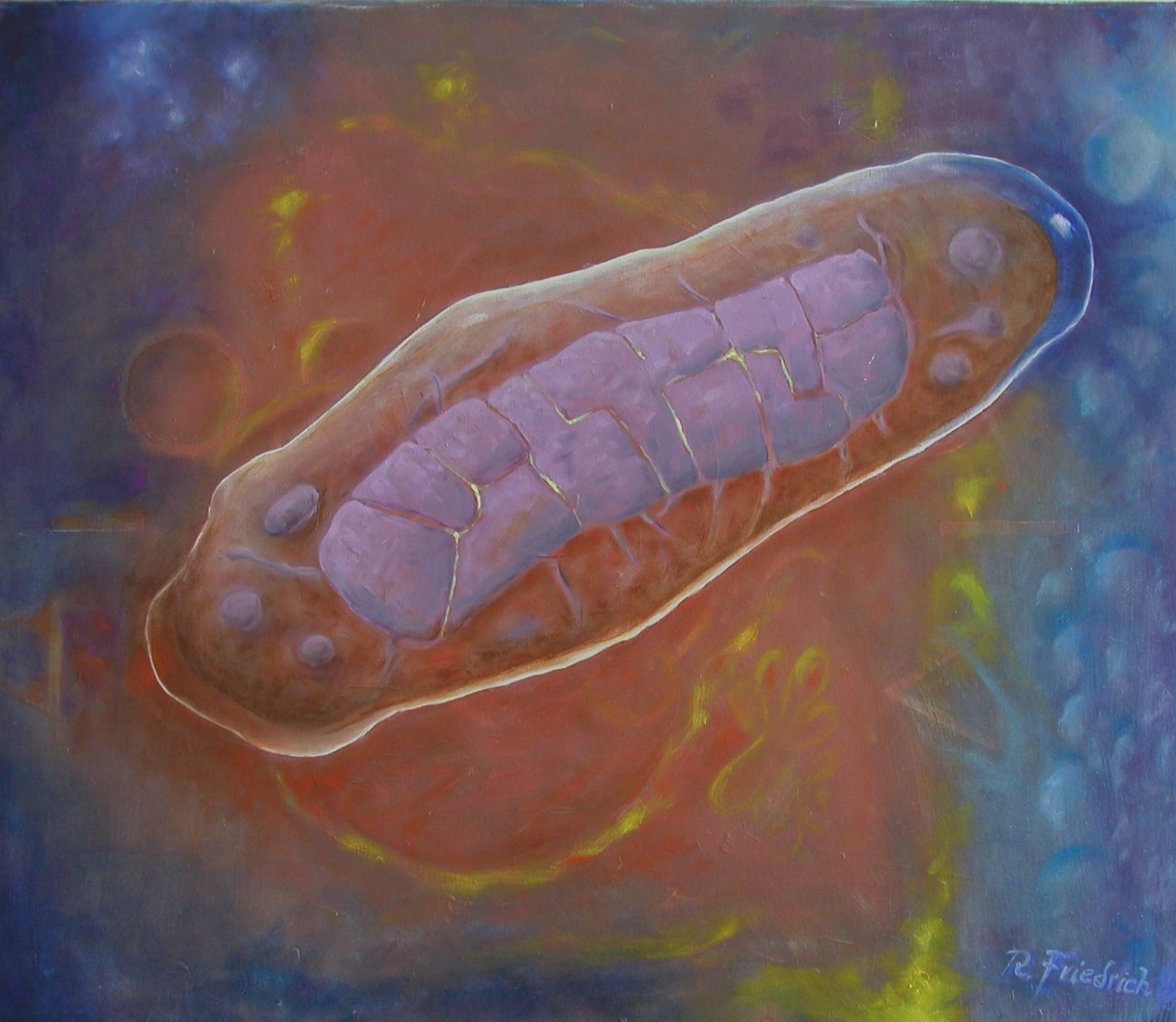
Mitochondrium 4 Oils on linen, 50×60 cm 2024
How do you make your art?
Today I only paint, on canvas or panels, in oils, sometimes with a draft drawing in acrylic. The Cambridge atmosphere lends itself to watercolours, which is well accepted.
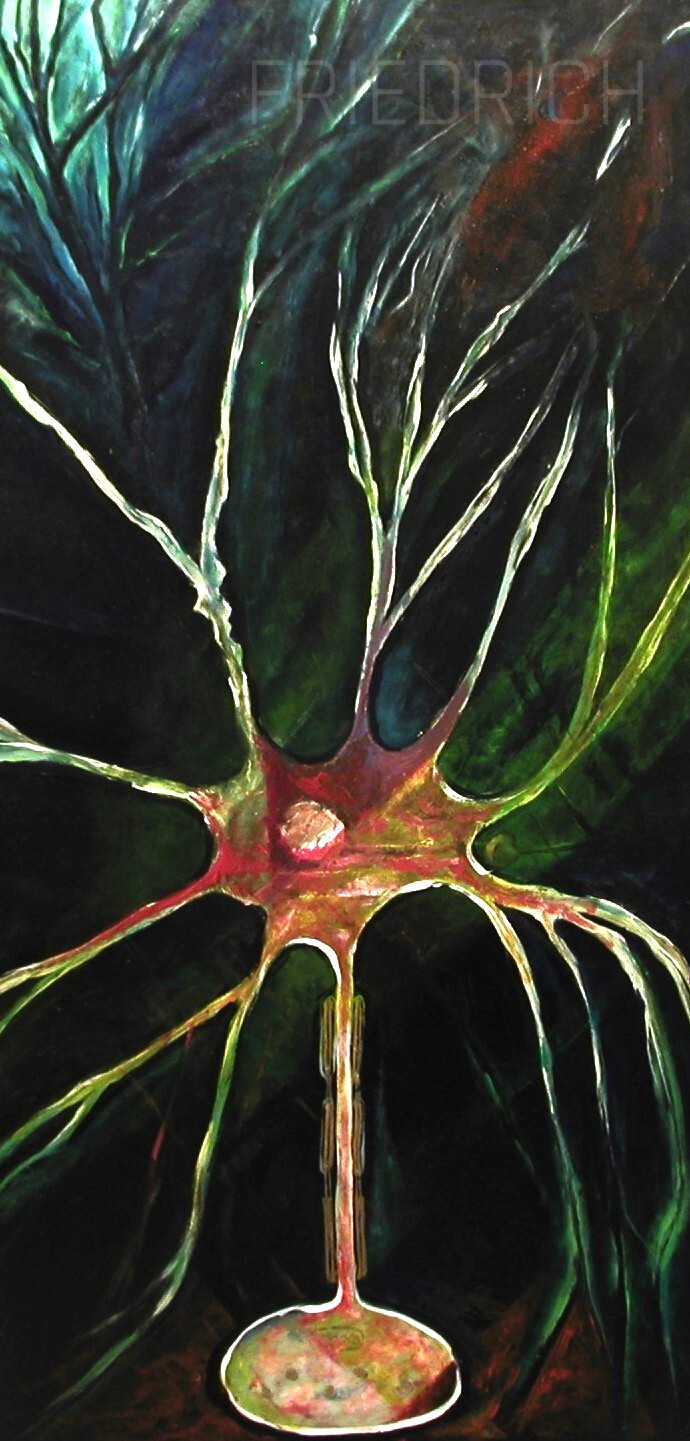
Neuron Acrylics and oils on panel, 70×135 cm 1985
Does your art influence your science at all, or are they separate worlds?
Whereas my scientific subjects definitely reflect my knowledge of cell biology, art has tuned my visual sensitivity to aesthetics in science and engineering, e.g. a factory with its piping systems or SEM photos of fungal hyphae and conidia. This has even, to some extent, determined significant professional choices I have made.
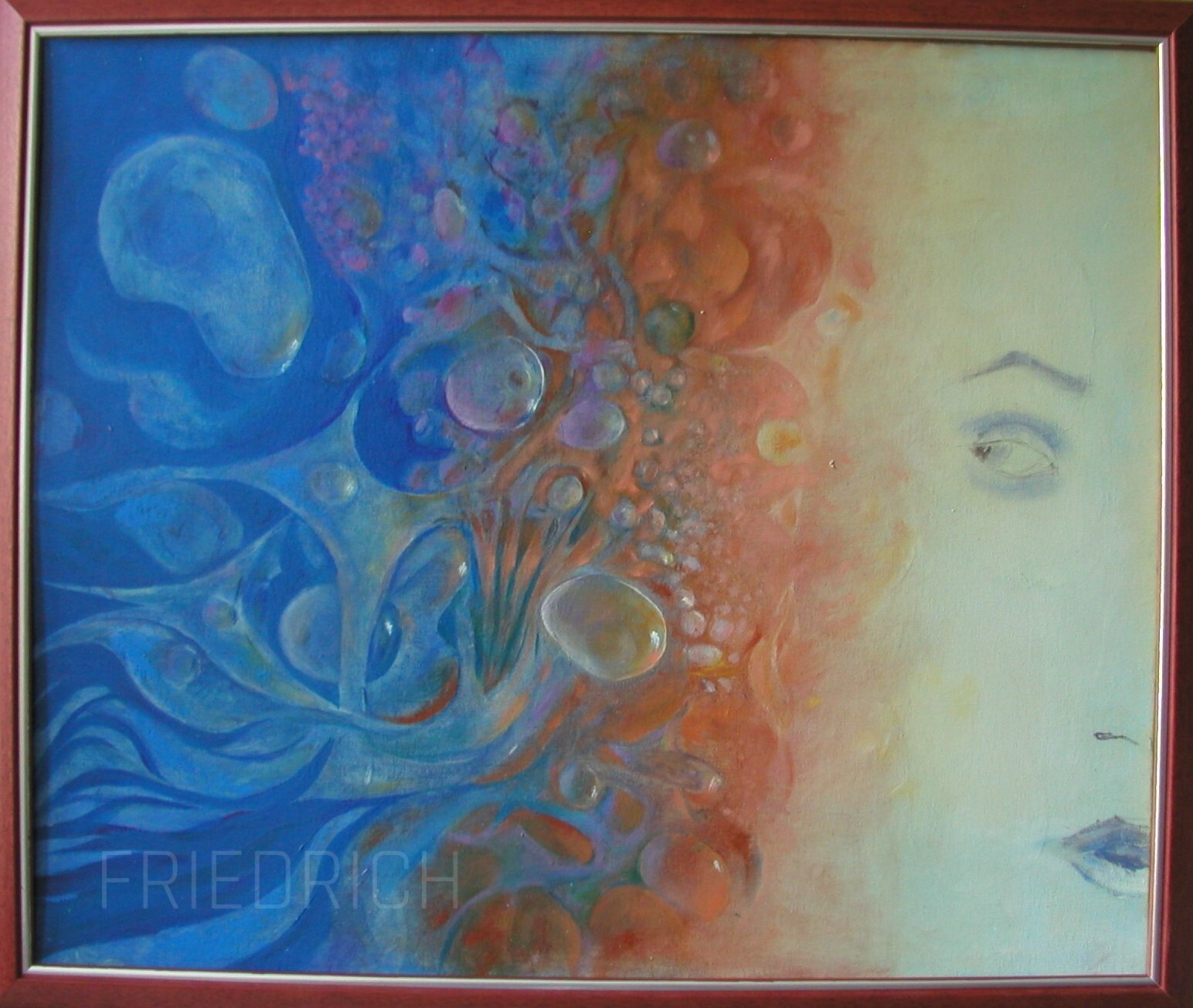
Compostion of the Self Oils on panel, 50×60 cm 2024
What are you thinking of working on next?
I will continue to expand on my scientific themes, one of which is the concept of evolution. But I also have a “macrobiological” subject, in which I seek to capture the essence of animals in their environment.
On the topic of plant development and chaired by Development Editor, Dominique Bergmann (Stanford University).
Wednesday 31 July – 16:00 BST
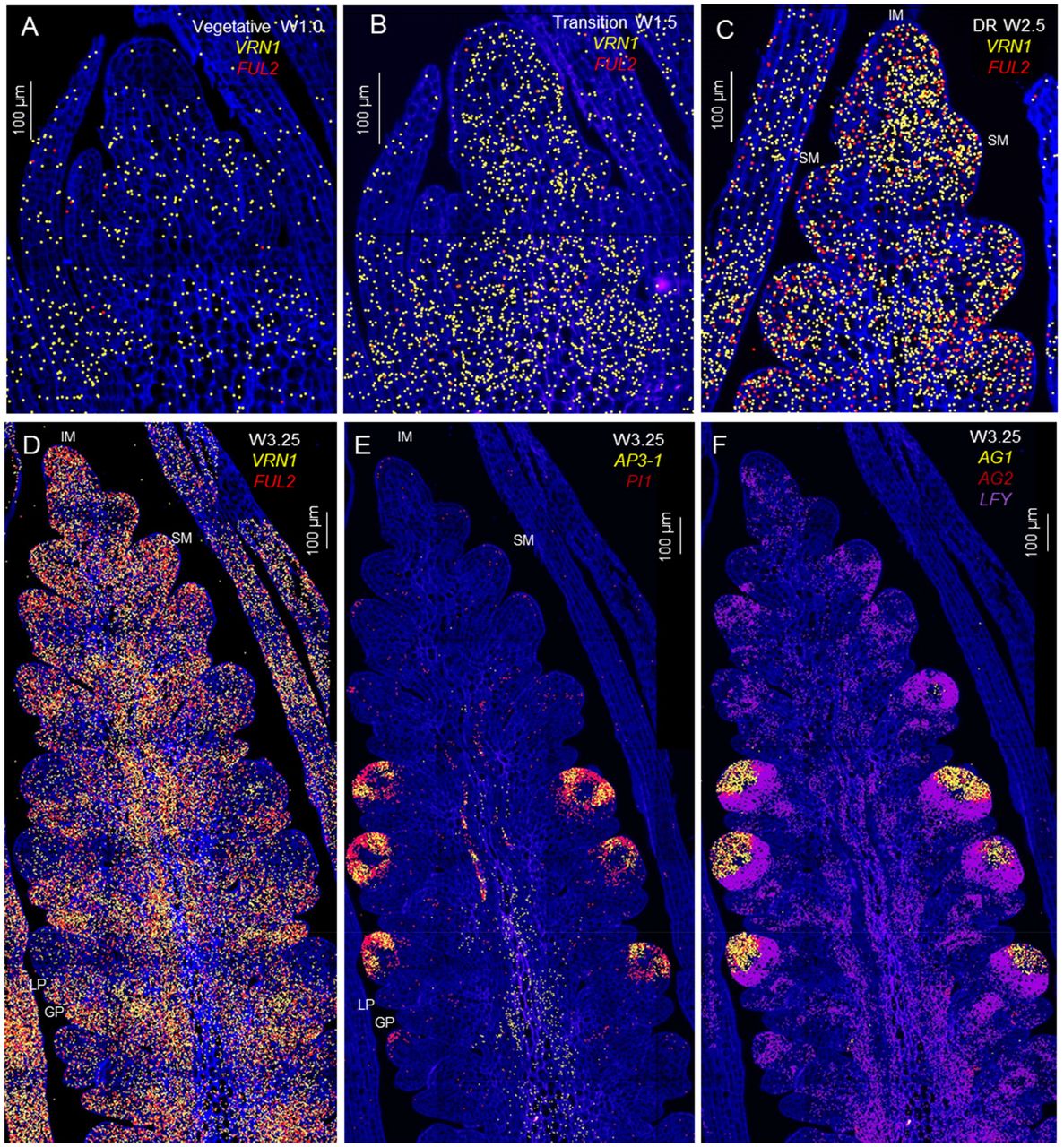
Martina Cerise (Max Planck Institute for Plant Breeding Research) ‘In plants, the organization of the apical stem-cell niche changes dynamically during the floral transition’
Vicky Spencer (Gregor Mendel Institute) ‘‘How to build a plant: Small meristems have big consequences’
Gwendolyn K. Kirschner (The James Hutton Institute) ‘Control of the root gravitropic set-point angle in barley’
At the speakers’ discretion, the webinar will be recorded for viewing on demand. To see the other webinars scheduled in our series, and to catch up on previous talks, please visit: thenode.biologists.com/devpres

 (2 votes)
 (No Ratings Yet)