“Shift work might be a real driver of health inequality; that these people are, as a function of their job, being forced to effectively mess with their biology”
Dr Priya Crosby
In the latest episode of the Genetics Unzipped podcast, we’re clocking in to chat about the genetics of circadian rhythms. How can molecules tell the time, why don’t we have drugs for jet lag yet and could a midnight snack stop malaria in its tracks?
Our November webinar will be chaired by Development’s Reviews Editor, Alex Eve, and features three early-career researchers studying the intersection between development and disease. The webinar will be held using Zoom with a Q&A session after each talk.
Tuesday 14 November 2023 – 14:00 GMT
Mauricio Rocha-Martins (Instituto Gulbenkian de Ciência) ‘How tissues orchestrate growth and morphogenesis: Lessons from the vertebrate retina’
Nicole Edwards (Cincinnati Children’s Hospital Medical Center) ‘Discovering the developmental basis of trachea-esophageal birth defects: evidence for endosomeopathies’
Cecilia Arriagada (Rutger’s University) ‘Role of mesodermal fibronectin in mechanotransduction during cardiac development’
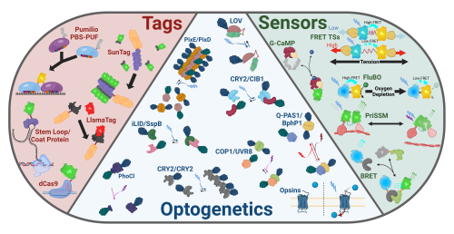
Writing in Nature, Bill Jia, Sean Megason, Adam Cohen, and colleagues ask the question: how does the heart go from silent to beating during embryonic development? Using genetically encoded sensors and optogenetic tools, they captured the very first heartbeat of a zebrafish embryo and dissected the biophysical basis for its timing and spatial structure. We chatted with first author Bill Jia to hear the story behind the paper.
How did you come to join the labs of Adam Cohen and Sean Megason?
Early in my PhD, I developed a fascination with how cells compute as a collective. Embryonic development jumped out to me as an incredible example of this phenomenon – cells must talk to each other and then make their own decisions based on the inputs they receive, yet the outcomes of these decisions are often patterns and functions at a level of organisation higher than the individual. The Megason lab’s goal is to identify the conceptual rules by which this comes to be, so it was a perfect fit. However, to understand the complex messages that cells send to each other, it is critical to find the channels through which this information is passed – what exactly are cells measuring? In developmental biology, a major emphasis has traditionally been placed on signal transduction of chemical morphogens. But this is just one of many processes that constitute cellular physiology, each with its own time and length scales over which it can transmit or process information. The Cohen lab has pioneered optical approaches to measure and perturb different aspects of cellular physiology. It also focuses on neuroscience, another discipline concerned with complex and collective computation which happens to exemplify the idea that patterns with different spatiotemporal scales can carry information in biology. I wanted to bring together the distinct strengths and shared interests of these labs. This has put me in a unique position to study developmental cell-cell communication and pattern formation in media that have historically been relatively inaccessible to biological inquiry – bioelectricity, second messenger signalling, and others.
Movie 1: Calcium dynamics across zebrafish gastrulation, convergence, and extension, recorded using the sensor jGCaMP6s.
How did the project get started?
We started out with a broad survey of different physiological dynamics in zebrafish development, using biosensors to look for patterns that might be correlated between cells across time and space. Our efforts imaging cellular calcium were particularly successful (Movie 1)! Consistent with previous work from the Miller lab and others (1), we found an enormous diversity of calcium transients in different cell types over early development. We struggled for a while to decide which of these patterns would be worth following up on because there were simply so many. Adam proposed looking at the first heartbeats because the purpose of the calcium dynamics in this system was known, so it would just be a matter of figuring out how they appeared. At first, I was concerned that too much would have been previously studied, but the literature suggested that there were still some major mysteries to be solved.
What was known about the emergence of the first heartbeat in vertebrate embryos before your work?
The question of how the heartbeat emerges can be broken down into a few pieces. At what time in development does it happen? What are the roles of different cells in the process? How does the heart first gain its regular timing, and how is its function coordinated across the entire tissue? It was already known that periodic electrical and calcium activity emerges in the heart much earlier in development than it is needed (e.g. for blood flow) (2). It was also previously suggested, based on surgical separation, dissociation, or genetic perturbation of heart morphogenesis, that most early cardiomyocytes had the capacity to beat on their own and that the fastest cell would synchronise the tissue (3–5). But reports differed on the exact details of these observations, which we think mainly owes to two reasons.
First, individual heartbeats are very fast compared to the progression of development – it is difficult to ascertain from a short recording of a few beats how far along the process any given animal is. As we saw in our measurements, there could be very large changes in the dynamics over very short developmental periods. Second, disruption of the tissue geometry by the methods described is also highly disruptive to electrophysiology, making any observed activity preparation-dependent. One simple and instructive example is to imagine a cell that expects to be coupled to its neighbour by gap junctions – after dissection of that interface, it now has hemichannels exposed to the extracellular fluid instead, which might completely change its electrical behaviour. Notwithstanding these potentially contradictory previous results, there was also no proposed mechanism for how the heart gains its timing, whose quantitative properties are integral to the organ’s function.
Can you summarize the findings of the paper?
Movie 2: Multiplexed recordings of the heart’s transition from silent to beating in zebrafish embryos, taken with the sensor jGCaMP7f (6).
First, we used long, multiplexed recordings of genetically encoded calcium sensors to efficiently capture the transition from silent to beating (including the very first heartbeat!) in many zebrafish hearts (Movie 2). This was a dramatic and sudden switch, which in combination with subsequent experiments to probe biophysical and molecular mechanism gave us confidence that something important had happened at that very moment. We then looked at the statistics of the beats after the first one and found that the trajectories of average beat rate and beat rate variability were quite stereotyped. This lent itself to a simple mathematical abstraction describing how oscillations can emerge from an apparently silent system, called a stochastic saddle-node on invariant-circle (SNIC) bifurcation.
This model has two important predictions – first, highly periodic oscillations can be driven by random noise; second, big changes in fast dynamics (beats) can emerge from very small changes in an underlying control parameter describing a decreasing distance between a resting state and a “threshold” for a single oscillation. This second idea, of small changes in one quantity resulting in big changes in another, is analogous to a miniscule change in temperature across the freezing point turning water into ice – which is why we describe it as a phase transition. Similar phenomena have been observed in tissue mechanical properties in development (7, 8), and perhaps more await discovery.
The abstract mathematical resting state and threshold described have a biological basis in the membrane voltage dynamics of many types of electrically excitable cells, including cardiomyocytes. We tested whether this was relevant in the earliest heartbeats by performing voltage imaging and perturbing various ion transporters. In a panel of several genes important in the adult spontaneous cardiac action potential, we found that only the L-type voltage-gated calcium channel (VGCC) was required for both voltage and calcium oscillations. This is consistent with the model – the voltage threshold for activation of the VGCC could be the threshold described in the equations.
We then used light-activated ion channels (channelrhodopsins) to see if we could optogenetically cross the predicted threshold before any endogenous activity could be observed. As the model predicted, the heart became increasingly easy to excite as the first spontaneous beat approached. Furthermore, local excitations could propagate across the tissue, explaining how many cells seemed to turn on all at once from the first beat (Movie 3) – the cells are primed and ready to respond to their neighbours.
Movie 3: Tissue-scale initiation of calcium activity (cyan) in the zebrafish heart cone (red), taken with calcium sensor jGCaMP7f.
Our observations on how the heart’s earliest temporal patterns were driven by noise and how the spatial patterns were always synchronised demanded a more careful analysis of functional specialisation in individual cells. If the threshold crossing is random, what permits a particular cell to do it? We know that the adult heart has a defined pacemaker region, and we knew from previous work where these cells ought to be at the time of the first heartbeat. Does the beat start in the same place every time, and is this where the pacemaker will be? How does the previously observed widespread spontaneity play into all of this? We performed imaging of the calcium activity together with markers defining cardiomyocyte subpopulations, showing that the spatial origin was indeed variable, but tended to start away from the future pacemakers. Using optogenetics we were able to establish that a fast intrinsic beat rate was in fact the biophysical mechanism that set the spatial origin.
Together our results provide a picture of how many aspects of heartbeat initiation fit together. Random noise allows the heartbeats to emerge from variable positions, but the cells are synchronised by frequency-based competition. This is likely implemented molecularly by a gradual increase in activation sensitivity of VGCCs. This process does not seem to be tightly coupled to the earliest steps of pacemaker development, but the biophysical mechanisms are sufficient to ensure early coordination and periodicity of the heartbeat.
Were you surprised to find that the individual heart cells abruptly start beating all at once?
Yes! Before we did the experiments, the analogy I had in my head was a swarm of fireflies. I was expecting to see individual cells blinking separately, with some sort of synchronisation process occurring gradually over time. How the heartbeat initiation could hypothetically play out depends on the relative ordering of the development of excitability, electrical coupling, and spontaneous oscillations – in my picture, the coupling would have had to emerge last. There is evidence that connexins are expressed very early on in heart development (9), so maybe the tissue-scale initiation ought to have been less surprising. But the dependence of the spatiotemporal dynamics on each of these features is quantitative, so someone who had started with that prior assumption could have just as likely been wrong – we had to do the measurements to find out!
Do you think this bioelectrical phase transition model is found across all developing vertebrate hearts?
It is very possible, given that many features of the transition have been suggested by other observations in different vertebrate models. The same L-type calcium channels are known to be required for early cardiac activity in mice (10), and similar observations of lability in the spatial origin of the heartbeat have been seen in chicks and mice (3, 10). One major way other species might differ is in the length scale of synchronisation. In animals with larger hearts, it may be possible to observe multiple regions of synchronisation with their own distinct origin points.
The paper involves optogenetic manipulations and live functional imaging, producing a lot of beautiful videos. Can you tell us more about the techniques and tools you used?
Many of our conclusions rested on measuring how the dynamics of the heartbeat changed in their response to fast perturbations over the slow timescales of development. To do this, it was essential to simultaneously manipulate and image the electrical activity without disrupting the heart’s developmental trajectory. A key enabler of these experiments was FR-GECO1c, a novel far-red calcium sensor developed by the labs of Robert Campbell and Yi Shen (11). This sensor offered spectral compatibility with several blue light-activated ion channels (channelrhodopsins), which allowed us to target the heart as accurately as light can be patterned. Using these tools, we reversibly stimulated or silenced the heart at scales of milliseconds and micrometres, while watching its response in real time. There has been a recent explosion in new optogenetic tools for manipulating and visualising cellular physiology beyond membrane potential, including activation of canonical signalling pathways (12). I think that these will push the field of developmental biology forward tremendously, as they open up causative studies of the spatial and temporal couplings that ultimately underlie the progression of organ patterning.
Can you postulate the function of early electrical activity even before the heart is connected to the circulatory system?
It could be a type of developmental checkpoint for later steps in heart development – now that the heart has established its basic electrical function, it is ready to perform later steps like specification of cell subtypes or the complex steps of morphogenesis involved in chamber formation. The distribution of spontaneity could be a mechanism of robustness in this process – if one cell that was supposed to drive the beat fails to develop, another one can take its place. One particularly interesting hypothesis is that calcium signalling downstream of the beats supports development (10, 13). Given that calcium is an important second messenger molecule for many cellular processes, it is unclear whether it is the calcium fluxes downstream of the beat that matter and whether these must be deconvolved from some other cellular calcium dynamics. This is a problem that applies to all second messenger signalling pathways, so the developing heart might be a useful model to teach us more about the general mechanisms of cellular computation.
Did you have any particular result or eureka moment that has stuck with you?
The success of the quantitative fit of our data by the SNIC bifurcation model was very gratifying. It’s really satisfying to me when important features of a complex biological process can be distilled down into a model that is simple, yet precise enough to be written as a mathematical formula. But one of my favourite things about this story was how interdisciplinary the findings are – I really believe there is a little something for everyone in there. It’s super fun to see feedback from developmental biologists, cardiologists, and physicists expressing excitement about completely different parts of the results.
And the flipside: were there any moments of frustration or despair?
At times I thought that our findings were too simple or obvious given what was already known about the developing heart. But something I’ve come to appreciate is that this is very subjective and depends strongly on how much one is willing to make assumptions based on what is already known.
What’s next for this story? And what’s next for you personally?
I think there are two major future directions for this story. The first is to understand the implications of the early self-organising activity for the heart’s development, as we discussed. The second is to understand in more depth the molecular basis of the predicted biophysical mechanism. How are the VGCCs being sensitised? Is it because of changes in channel expression levels, relative intracellular and extracellular calcium levels, changes in resting potential by modulation of other ion pumps and channels, or something else? What is the source of the noise that seems to drive the timing? Personally, I am nearing the end of my PhD. I hope to continue along the lines of the first direction – how do properties of biological electricity instruct cell decision making and tissue patterning in development? I want to do so by opening my own lab but am also thinking about postdoctoral positions that might allow me to pursue these questions.
References
1. S. E. Webb, R. A. Fluck, A. L. Miller, Calcium signaling during the early development of medaka and zebrafish. Biochimie. 93, 2112–2125 (2011).
2. W. Rottbauer, K. Baker, Z. G. Wo, M.-A. P. K. Mohideen, H. F. Cantiello, M. C. Fishman, Growth and Function of the Embryonic Heart Depend upon the Cardiac-Specific L-Type Calcium Channel α1 Subunit. Dev. Cell. 1, 265–275 (2001).
3. T. Sakai, T. Yada, A. Hirota, H. Komuro, K. Kamino, A regional gradient of cardiac intrinsic rhythmicity depicted in embryonic cultured multiple hearts. Pflüg. Arch.437, 61–69 (1998).
4. J. F. Reiter, J. Alexander, A. Rodaway, D. Yelon, R. Patient, N. Holder, D. Y. R. Stainier, Gata5 is required for the development of the heart and endoderm in zebrafish. Genes Dev.13, 2983–2995 (1999).
5. R. L. DeHaan, Synchronization of pulsation rates in isolated cardiac myocytes. Exp. Cell Res.70, 214–220 (1972).
6. B. Z. Jia, Y. Qi, J. D. Wong-Campos, S. G. Megason, A. E. Cohen, A bioelectrical phase transition patterns the first vertebrate heartbeats. Nature, 1–7 (2023).
7. A. Mongera, P. Rowghanian, H. J. Gustafson, E. Shelton, D. A. Kealhofer, E. K. Carn, F. Serwane, A. A. Lucio, J. Giammona, O. Campàs, A fluid-to-solid jamming transition underlies vertebrate body axis elongation. Nature. 561, 401–405 (2018).
8. N. I. Petridou, B. Corominas-Murtra, C.-P. Heisenberg, E. Hannezo, Rigidity percolation uncovers a structural basis for embryonic tissue phase transitions. Cell. 184, 1914-1928.e19 (2021).
9. N. C. Chi, R. M. Shaw, B. Jungblut, J. Huisken, T. Ferrer, R. Arnaout, I. Scott, D. Beis, T. Xiao, H. Baier, L. Y. Jan, M. Tristani-Firouzi, D. Y. R. Stainier, Genetic and Physiologic Dissection of the Vertebrate Cardiac Conduction System. PLOS Biol.6, e109 (2008).
10. R. C. Tyser, A. M. Miranda, C. Chen, S. M. Davidson, S. Srinivas, P. R. Riley, Calcium handling precedes cardiac differentiation to initiate the first heartbeat. eLife. 5, e17113 (2016).
11. R. Dalangin, M. Drobizhev, R. S. Molina, A. Aggarwal, R. Patel, A. S. Abdelfattah, Y. Zhao, J. Wu, K. Podgorski, E. R. Schreiter, T. E. Hughes, R. E. Campbell, Y. Shen, bioRxiv, in press, doi:10.1101/2020.11.12.380089.
12. H. M. McNamara, B. Ramm, J. E. Toettcher, Synthetic developmental biology: New tools to deconstruct and rebuild developmental systems. Semin. Cell Dev. Biol.141, 33–42 (2023).
13. N. D. Andersen, K. V. Ramachandran, M. M. Bao, M. L. Kirby, G. S. Pitt, M. R. Hutson, Calcium signaling regulates ventricular hypertrophy during development independent of contraction or blood flow. J. Mol. Cell. Cardiol.80, 1–9 (2015).
We are pleased to invite all members of the biomedical community to:
Medicine at the Crick: What can development tell us about disease?
Thursday 2nd November 2023, 14:30-18.00
Free hybrid event taking place virtually and in person, organised by The Francis Crick Institute, London
Our Medicine at the Crickevent series showcases major advances in biomedical science and raises awareness of the medical implications of major scientific advances amongst the Crick and wider UK biomedical community. Each event comprises a series of talks and a Q&A panel discussion, followed by a networking and drinks reception for in person attendees.
More about our next event
Hosted by Alex Gould, this 12th edition of Medicine at the Crick will focus on recent advances in our understanding of how embryonic development influences adult disease.
The early-life environment is well established to impact upon infant health and disease. It is less widely known that it also influences the risk of adult diseases manifested many decades later. Developmentally “programmed” adult diseases are diverse and include type 2 diabetes, asthma, cardiovascular disease, neurodegenerative conditions and even some cancers. Research in this area, the developmental origins of health and disease (DOHaD), is clinically important but surprisingly still not on the radar of many developmental biologists. This Medicine at the Crick event aims to raise awareness of DOHaD research. It will highlight emerging organogenic and epigenetic links between embryonic development and adult disease. It will explore how diseases such as cancer may involve inappropriate reruns of developmental genetic programmes. Key impacts of DOHaD research on public health policy will also be discussed.
The panel discussion will be followed by a networking and drinks reception for in person attendees until 19:00.
Registration
Please find attached a poster for the event. Visit our webpages for further details including a more detailed programme, andregister via Eventbriteto order a free virtual or in person ticket for the event.
Please send any questions to: medicine-at-crick@crick.ac.uk and feel free to circulate this information among any colleagues who may be interested.
We look forward to welcoming you to our Medicine at the Crick event.
“We can’t rely on studying flies and mice and fish,” said Katherine Brown, Executive Editor of Development, in her opening remarks at the journal’s annual meeting. “We really need to be expanding our repertoire.”
From Sep. 17–20, I joined nearly 100 developmental biologists at the Wotton House to learn from researchers working with unconventional experimental organisms in cell and developmental biology. In those four days, we caught glimpses of how these emerging systems can bridge gaps in scientific knowledge and between scientific disciplines. The issues discussed were complex and unlikely to be settled in a single conference. But this seminal meeting signaled a readiness for the field of developmental biology to 1) broaden its taxonomic sampling, 2) educate scientific gatekeepers to the benefits and challenges of working with unconventional systems, and 3) expand its vision to include the impacts of climate change.
We need broader taxonomic sampling
“Can we even say we know what a eukaryote is? I’m not sure.”
Iñaki Ruiz-Trillo, Institut de Biologia Evolutiva, CSIC, Spain
Researchers who are devoted to unconventional and emerging organisms are especially sensitive to the need for wider taxonomic sampling. Attendees of the meeting represented nearly 50 species ranging from single-celled choanoflagellates to cnidarians to honeydews to eusocial insects to mollusks to geckos. Several talks ended with speakers thanking their experimental organisms — an overt acknowledgment of how important biodiversity is to unravel the biological mechanisms that contribute to development.
And yet, despite the “biodiversity” buzz word percolating through the scientific literature, the classical models of biological research are still primarily limited to just three percent of eukaryotic lineages: animals, fungi, and plants.
“Can we even say what a eukaryote is?” Iñaki Ruiz-Trillo asked in the opening session. “I’m not sure when it’s three percent.”
We were reminded over and over how unconventional organisms show us what we don’t know. While knowing the limitations of our knowledge is important for intellectual growth, it’s a tough premise for convincing funding agencies and taxpayers to support basic research that is inherently challenging and at high risk of failure. One way to overcome that hurdle is to reframe the discussion.
“You don’t ask: ‘What can this model tell me?’ but ‘What can I ask this model?’” said Ruiz-Trillo, highlighting how research should start with the biological question, not necessarily the organism. This idea was reiterated by evolutionary biologist Kim Cooper at the meeting when she described the extreme skeletal morphology of jerboas (affectionately referred to as “potatoes on sticks”) to probe deeper into bone development.
“Unconventional organisms are important,” said Ruiz-Trillo. “They allow us to see things we could not see before. They allow us to see things we didn’t even know we could not see before.”
The lack of tools available to researchers studying unconventional systems was a source of commiseration with attendees. But the outlook was more optimistic than grim. As Magdalena Bezanilla reminded us: “We’re a small, robust community. But we share.”
We need to educate the field about challenges inherent with unconventional systems
“My least favorite beginning of any [reviewer’s] comment is, ‘Why don’t you just… ?'”
Kim Cooper, UC San Diego, USA
As a general rule, researchers working with unconventional systems have trouble gaining traction with researchers studying more traditional model organisms. Unconventional systems lack resources and are riddled with unknowns — that’s all part of the intrigue of basic science research. But it’s also extremely frustrating when, after sharing exciting results with reviewers, our manuscripts come back with follow-up experiments that just aren’t possible in certain unconventional systems.
“My least favorite beginning of any [reviewer’s] comment is, ‘Why don’t you just… ?’” said Cooper.
Evolutionary germ cell biologist Cassandra Extavour reminded the professional scientists at the conference that they are the reviewers. It’s easy to forget. “When we’re reviewing a paper, we are a helpful colleague providing constructive input into the field. But when we’re an author, the reviewers are these hateful aliens who hate us,” she said, prompting laughter from the audience. “But it’s us, right? As reviewers, I would ask us all to remember that we’re not trying to apply the standards of one field to another unnecessarily but asking, what are the insights that this paper offers?”
Extavour went on to argue that we must educate ourselves to the standards of data production and presentation and analysis in different fields so that we can assess author claims. It’s simply not reasonable to expect mouse techniques in a paper about choanoflagellates. Reviewers need to be asking: 1) Are the data being rigorously applied? 2) Are the authors’ conclusions supported by the data they provide? and 3) Do the authors’ conclusions or insights match the vision or the mission of the journal?
Abouheif suggested that journal editors can help curb some of those unrealistic expectations from reviewers. “You have to kind of let some things go,” he said. “It’s hard to find the sweet spot. But yeah, we have to advocate for it.” Because unconventional research systems have a lot to offer.
We can (and should) contribute to climate change research
“Developmental systems may promote phenotypic innovation and complexity in the face of climate change.”
Ehab Abouheif, McGill University, Canada
One of the motivations for this year’s Development meeting was to gather developmental biologists together to discuss what role, if any, the field of developmental biology should have in addressing climate change. The fields of developmental biology and climate research might seem unrelated at first glance. But everyone agreed that climate change is multi-faceted and spans scientific fields. We need all hands on deck. The main question, then, is what can we, as developmental biologists, contribute?
“You have to be comfortable with being a jack of all trades but the master of none,” Ehab Abouheif said, reminding us that working with non-model organisms is integrative by nature. There’s no simple answer to solving climate change, meaning that multi-pronged approaches are warranted.
“We don’t know what the solution is,” said plant developmental geneticist Michael Raissig. “So we have to look everywhere.”
Loss of biodiversity was brought up, tempered somewhat by Abouheif, who said, “Developmental systems may promote phenotypic innovation and complexity in the face of climate change.” He went on to say that ecologists may not typically think of phenotypic predictive theories. But developmental biologists are constantly teasing apart what genotype results in adaptive phenotypes, suggesting that developmental biology may have a lot to say in the coming years.
Raissig pointed out that three challenges of climate change are catastrophic events, biodiversity loss, and agriculture. But there seems to be a general “plant blindness” in society, said Raissig. “I would argue it’s the photosynthetic organisms we should focus on,” he said, bearing in mind his biased perspective as a plant biologist.
The panelists discussed the responsibility developmental biologists have in addressing climate change, raising the question of whether we can afford curiosity-driven research. Cooper pointed out that developmental biologists’ contributions can extend beyond the lab bench. Most academic researchers have teaching responsibilities, she said, with a (generally) captive audience eager to learn and contribute to society. Discussions of how climate change has shaped earth’s biosphere over eons could be leveraged in the classroom.
“Basic, fundamental science is important for society,” Abouheif said. I doubt anyone at the meeting disagreed. But how do we share that with decision makers who may not be as gracious with so-called “basic” science for its lack of apparent utility? Joyce Yu recently shared some thoughts on this matter. Check out what she has to say about how to more effectively communicate basic science.
“Communicating what we do and how we do it can be really challenging,” said Cooper. “But it’s rewarding, especially when you’re the most different speaker at a meeting. It’s like a coffee break in the middle of a session.” This year’s Development meeting had the buzz of a giant, fun coffee break where we could share our woes and our triumphs from the lab bench. Now we can get back to work, energized and refreshed.
“Most of our genome – a staggering 2 metres of DNA in every cell – is packaged into X-shaped chromosomes. And then there’s the weird stuff.”
Dr Kat Arney
In the latest episode of the Genetics Unzipped podcast, we’re exploring the weird and wonderful world of extrachromosomal DNA – what it is, what it does, and why it breaks the normal rules of inheritance.
If you enjoy the show, please do rate and review on Apple podcasts and help to spread the word on social media. And you can always send feedback and suggestions for future episodes and guests to podcast@geneticsunzipped.com Follow us on Twitter – @geneticsunzip
In this SciArt profile, we caught up with Ana Beiriger, a medical illustrator and a developmental biologist. We approached Ana after learning that she was the illustrator of the poster for the 2023 Society for Developmental Biology (SDB) Annual Meeting!
Poster illustration for the SDB Annual Meeting. Created using Procreate and Adobe Illustrator. 2023.
Can you tell us about your background and what you work on now?
My background is pretty solidly in developmental biology. I received my bachelor’s degree from the University of Illinois in 2013, where my first independent research project focused on comparative limb development in marsupials. That had me hooked, and I went on to earn my doctorate in developmental biology from the University of Chicago in 2020. In my doctoral work, I studied the birth and migration of cranial efferent neurons in the zebrafish, using lightsheet microscopy and 2-photon photoconversion techniques to characterize the development of the facial branchiomotor (CNVII) and octavolateral efferent neurons (CNVIII).
Towards the end of my doctorate, I found out about the field of scientific visualization from a colleague. I had always loved the arts and was already doing a lot of illustration work for my labmates, helping them out with figures for review articles and talks. The process of taking a complicated scientific topic and boiling it down to a clear visual message was both challenging and rewarding, and I loved the collaborative nature of the work. It took a lot of reflection, but after a decade in research I decided to pivot to something new.
After my doctorate, I did a deep dive into scientific art at UIC’s Biomedical Visualization (BVIS) program, building my skills in digital illustration, graphic design, storyboarding, and 2D/3D animation. I now work as a project manager for a scientific visualization company, occasionally taking on freelance projects to make graphical abstracts and illustrations for journal articles. It’s a lot of fun, but old habits die hard, and embryos are (predictably) still one of my favorite things to illustrate.
Selected stills from an animation on neurocristopathies. Created in Autodesk 3ds Max, Zbrush, and Adobe After Effects. 2022.
Were you always going to be a scientist?
I always figured I’d do something related to medicine or biology. When I was four, I wanted to be a “bone doctor” and help people heal from fractures. While I didn’t quite go the medical route, I did get to work on long bone development many years later, and I think four-year-old me would have been pretty excited about that.
Procedural illustration describing a suturing technique used for reconstruction of the fibularis tendon in the ankle. Created using Procreate and Adobe Photoshop. 2021.
And what about art – have you always enjoyed it?
I have loved the arts ever since I was very young. I’d taken drawing and painting classes on and off over the years but never felt like I had quite enough time for them among my other studies. It wasn’t until I realized there was a way to combine my love of art with science that I really gave myself permission to pursue the arts. During the last two years of my doctoral work, I spent mornings in the lab, afternoons writing, and evenings in the art studio. It was a hectic time, but perhaps one of the most personally fulfilling for me.
Illustration of a T6 vertebra. Pencil on toned paper. 2020.Illustration of a bird skull. Pencil on toned paper. 2018.
What or who are your most important artistic influences?
This is a hard question because there are so many artists that I admire! I’ve always adored expressionists like Marc and Kandinsky (in the early years) for their use of color, and more abstract artists like Moholy-Nagy and, yes, Kandinsky again (in the later years) for their sense of balance and design. I do also listen to a ton of music, and I pull a lot of indirect inspiration from reading about the techniques musicians use to push themselves outside of their comfort zone. Things like rearranging elements at random to see if you can “break” the composition in interesting ways, or leaning into technical errors to create new textures… they’re all methods that work just as well in the visual arts as in music, and can help get the creativity flowing again when you’ve been working on the same piece for hours!
Infographic describing a potential drug binding pocket within CFTR and its role in potential treatment for cystic fibrosis. Created using Visual Molecular Dynamics, Autodesk 3ds Max, and Adobe Illustrator. 2021.
How do you make your art?
For scientific art, my process always starts with collecting research articles and reading as much as possible about the subject. Once I have a good handle on the science, I transition into the sketch stage. I’ll generally develop 2-3 sketches that tell the scientific story in a few different ways. This part is the most challenging, but is a lot of fun in projects where I’m working directly with another scientist, as we can really talk shop about their project during this stage. Once I have a sketch that is clear, accurate, and reaches their communication objectives, I’ll start on a color version.
Most of my work starts with pencil and paper, as I’m still the most comfortable working in traditional techniques. As the sketch gets more concrete, I’ll bring it into digital programs like Procreate, Adobe Illustrator, and Photoshop to refine. For molecular illustrations specifically, I’ll often work directly with protein sequence and crystal structure data, using 3D rendering software including Visual Molecular Dynamics, Blender, and 3ds Max to create highly accurate surface representations.
Selected stills from an animation on DNA replication stress in cancer. Created with Autodesk 3ds Max, Visual Molecular Dynamics, Zbrush, and Adobe After Effects. 2021.
Does your art influence your science at all, or are they separate worlds?
I would say that it’s more the reverse – my science influences my art. There’s a similar process of experimentation, iteration, and gradual refinement, as you hone in on whatever you’re trying to study or portray. I’m constantly trying out new techniques or software in my art, too, and the process of learning them and figuring out your own optimal way of using them is really not unlike developing a new protocol in the lab. It feels strange to say, but on the whole, I have been surprised by how many of the skills I learned as a scientist translate to my art.
What are you thinking of working on next?
I’ve been working pretty exclusively on illustration and design for the last year. However, I also love 3D animation and am hoping to dip my toes back into it soon!
This summer I was privileged with the opportunity to conduct research at the Francis Crick Institute in the Mathematical and Physical Biology Laboratory, which studies how structure and organisation emerge in living systems using mathematics and computational analysis.
Integral feedback control is a type of control strategy used in engineering or automation that utilises accumulated error over time to make gradual adjustments to a system’s output signal. As a mechanical engineering student, it would be more common for me to see integral feedback control in thermostats, cruise control systems in cars, or other mechanical systems where a system must be restored back to an equilibrium without oscillating. However, the integral feedback control mechanism is also observed in multiple biological systems, including in one model used to explain morphogen gradient scaling.
Morphogens are signalling molecules that are produced in a restricted region of a tissue and spread away from their source via diffusion to form a concentration gradient [1]. The shape of the morphogen gradient dictates cell fate in a concentration-dependent manner (Fig. 1a). Although individuals of the same species can differ in size during development, their proportions remain approximately constant. This requires that the length scales associated with morphogen gradients increase proportionally with system size, and is referred to as morphogen gradient scaling [2].
Figure 1: (a) Schematic of morphogen-mediated patterning. Morphogens diffuse into the tissue from a source region on the left, and their diffusion and degradation forms a concentration gradient. Different morphogen concentrations result in different cell fates. (b) Schematic of a novel version of the expansion-repression model, showing that morphogen signalling represses the production of the expander and that the expander in turns expands the morphogen gradient. (c) The equations describing the evolution of the morphogen and expander concentrations according to the expansion- repression model in (b) with respect to space (x) and time (t). The diffusion terms (orange) include the diffusion constants DM and DE, the degradation terms (green) include the degradation rate constants and k and μ, and the production terms (blue) include the production rate constants νM and νE. h is a Hill coefficient, θ ( x ) is the Heaviside step function, and δk, μand m are constants.
One mechanism that may be used to achieve morphogen gradient scaling utilises an integral feedback control mechanism, and is termed the “expansion-repression” model [2]. In this case, the shape of the morphogen concentration gradient that prescribes patterning is regulated by a diffusible molecule called the “expander”, which aids the spreading of morphogens either by enhancing their diffusion or by protecting them from degradation (Fig. 1b). In turn, the production of expander molecules is repressed by morphogen signalling. This morphogen-expander feedback is the same type of integral control feedback observed in other biological and engineering systems. The behaviour of morphogen and expander molecules can be expressed mathematically (Fig. 1c), and these equations can be solved numerically. Simulations indicate that the novel formulation of the expansion-repression model shown in Fig. 1b can become unstable at certain system sizes as a result of the integral feedback control mechanism, which results in oscillations in the morphogen concentration that continue forever.
During my time with the Mathematical and Physical Biology Laboratory, I adapted code written in Julia to investigate the instabilities that arise for a morphogen-expander system (Fig. 1b,c) at different system sizes. I calculated the critical length of the system, which is the system size below which normal patterning can be achieved for biological systems, and above which the system becomes unstable. In addition, by varying system parameters such as the diffusion, degradation rate or production rate constants for either the morphogen or expander molecules, we began to investigate what may cause the morphogen-expander system to become unstable. For example, increasing the morphogen diffusion constant causes the critical length of the system to vary non- monotonically (Fig. 2). This means that for certain values of the diffusion constant the system becomes unstable at smaller system sizes, and that there is a value of the diffusion constant for which the system is at its least stable. Similar changes can be observed in the rate at which the system approaches the instability, characterised by the value of the critical exponent β (Fig. 2).
Figure 2: The critical length for the morphogen-expander system decreases rapidly with increasing morphogen diffusion constant until it reaches a minimum value, then it increases steadily. This suggests that there is a value of the diffusion constant for which the system is at its least stable. The critical exponent, β , tends to follow a similar trend.
This project has allowed me to build on my skills in programming and data analysis, as well as gain a better understanding of essential research skills such as presenting research and keeping a record of my learning. I am immensely grateful to have had the opportunity to work with a diverse range of scientists, including both biologists and physicists. I enjoyed this diversity and how fascinating this made lab meetings. It was incredible to see how people from different academic backgrounds come together to explore scientific problems. This has really motivated me to pursue similar interdisciplinary research in the future.
I would like to thank Dr Lewis Mosby for his incredible supervision, as well as Dr Zena Hadjivasiliou and the entire Mathematical and Physical Biology Laboratory for their support and for creating such a fascinating experience.
I look forward to continuing a career in research and I urge other undergraduates to apply to the Francis Crick Institute summer student training programme. I would like to thank the Francis Crick Institute for hosting me and the Medical Research Foundation Rosa Beddington Fund for supporting my project.
– Jan L. Christian. Morphogen gradients in development: from form to function. Wiley Interdiscip. Rev. Dev. Biol., 1(1):3-15, 2012.
– Danny Ben-Zvi and Naama Barkai. Scaling of morphogen gradients by an expansion- repression integral feedback control. Proc. Natl. Acad. Sci. U.S.A., 107(15):6924–6929, 2010.
Enthusiastic about science communication and looking for a chance to broaden your writing experience alongside your research activities? The Node, our community site for developmental and stem cell biologists, is looking to appoint three correspondents who will play a key role in developing and writing content over the coming year.
As part of a small cohort, you will have the chance to engage with fellow researchers and scicomm enthusiasts as you work together to plan and generate fresh content. You will also gain insight into the publishing industry through meetings with our in-house Editors, Community Managers and Science Communications Officer, and receive regular feedback on your writing. You can find a writeup on our workshop on conducting interviews on our sister site, preLights.
We will help raise your profile as a researcher and science communicator and are also happy to support you by contributing towards conference attendance costs relating to the role, providing reference letters, or in other ways.
You will be expected to contribute around six posts over the course of the year – this could involve creating your own blog series around a theme of your choice, reporting on the latest exciting developments in developmental and stem cell biology, interviewing inspiring scientists, or writing about conferences and other events. We are also open to any other ideas you might have as we would like to shape a programme that both appeals to your interests and benefits the research community.
Please note, we are also recruiting correspondents for FocalPlane, so when applying you will have the option of choosing to apply for the Node, FocalPlane or both.
We encourage applications from all individuals regardless of sexual orientation, gender identity or expression, religion, ethnicity, age, neurodiversity or disability status. We also welcome applicants from a range of geographic locations.
Please get in touch with us if you have any questions about the programme at thenode@biologists.com
A recent paper was published in Developmental Cell, titled “Mitochondrial leak metabolism induces the Spemann-Mangold Organizer via Hif-1α in Xenopus”1. The researchers find that a mitochondrial membrane leak actives Hif-1α, which is sufficient to induce the Organizer and activate Wnt reporters. They propose that mitochondrial leak metabolism could be a general mechanism for activating Hif-1α and Wnt/β-catenin signalling. We caught up with the first author, Alexandra MacColl Garfinkel, PhD, to learn more about the story behind the paper.
How did you come to join the Khokha lab?
When I got to Yale University to start my PhD, I wanted to work in a setting that empowered independent and explorative science and used a well-established model and experimental framework. The Khokha lab provides such a wonderful space to do this. By utilizing available human congenital heart defect databases, and the well-established Xenopus model organism, students and post-docs can identify potentially important genes for human development and quickly screen for functional conservation in Xenopus. I was also deeply motivated by the underlying philosophy of Dr. Khokha’s lab, which is rooted in a commitment to provide discoveries made in the lab to physicians who work directly with families struggling with the realities of birth defects. Discoveries made in the lab can provide information to families about why this type of defect occurs and can inform genetic testing that is done before conception or for in vitro fertilization procedures.
Because most genes do not have well defined roles in development, this lab framework leads to an amazing diversity of focuses and discoveries. This can be challenging, as everyone in the lab is working on something different, learning as they go; but it’s also an incredible asset. Everyone uses the same tools to begin their projects, which means a lot of intra-lab collaboration. It also breeds an incredibly supportive learning environment. Everyone becomes a teacher in the lab, and lab meetings are exceptionally diverse and interesting. It is also an environment that encourages inter-lab and inter-departmental collaborations. As students and post-docs delve deeper into the molecular mechanisms of the gene, pathway, or cellular process they are studying, reaching out to experts to learn new techniques and theories becomes crucial! This really attracted me to the lab.
How did the project get started?
My project began with the study of the gene LRPPRC, identified in a patient with congenital heart defects. I quickly found data in the literature that patients with mutations in LRPPRC are often diagnosed with French Canadian Leigh Syndrome or related metabolic diseases. However, I also found that some of these patients have a variety of structural birth defects. LRPPRC is a known RNA binding protein critical for the maintenance of the electron transport chain (ETC) and ATP synthase. So, the metabolic phenotypes made sense. But based on the other patient data, I was interested in a possible role of LRPPRC in embryonic patterning.
By using the CRISPR/Cas9 gene editing system, I knocked down this gene in Xenopus and found a significant number of heart and craniofacial defects, as well as more severe neural tube patterning defects. The neural tube and broad patterning defects suggested an early role for lrpprc in development. I looked at patterning markers throughout development and eventually found that the earliest defects occur at gastrulation. These gastrulation defects are caused by abnormal mesoderm specification, specifically the expansion of the Spemann-Mangold Organizer. I was shocked. I had expected to find a loss-of-function phenotype, associated with the disruption of mitochondrial ETC and ATP synthase functions. I had assumed that mutating this gene would cause a loss of cell fate specification or even cell death due to a decrease in energy production or increase in autophagy. But instead, I found this incredible shift in mesodermal cell fate specification, suggesting there were metabolic mechanisms driving cell fate decisions.
What was known about the establishment of the Spemann-Mangold Organizer and the role of mitochondria metabolism in Organizer specification before your work?
The Organizer is an evolutionarily conserved tissue that is named for its ability to “organize” pluripotent embryonic stem cells into the structures of the primary body axis. In 1924, Hans Spemann and Hilde Mangold first identified the Organizer and showed its capacity to instruct the fate and organization of nearby cells. By transplanting a pigmented newt embryo “organizer” into a non-pigmented newt embryo, they were able to track the transplanted cells and confirm their capacity to organize the host-tissue into a second, ectopic, embryonic body.2
Since the advent of genetic cloning, a handful of signaling pathways have been investigated for their role in establishing the Organizer. In particular, the canonical Wnt/β-catenin signaling pathway stimulates the expression of key Organizer genes. β-catenin is stabilized in the dorsal region of the developing embryo. It drives Organizer gene expression there and eventually establishes dorsal and neural cell lineages. However, before the field of developmental biology exploded with genetic tools, developmental biologists in the early 20th century thought that metabolism could be involved in the Organizer’s function. This has mostly been forgotten, as the progression of developmental biology became increasingly defined by the identification of powerful genetic signaling cascades. Looking back though, it is interesting to see how even in the 1930’s and 1950’s this idea was percolating.
At the time, developmental biologists were trying to understand what signal could emanate from the Organizer to communicate with surrounding cells and instruct them so thoroughly. We now know that BMP inhibitors are expressed in the Organizer and are secreted to nearby cells. However, these early studies hit upon something intriguing. Although controversial due to the difficulty of reproducibility, some scientists including Norma Ornstein and John Gregg found that dorsal explants from amphibian embryos consume more oxygen than ventral explants3. There was a lot of speculation about how this might be related to signaling from the Organizer, but not about how this might relate to the establishment of the Organizer itself.
Excerpts from Norma Ornstein and John Gregg’s 1952 paper titled “Respiratory Metabolism of Amphibian Gastrula Explants.” Their experiments showed a respiratory gradient that is highest in dorsal Organizer explants of gastrula embryos.
Can you summarize the paper’s key findings?
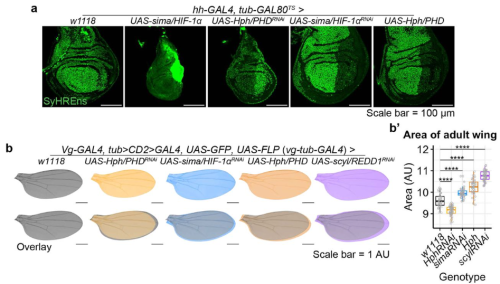
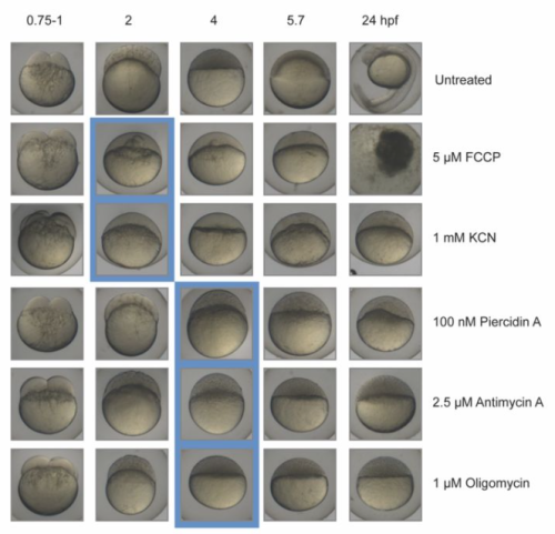
Disrupting oxidative phosphorylation, by genetic or environmental manipulation (hypoxia, Oligomycin), results in the expansion of the Organizer by upregulating Hif-1α.
Hif-1α overexpression leads to an expansion of the Organizer; knockdown of Hif-1α inhibits the expansion of the Organizer induced by mitochondrial manipulations or β-catenin overexpression.
Hif-1α acts downstream from β-catenin to establish Organizer gene expression and can induce Organizer specification even in the context of β-catenin knockdown.
Respiration in the Organizer is significantly higher than in the ventral mesoderm, suggesting Hif-1α is activated there despite functioning mitochondria. In fact, a functional ETC seems to be necessary for Organizer specification in wildtype embryos. This is due to the c-subunit of ATP Synthase acting as a mitochondrial membrane leak, which drives up ETC activity and increases oxygen consumption.
Targeted expression of the c-subunit of ATP Synthase can induce an ectopic Organizer in any region of the embryo and can result in a second embryonic head and body axis. This effect is dependent on Hif-1α and occurs downstream of β-catenin.
Overall, our work suggests that mitochondrial leak metabolism could be a general mechanism for activating Hif-1α and Wnt/β-catenin-target gene expression. It is also incredibly cool to answer an almost 75-year-old question and finally know why the Organizer uses more oxygen than other tissues!
(1) Regional oxygen consumption measured in whole, live embryos, the Organizer tissue is highly aerobic, consuming 20% more oxygen than ventral mesoderm. (2) Overexpression of ATP Synthase c-subunit mRNA is sufficient to induce a second Organizer and body axis.
Were you surprised to find that Hif-1α is sufficient to expand the Organizer cell fates in normoxia?
When I first overexpressed Hif-1a, and saw such a clear expansion of the Organizer, I was stunned and relieved. It was a major puzzle piece falling into place. I had been trying to make sense of why β-catenin was not increased in lrpprc CRISPR mutants. When you lose β-catenin, you lose Organizer specification completely, and when you overexpress it, you get an expansion or duplication of the Organizer, so I was expecting β-catenin to be driving the phenotype in the mutants. Because of the role of Lrpprc in mitochondrial maintenance, I had made a list of possible metabolic proteins to manipulate to see if I could recapitulate this phenotype. With the Hypoxia data we obtained in collaboration with Dr. Andrea Wills at the University of Washington, and from helpful conversations with her, I started with Hif-1α and saved a lot of time!
In terms of being surprised about the normoxia situation – honestly, I think I was so focused on the data, and focused on getting to a point where I deeply trusted my findings, that it didn’t strike me as that odd. There is something magical about being a PhD student setting out into a new field, especially because you come in with very few pre-conceived notions. I read so much about Hif-1α in a range of contexts and from what I learned it seemed pretty reasonable that something could be going on to stabilize HIf-1a in the embryo. In the end, biology is what it is, whether we understand the nuances or not. In the case of Hif-1a, I trusted the data, and used the amazing work already published by others to start making sense of the results!
Can you postulate about how Hif-1a is activated?
I have a few ideas about this. Hif-1α is dynamically regulated by so many signaling pathways and molecules. Obviously, you have the oxygen piece, then there are other factors like a-Ketoglutarate vs Succinate/Fumarate, pyruvate, and lactate, that are involved in regulating the activity of the prolyl hydroxylase domain (PHD) proteins that target Hif-1α for degradation; there are genetic regulators and growth factor signaling pathways that interact with Hif-1α. Then there are the direct interactions that have been described between Hif-1α and β-catenin, TCF/LEF, YAP1, and more. I think that in the context of mitochondrial membrane leak, regional increases in lactate and pyruvate could result in their competitive binding to PHDs and inhibit Hif-1α degradation. It is also possible that the increase in oxygen consumption produces high ROS levels which can inhibit PHD activity. Additionally, I saw an increase in regional mRNA levels in the Organizer and dorsal ectoderm, suggesting possible regulation at the transcription and/or translation levels. We are currently exploring these possibilities and I am excited for what is to come.
Did you have any particular result or eureka moment that has stuck with you?
Oh my, I feel like there were so many. During this work, I often felt like I was in the dark, following the dim light of my data, which sometimes felt like it was taking me in circles. I had a LOT of opportunities to practice trusting my data, even though I couldn’t see where it was leading me. And then there would be these flares of light, from a new result, or from reading a paper, and something would click, and I would jump 10 steps ahead. The whole project was truly an incredible learning experience.
I know I should probably stick to one, but I really couldn’t choose between these!
I have to say again, that the effect of overexpressing Hif-1α in the early embryo and getting such a striking Organizer phenotype was profound. It got us on track to begin understanding the mechanism. For me, it was the western blot I did to confirm Hif-1α is upregulated in lrpprc CRISPR mutants that really changed the game! That was when I did the experiments to confirm that losing Hif-1α rescues the lrpprc Organizer phenotype and I felt like I could begin sinking my teeth into the problem of what Hif-1α is doing early in development.
Seeing that Hif-1α can rescue the Organizer in β-catenin depleted embryos – I was absolutely shocked.
Going to London to perform regional Oxygen-consumption experiments in live embryos was such a privilege and a treat, then to find that the Organizer consumes more oxygen than other tissue blew me away. At the time I was such a novice in thinking about metabolism. Thank God for Dr Jonas and Dr Alavian – they laughed kind heartedly at my look of utter astonishment when we saw the result. They suspected what was going on immediately and helped me frame the concept of physiologically useful mitochondrial membrane leak. Seeing that the c-subunit leak could induce an ectopic body axis and Organizer was an incredible finale to this work.
In the context of β-catenin depletion, overexpression of Hif-1α can re-establish the Organizer.
And the flip-side: were there any moments of frustration or despair? What got you through?
Again, so many. Especially early on. I got the lrpprc ‘expanded Organizer’ phenotype and was so stumped because β-catenin wasn’t up. It took patience, creative experimenting, reading, and good conversations. When I read the 2017 papers that came out of Dr. Olivier Pourquié’s4 and Dr. Alexander Aulehla’s5 labs on glycolysis and cell fate specification during somitogenesis, it was huge. I finally felt like someone else was in the dark with me, shedding light on what could be going on. Even though our paper doesn’t focus on glycolysis, just being able to read about metabolism being involved in developmental cell fate and patterning was so helpful. Up until then, there was so little out there! Building out a new field of intersectional biology is difficult. It can feel lonely and frustrating, but also so exciting. I am deeply grateful for the people I’ve found along the way who have been exploring and defining the burgeoning field of developmental metabolism. For example, I just went to an EMBO Workshop at the EMBL in Heidelberg called “Developmental metabolism: Flows of energy, matter, and information.” It was incredible to see the diversity of backgrounds and fields represented by this excited group of people, all of them ready to collaborate to continue building out this field.
What is next for this story?
I am currently working with Dr. Jonas in the Endocrinology Department at Yale Medical School, delving deeper into the molecular mechanisms of how c-subunit, Hif-1α, and β-catenin collaborate to regulate embryonic patterning using mice and human stem cell models. I love this work and am so excited to continue learning about the mitochondria and metabolism side of things from Dr. Jonas. She has an incredible lab and skill set and I have learned so much from her already. In my own lab in the future, I hope to use my discoveries as a proof-of-concept to define other mechanisms that lie at the intersection of metabolism and known developmental pathways. I believe that exploring this intersection is crucial for the field developmental biology, but also for our understanding of how complex multicellular life evolved and diversified. I am so excited for what’s to come and to continue building and investigating the field of developmental metabolism!
Cited in this article:
1. MacColl Garfinkel, A., Mnatsakanyan, N., Patel, J.H., Wills, A.E., Shteyman, A., Smith, P.J.S., Alavian, K.N., Jonas, E.A., and Khokha, M.K. (2023). Mitochondrial leak metabolism induces the Spemann-Mangold Organizer via Hif-1α in Xenopus. Dev Cell. 10.1016/j.devcel.2023.08.015.
2. Spemann, H., and Mangold, H. (1924). über Induktion von Embryonalanlagen durch Implantation artfremder Organisatoren. Archiv für Mikroskopische Anatomie und Entwicklungsmechanik 100, 599-638. 10.1007/bf02108133.
3. Gregg, J.R., and Ornstein, N. (1952). Anaerobic Ammonia Production by Amphibian Gastrulae Explants. Biological Bulletin 102, 22-24. 10.2307/1538619.
4. Oginuma, M., Moncuquet, P., Xiong, F., Karoly, E., Chal, J., Guevorkian, K., and Pourquie, O. (2017). A Gradient of Glycolytic Activity Coordinates FGF and Wnt Signaling during Elongation of the Body Axis in Amniote Embryos. Dev Cell 40, 342-353 e310. 10.1016/j.devcel.2017.02.001.
5. Bulusu, V., Prior, N., Snaebjornsson, M.T., Kuehne, A., Sonnen, K.F., Kress, J., Stein, F., Schultz, C., Sauer, U., and Aulehla, A. (2017). Spatiotemporal Analysis of a Glycolytic Activity Gradient Linked to Mouse Embryo Mesoderm Development. Dev Cell 40, 331-341 e334. 10.1016/j.devcel.2017.01.015.