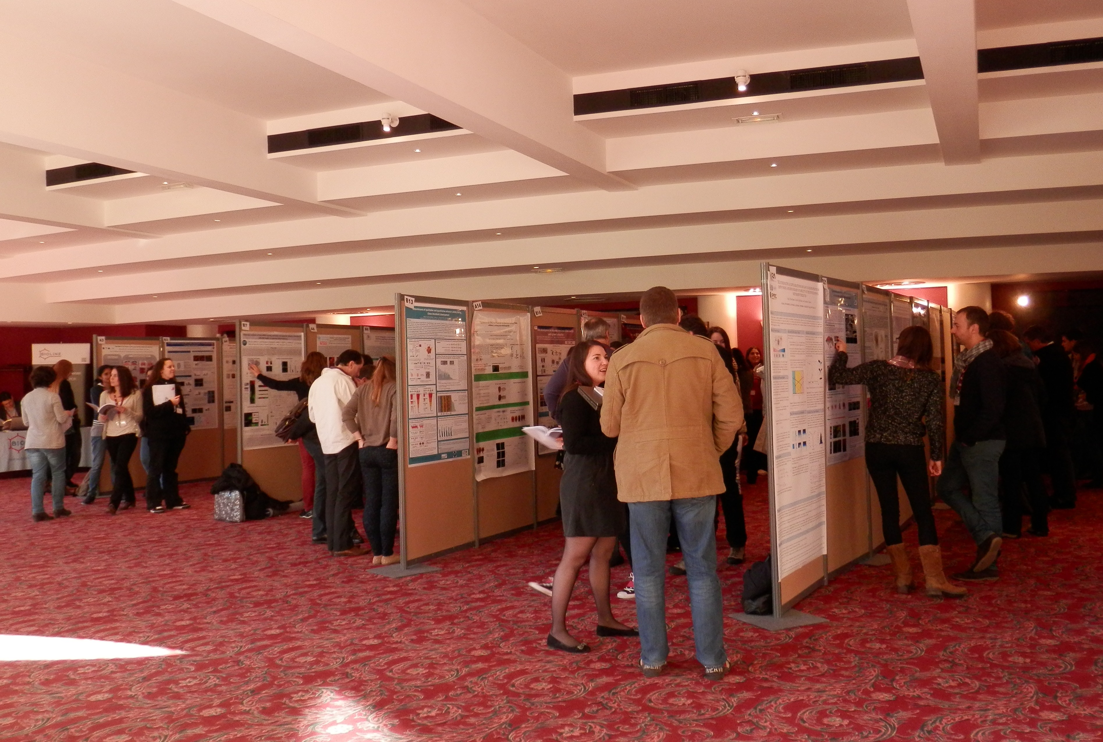
What is a science exhibition?
These are publicly accessible exhibitions that hold stalls designed to communicate specific areas of science to a lay audience. They tend to vary in terms of their content and the groups of people they are intended to engage with but will usually involve a series of organised events and stalls that communicate specific areas of science. One of the most widely known and well attended of these exhibitions is the The Royal Society Summer Science Exhibition. This is a week-long event with scientists from across the UK showing off some amazing work from all fields of science at the Royal Society in London. My personal highlights from this year were learning that a t-rex had feathers, that I’m not smarter than a zebra fish and just what on earth the Higgs boson actually is.
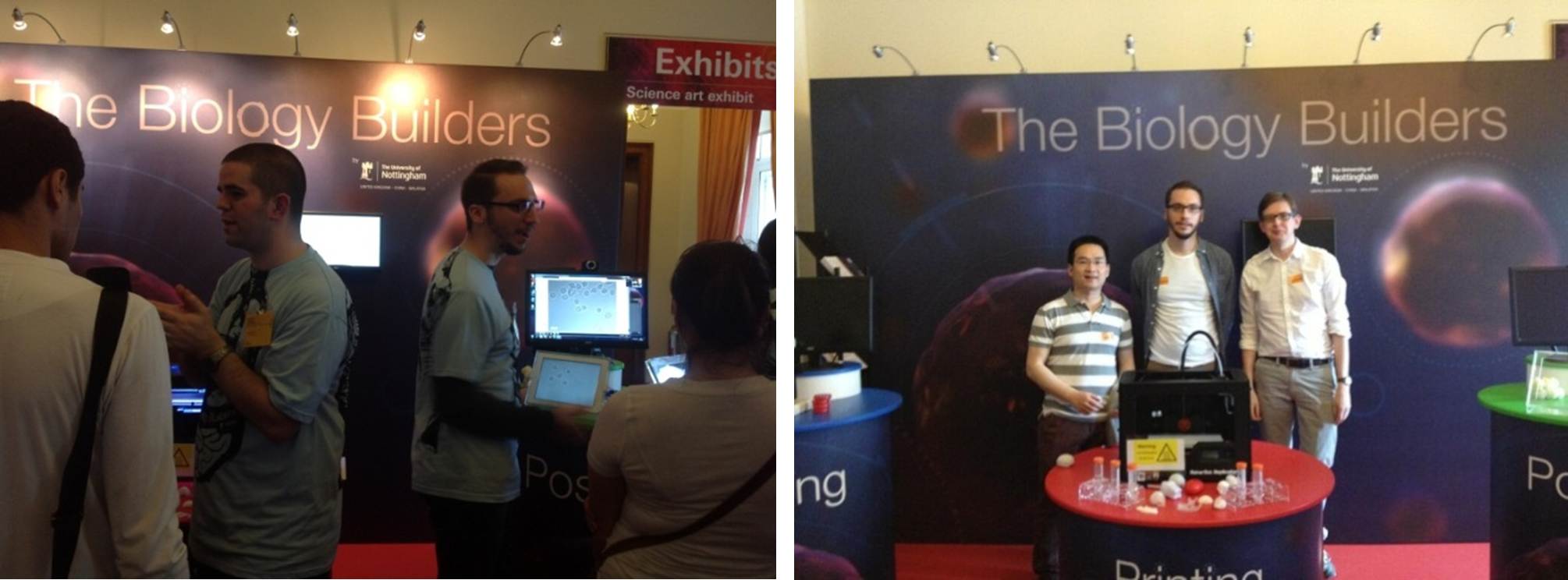
We were lucky enough to be granted the opportunity to show some of our work at this year’s exhibition and our stall was:

http://sse.royalsociety.org/2013/exhibits/biology-builders/
These types of exhibitions are designed to be fun, interactive and educational, demonstrating the amazing range and quality of science being performed in the UK. It is not only the physical stalls themselves that the public come to see but you, the person actually doing the work. Putting a face to the science is the best way to showcase it and the interactions that you have with the public can be some of the most rewarding you will ever have in your carrier.
Why are these events important?
First and foremost these events are about feeding back to the people who fund your work, the general public. The majority of the work we do as academic scientists is funded from public money in the form of taxation or donation and as the financiers these people have the right to know what their money is being spent on; and science exhibitions are designed to do just that. Communicating science to the general public is also becoming increasingly important to funding bodies and many are beginning to require public engagement strategies to be written into grant applications.
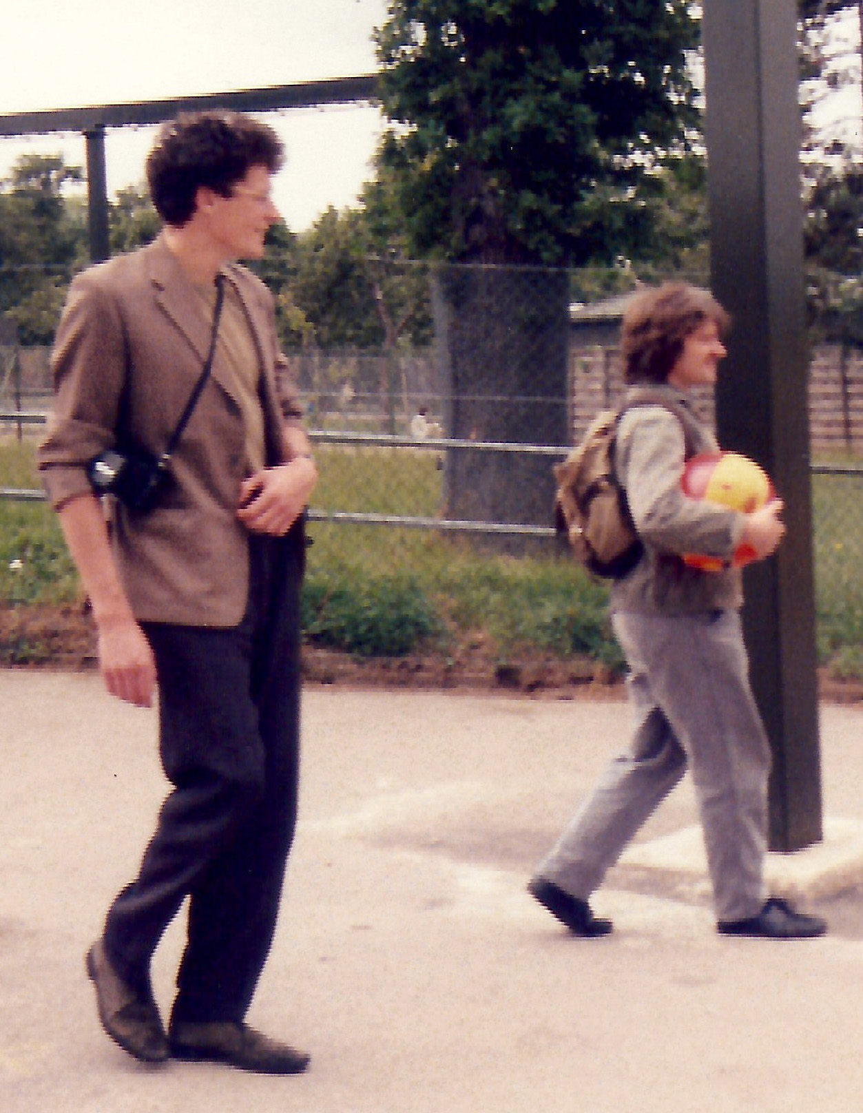
The personal experience of doing these events is highly rewarding and all members of our stall team thoroughly enjoyed the experience. Speaking with the public can give you a fresh perspective on what you do as well as reinforce to you as a person how important and valuable your work really is. We even had staff and students fighting over shifts so they could spend as much time on the stall as possible.
Some of the most valuable interactions we experienced involved young people and school groups. These young adults showed an incredible level of knowledge and enthusiasm not only for the science we discussed but us as scientists. This event provided an amazing opportunity to educate and inspire the next generation of scientist, breaking down the stereotypes of who scientists really are and what we do.
Getting started
The Royal Society Summer Science Exhibition is one of the biggest and most prestigious science outreach events in the UK and so when we first began to plan our stall we realised how important it would be to get experience with these types of events. We therefore decided to show a very simple stall at our Universities annual public outreach day called Mayfest. The experiences we gathered from this provided invaluable feedback on the basic design of our stall at the Royal Society and enabled us to try out a variety of different interactive elements to all age groups.
Even if you intend to only hold a small stall at such a local event it is always worth gaining some practical experience in speaking to the general public about your science before you hold your stall. Most organisations have some form of public outreach experience and it is worth seeking out those who run these activates within your institution. The best advice would of course be from someone who has run a stall of this kind but any advice or guidance they can offer would be invaluable.
There are many different aspects to running a stall of this kind ranging from design and construction to safeguarding vulnerable people during interactions with the public. Form an enthusiastic and hardworking team of people who work well together and most importantly plan meticulously. We were lucky enough to have a healthy budget for our exhibition stand but you can run a stall with almost any budget – you will amazed what you can beg and steal – but you can also seek funding from external sources.
Here are some links to UK based grant opportunities:
Welcome Trust: http://www.wellcome.ac.uk/Funding/Public-engagement/index.htm
EPSRC and BBSRC: No longer have separate calls as they now require engagement within existing grant calls so speak to PI’s within your organisation about how a stall could satisfy their grant stipulations. They may be persuaded to part with some cash as a result.
Science and Technology Facilities Council: http://www.stfc.ac.uk/1780.aspx
Society for Applied Microbiology: http://www.sfam.org.uk/en/grants–awards/public-engagement-grant.cfm
The Physiological Society: http://www.physoc.org/public-engagement-grants
Institute of Physics: http://www.iop.org/about/grants/outreach/page_38843.html
British Society for Plant Pathology: http://www.bspp.org.uk/funds/promotion.php
British Ecological Society: http://www.britishecologicalsociety.org/grants-awards/outreach-grants/
Biochemical Society: http://www.biochemistry.org/Grants/EducationalGrants/ScientificOutreachGrants.aspx
Designing and building the stall for your audience
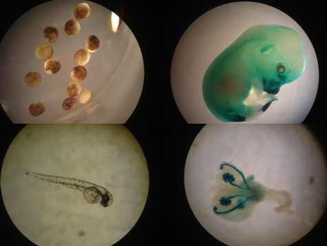
When we sat down and began to decide what the stall would be like the central questions we asked ourselves were; ‘what message are we trying to convey’ and ‘who are we delivering this message too’. The Royal Society exhibition covers all age ranges, from parents with very young children through to informed retired professionals visiting for the day and guided school groups. In terms of our message we had quite a complicated idea that would use three current technologies to demonstrate biological complexity during early development and how this could be rebuilt in a laboratory for future clinical therapies. You could have designed an entire stall around each of these but we decided to simplify and link them into a cohesive and exciting experience for the visitor.
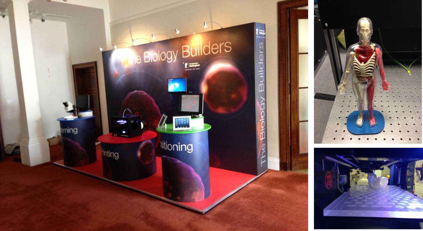
Our stall ultimately consisted of a 3D printer in the central podium that produced large scale biological structures, allowing visitors to see the machine in action and interact with the printed objects. The podium to the left of the printer consisted of a microscope that allowed the visualisation of stem cells patterned using protein stencil technology. The third and final podium showed a live link to our laboratory in Nottingham allowing member of the public to position live stem cells using an optical tweezer system with the click of a mouse.
Once we knew who we were pitching at and the message we wanted to convey the next question was ‘how do we design the stall to deliver this message to all age ranges’. This was a real challenge particularly as the work we were trying to show covered everything from the physics of lasers to what a stem cell is. We rapidly realised that you cannot cover everything and that it may be necessary to sacrifice detail so that all visitors can have a fun and interesting experience whilst leaning something new about our science. Consequently the basic design was very simple with three podiums displaying the three technologies we were displaying with more detailed, simpler interactive objects to hand so that we could adapt to all age ranges and leaning abilities depending on need.
Would we recommend running a stall?
Absolutely! When we started this process we had never run a stall of this kind and the majority of the people who ultimately staffed the stall had never done any form of public engagement. Everyone thoroughly enjoyed the experience and if we can do it anyone can, so we would absolutely encourage all scientists to participate in science exhibitions.
Also read Glen’s outreach activity suggestion- Using modified ping-pong balls to demonstrate early embryogenesis and embryonic stem cell activity
 This post is part of a series on science outreach. You can read the introduction to the series here and read other posts in this series here.
 (1 votes)
 Loading...
 (No Ratings Yet)