The community site for and by
developmental and stem cell biologists

Six weeks in Woods Hole

Posted by , on 23 July 2013

Six weeks of Science.

Forty-four days of lectures, discussions and (sometimes crazy) experiments at the bench.

One thousand fifty-six hours in Woods Hole, attending to the 125th Embryology Course organized by MBL.

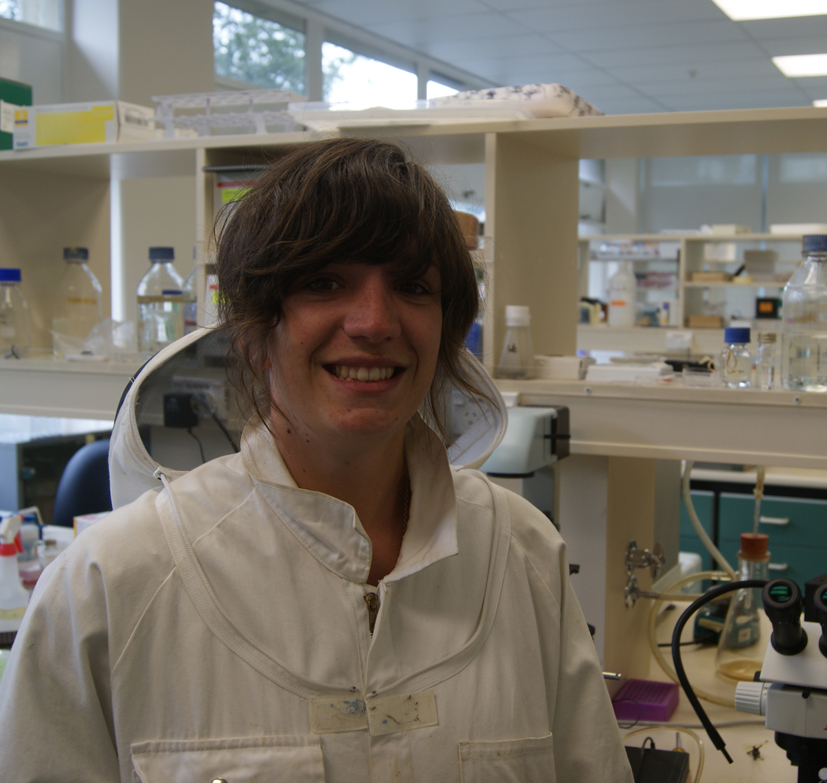
I look at the pictures taken during this period and I see a time lapse of the development of 24 students who discovered new models, techniques and perspectives to look at their research and at the world.

Which is the answer at the question “What is the Embryology Course in Woods Hole?”

The Embryology Course in Woods Hole is lectures in the auditory room with major experts in the field of development, it is the possibility of getting into a true scientific discussion after the lecture, it is the experiments in the laboratory at each time of the day and night, it is sharing of knowledge and skills and, much more than this, it consists of a continued cultural exchange thanks to the thirteen Countries represented by the 24 students.

During the Embryology Course we had the unique opportunity of using invertebrate and vertebrate models. After sea urchin, nematodes and arthropods (weeks 1 and 2) and then Xenopus, zebrafish, chick and mouse (weeks 3 and 4), we could approach to Cnidaria, Planaria, Spiralians, Ctenophores, Ascidians and Annelids. Most of these animals are non-model organisms for developmental biology and most of us had never worked on them. The laboratory was full of these organisms with uncommon shape and different colors. The week 5 was dedicated to regeneration and transplant. We evaluated the regeneration ability of the anemone Nematostella, of different species of Planaria and we transplanted tissues from one labeled Hydra to another. We spent days doing movies of the ascidian metamorphosis and of fertilized worm egg development. After each experiments, we did antibody staining or in situ hybridization and we spent hours and hours facing the microscope in order to capture our results. Surprisingly this life-style is not hard, your brain is always activate and your body full of adrenalin, and like playing children, we were never ready to go to bed.

But the Embryology Course is also outside the laboratory. On July 4th all Woods Hole was involved in the parade and the Embryology class bring colors and laughs in the street. During these weeks we train both the mind and the body. With great pleasure I can state I belong to the class, that, after many years, won the Softball match “Embryology vs. Physiology”. We were a real good team not only on the softball field but also in the kitchen. In the laboratory we share our skills and in the kitchen we shared the typical foods of our country. On the table there was pasta, salads, sushi, tandoori chicken, guacamole, turkish salad, chocolate cakes and sweets with peanut butter.

And finally, it is arrived, the dreadful final week 6, the last six days in this Wonderland of Science. Alice, the time is over, and you must wake up, even though there are nor Queen neither playing cards running after you-. The time for the cleaning up of your bench is arrived, suddenly.

Thank you Embryology Course, thank you all the student, thank you the faculties, thank you the TAs, thank you MBL, thank you all the people that makes it possible each year and thank you Woods Hole, the watering landscape of this great experience.

I am thinking that exactly one years ago 24 students were leaving this place with our same feelings, scientifically and personally enriched as we are now. How many Embriology Course students look at this sea and say goodbye to this shore? But I hope. I really do hope that the bonds that link us will be more strong than the distance and the time. And maybe one of us will get back as a lecturer or invited speaker for the 150 year Anniversary Symposium.

Above all, I hope that this Course will be organized over and over, because it is a unique occasion for improving knowledge, acquiring new techniques, working with a vast choice of models, interact with experts in the different fields of development.

During the 2013 Embryology Course, we won several times. We won in the softball match and we won as developing researchers. May those will attend to the 2014 Embryology Course feel grateful and satisfied as I feel now.

Thumbs up (7 votes)
Loading...

Tags:
Categories: Events

Navigate the archive

Use our Advanced Search tool to search and filter posts by date, category, tags and authors.

ESF QuanTissue Workshop

Posted by , on 22 July 2013

The 8th European zebrafish meeting first started with the “ESF QuanTissue” workshop, a 4 hour session where speakers showed current research about basic mechanisms that regulate cellular behavior during morphogenetic processes. Talks were given by outstanding scientists studying in detail mechanistic behind epiboly, rhombomeres and MHB formation, proneuromast assembly, somitogenesis, and tracheal morphogenesis.

In the first talk, Carl-Philipp Heisenberg showed how the Enveloping Cell Layer spreads during epiboly to completely engulf the embryo at the end of gastrulation. Using laser cuts, cell ablation and embryo deformation to induce ectopic tension, he dissected the role of orientation of the mitotic spindle as a key regulator of this process. Then, Guillaume Salbreux and Philippe-Alexandre Pouille, talked about physics behind epiboly, how pulling forces and friction govern the movement of cells during early gastrulation and mediates shape changes.

Next, Cristina Pujades finely showed the role of apical actomyosin cables in cell segregation during rhombomeres boundary formation. Acting as barriers, these cables prevent cells to intermingle between adjacent rhombomere domains. Before the coffee break, Jordi Casanova (the fly “intruder”), showed his beautiful data about how tracheas are formed by migration and cell intercalation during development, a process that require modification of cadherin accumulation to change cell shape and allow cells to execute their normal behavior.

Later on, Mansi Gupta explored the relevance played by the extracellular matrix on the establishment of gradients of secreted factors (like fgf8) during gastrulation. Heparan sulfate molecules perform an active role in gradient formation regulating fgf8 diffusion from it source. Then, Virginie Lecaudey, showed her fantastic work related to dissect signaling pathways required for rosette formation during proneuromast assembly. These rosettes are formed by constriction of the apical side of epithelial cells mediated by fgf signaling. She showed that shroom3 is essential for rosette formation and that this gene act downstream of fgf signaling to promote apical constriction and hence rosette formation in the proneuromast.

Finally Daniele Soroldoni, showed amazing data related to the role of gene expression waves during somitogenesis. He visualized the activity of the segmentation clock and quantitatively compared the rate of somite formation and the release of gene expression waves in the pacemaker region. Surprisingly, he found that the period in the pacemaker region is different to the period of somitogenesis.

Overall, the workshop was very interesting, with high quality talks. The mechanistic behind different processes during development is getting unraveled.

This post is part of a series of posts on the 8th european zebrafish meeting.

Thumbs up (13 votes)
Loading...

Tags: , , ,
Categories: Events

ERC funded Post-doctoral position: Pasteur Institute, Paris

Posted by , on 19 July 2013

Closing Date: 15 March 2021

How stem cells contribute to tissues during growth and regeneration remains largely unknown in vertebrates. Skeletal muscle provides an excellent paradigm to investigate this issue since powerful transcription factors that play important roles in specifying and determining skeletal muscle identity have been identified. The Tajbakhsh lab has a wide range of genetically modified mice and has been investigating this question in the context of developmental and regenerative biology.


Project: Mechanisms regulating asymmetric and symmetric cells divisions in adult muscle stem cells in the mouse. This project follows from previous work in the laboratory demonstrating asymmetric segregation of DNA strands to daughter cells, and direct association of this phenomenon with daughter cell fates (Nature Cell Biology 2006, Nature Reviews Molecular Cellular Biology, 2009; Cell, 2012).


Funding: ERC Advanced Grant. Funding is available for the postdoctoral position for 3 years with the possibility of an extension. Applicants willing to apply for their own independent funding, however, are encouraged to do so.


Requirements: A doctoral degree, strong experience in cellular and molecular biology and microscopy. Proficiency in English is required. Outstanding, highly motivated candidates who are creative minded and independent are encouraged to apply.


Application process: Please send one PDF file to shaht@pasteur.fr with the following:

–  cover letter

– concise summary of previous research activities

– curriculum vitae including the publication list and contact details for 2 referees


Prof. Shahragim Tajbakhsh

Stem Cells and Development

Dept. of Developmental & Stem Cell Biology

Pasteur Institute

25 rue du Dr. Roux

75015, Paris, FRANCE

E-mail:   shaht@pasteur.fr

Thumbs up (No Ratings Yet)
Loading...

Categories: Jobs

Post Doctoral Research Associate Position available in Tooth EvoDevo in UK

Posted by , on 18 July 2013

Closing Date: 15 March 2021

Postdoc: USheffield.SharkEvoDevo

A Post-doctoral Research Associate position is available in evolutionary developmental biology of shark and ray dentitions, Department of Animal and Plant Sciences, University of Sheffield, UK. This is an ideal opportunity for an ambitious Post-doctoral Research Associate to work in the Laboratory of Gareth Fraser at the interface of evolutionary and developmental biology. This project aims to explore novel morphological and genetic methods to understand the evolution and development of tooth patterning and functional mechanisms responsible for tooth regeneration in a comparative framework in sharks and rays. The work will be performed in a large, high profile 5* research department with a strong commitment to research-led teaching. The position is funded by the Natural Environment Research Council (NERC) in collaboration with the Natural History Museum (Dr. Zerina Johanson), King’s Collage London (Prof. Moya Smith) and Birkbeck, University of London (Dr. Charlie Underwood) to study the evolution of chondrichthyan dentitions. The overall aim of the project is to use an integrative evo-devo approach to study the dentition in a major group of jawed vertebrates, the chondrichthyans.

Potential candidates will have a PhD or equivalent experience in evolutionary or developmental biology, preferably in vertebrates, and a track record of publishing in leading specialist or multidisciplinary journals. A strong interest and/or background in evolutionary biology, palaeobiology, developmental biology or regenerative biology would be desirable. The post is fixed-term for 2 years with the potential for extension.

To apply please follow the current vacancies page at: http://bit.ly/1atNZan For informal enquiries about this position and the Department of Animal and Plant Sciences at the University of Sheffield, please contact Dr. Gareth Fraser at: g.fraser@sheffield.ac.uk or 0114 222 4317. Gareth J. Fraser, Ph.D Lecturer in Zoology Department of Animal and Plant Sciences Alfred Denny Building University of Sheffield Western Bank Sheffield S10 2TN UK Phone: +44(0)1142224317 Email: g.fraser@sheffield.ac.uk Website: Fraser Lab Gareth Fraser

Thumbs up (No Ratings Yet)
Loading...

Categories: Jobs

“Working with Zebrafish Genome Resources” Workshop during the 8th European Zebrafish Meeting

Posted by , on 18 July 2013

This was my first time in lovely Barcelona. I travelled from London the day before the zebrafish meeting kicked-off (in order to be well rested and alert for all the talks and posters!). I attended the “Working with Zebrafish Genome Resources” workshop and I have to say it was absolutely helpful for any scientist working with fish (although the same principles taught can be easily applied to other model organisms). The workshop was organized by the Wellcome Trust Sanger Institute crew, which are experts in the field and provide most of the genomics resourced we use everyday in our work.

It started at 10.30 on the first day of the meeting. The workshop was composed of several modules designed to get an overview of each topic with examples explained by the trainers (which we could follow in our own computers) and with proper discussion of theory and methods. After a general introduction, we started with the hands-on learning.

Even if I knew some things about the topics, I was amazed by such an amount of options for analyzing sequences and how useful it is to handle the available genomic tools in a proper way. The first talks were about manual genome annotation and de novo analysis of sequences, covering some basic resources to access sequence information using Entrez and UniProt, Vega browser and some tools for alignment and open reading frame finders; it was all complemented with hands-on worked examples. Even if some of us were familiar with these resources, it was very helpful to understand the theory behind.

The second part was a deep swimming into genome browsing, using Ensembl, NCBI map viewer and UCSC genome browser. We got a nice idea about why it can be useful to look at the whole genome, and we were demonstrated some of the features and applications of this potent tools. It included more worked examples, which nicely complemented the theory about search and retrieve across the whole genome, basic comparative analysis, features around specific genes and chromosomal regions. Overall it was quite useful.

The third module was to learn how to explore gene function and disease, and sequence variation. A useful search for polymorphisms and their consequences on transcripts and proteins was presented, and how biological pathways associated with those gene products can be altered in several conditions. Even if this module was focused on zebrafish data when possible, disease database were primarily concerned with humans, as there is more sequence variation information for our specie than for zebrafish. However this results in a fantastic way to reinforce the importance of favourite model organism for human disease.

The final module was based on deep comparative sequence analysis. We were shown how to retrieve sequences from different organisms to identify putative homologous genes and compare genomic contexts for potential regulatory function. This is a powerful tool when we want to model human diseases and also for generating transgenic animals. The instructors were very helpful when we got our own genomic problematic sequences and were happy to help us analyzing them.

After this module finished, it was time to register in the zebrafish meeting and the garden party was about to start. The enormous effort of the speakers for organizing this workshop (and patience for helping us one by one!), together with the meeting organizers, made possible a successful experience that will have a good impact in the scientific career of those who attended. The instructors said the workshop would be organized again during the zebrafish meeting in Madison on next year and I really recommend registering on it. Thumbs up for the workshop!


This post is part of a series of posts on the 8th european zebrafish meeting.

Thumbs up (6 votes)
Loading...

Tags: , , , ,
Categories: Education, Events

A fantastic trip in Sydney, supported by a Travelling Fellowship

Posted by , on 16 July 2013

With the support from The Company of Biologists, I was lucky to obtain the opportunity to visit Macquaire University in Australia for approximately two months. It was a fantastic trip, and I have made a lot of progress which I think will contribute enormously to my future research-career plans.

I am a master degree student, studying in Jiangxi Agricultural University, China. My research field mainly focuses on the biology of honey bees. In order to apply for the Travelling Fellowship, we designed a research plan on honey bees, aiming to determine whether there is a sub-caste of worker bees who specifically feed queen larvae. We started and finished this project during this trip, and we fortunately obtained very interesting results from our experiments.

First, the background of this study. Whether honey bee young larvae develop into queens or workers depends on the amount and type of food received during early larval development. For instance, the food can differ in  the content of sugar, proteins, vitamins and microRNA. In addition, many previous studies indicated that the division of labor of honey bees depends on the requirements of its social environment. Our previous study on the genetic differentiation between nurses attending worker larvae (AWL) and others attending queen larvae (AQL) revealed that hundreds of genes were significantly differentially expressed between them as well.

However, the results of the behavioral experiment that we completed in this trip indicated that there were no special royal nurses in the honey bee colony when the queen larvae suddenly emerge. Nurses attended both worker larvae and queen larvae. Our results also revealed that there was no difference in age between AQL and AWL. The only difference detected was that AQL were significantly more active in term of attending activities, and needed a short break (couples of minutes) before attendance, whereas AWL could keep on attending worker larvae without any break. These behavioral differences suggest that producing Royal Jelly (which is specifically fed to queen bees and is more nutritive than Workers Jelly), is possibly more labour intensive, but bees are not required to change roles to achieve this. This study highlights a novel model for the interaction between the environmental factors and the division of labor in eusocial insects. Therefore, we aim to publish these results soon, and hope it could be accepted by the journal Molecular Ecology.

Furthermore, during this trip, I have found some differences between the scientists from Australia and China. For example, people from China and Australia do their scientific work in different ways. Chinese researchers consider more the results that they can obtain in their scientific work, whereas Australian focus on the processes of their work more. This difference may reflect different cultures. I have decide to make this difference known to chinese people that I interact with in my future career, as it may help them understand more easily the scientific work outside China, and communicate with scientists of other nations more efficiently.

My English has also improved rapidly during this period studying in Australia, which is enormously helpful for my further career. Thus, I thank the Company of Biologists for giving me a chance to study overseas: thank you! I hope the Travelling Fellowship program can help more people wherever he/she comes from, and more and more Chinese students or young scientists can be supported in the future.


work in Macquiare University

Thumbs up (4 votes)
Loading...

Tags: , , ,
Categories: Funding, Research

An interview with Maria Leptin

Posted by , on 16 July 2013

Maria Leptin is a successful group leader in the developmental biology and immunology fields, running labs in both Cologne and Heidelberg. In 2010 she also became the director of the European Molecular Biology Organisation (EMBO). The Node interviewed Maria at the recent International Society for Developmental Biology (ISDB) meeting, and we talked about her career, her vision for EMBO, and the current economic situation in Europe.


Photo of Maria LeptinHow did you first become interested in biology? Was there someone who inspired you?

Biology at school was fun. We had a fantastic teacher, a skinny little old lady, who was very original, and very enthusiastic. But I was not trained to be a research scientist. My degree was in mathematics and biology, with the aim of becoming a teacher. I only decided to become a scientist when I realized that I couldn’t stand the idea of having to go to school for another 40 years! I decided to do research because of a practical at the University of Heidelberg where I was studying, taught by scientists from the Basel Institute of Immunology. It was very intellectually stimulating, much more than my other courses, and I really enjoyed it.


You did your PhD in immunology but then later on switched to developmental biology. Was this a challenge, and do you have any advice for graduate students considering changing fields?

If you do high-level science, everything is a challenge. That’s why it’s fun! I benefited a lot from the change. The step from PhD to postdoc is the one time when you can handle that challenge best: your mind is fresh and open, and you don’t have the pressures that you will have later. And it is good to learn how to think in different ways. I have been able to return to immunity later more easily than someone from a purely developmental biology position. Changing fields definitely opens your horizons. No one looks backwards in science, so you must explore, go to new places, work with new organisms and on different problems…


As you mentioned, you later returned to immunology. What are the challenges of working simultaneously in two fields that don’t overlap much?

I had intended to leave development and cell biology behind, because I got bored of it. But then that research suddenly picked up again. In addition, it is more feasible for me to get funding for my developmental biology work: it is very hard to get funding for a project when you don’t have prior publications, or results on which to base your grant.

One major challenge is how to find the time to think about all the ongoing scientific projects productively and critically. You have to think very deeply about your work, and I cannot spend enough time doing this for all my projects. But I have good people in my lab, and we do the thinking together. And both topics are fun, so I find it hard to give either up.


You became the director of EMBO in 2010. What do you think are the strengths of this organization, and what objectives did you set yourself at the beginning of your directorship?

The biggest asset of EMBO is its members: 1400 committed, leading scientists who are prepared to help the organisation. They do the interviews for the young investigator awards, the selection for courses and workshops, and for the postdoctoral fellowships. Their expertise drives everything at EMBO.

The other thing that is really special is that EMBO is non-national, not populated by country or society representatives, but rather by individuals who are there for their excellence. The absence of national bias is a huge strength, and creates a body of experts that could do a lot more to advise the central European policy makers and individual countries.

Our real aim is to serve the community of European researchers, and make sure that Europe is a fantastic place for doing science. Our training, courses and workshops are important for that, and so is the general network building- creating a European identity among researchers, rather than just national identities.

Another important area where we have done a lot of work is with the journals. We have have tried very hard to make the publishing process fairer and better for authors: by improving the refereeing process and making it transparent, by helping protect authors against being scooped and so on…


Europe is undergoing a period of great economic recession, with unfortunate consequences for research funding. How can EMBO and its members face these challenges?

We need to apply for our next budget by 2015. We are talking to the delegates of our governing body, EMBC, and they all appreciate the need for continued or even increased funding for EMBO. But they will go back to their governments and say ‘Give them some money’, and the governments are going to say ‘Where from?’. It is really hard, and everyone is just hoping that the economic climate will change for the better. What can the individuals do? Talk to politicians, talk to funders, and stress the importance of what EMBO is doing for Europe.


Besides researching in two different areas, in two different model organisms [Drosophila and zebrafish], in two different cities and being the director of a major international organization, you are also a mother. Balancing your career and your family responsibilities must be very challenging. Do you have any suggestions for other female scientists?

I am through the hard bit: both kids are grown up and at university. I take time off when they come home, but otherwise I am back to 80-hour weeks. That was of course not possible when they were still at school. My husband was incredibly supportive. In fact, it is wrong to say supportive- we just did it together. We had help throughout with nannies and kindergartens, and I never thought it was appropriate to complain ‘my whole salary goes on childcare’. You compromise: I didn’t go to the cinema for about 10 years, and I wore jeans and t-shirts for years.

I also didn’t listen much to what other people thought was right- I knew the kids didn’t need me 24 hours a day. And they are now two beautiful, balanced young men, so what I was told I was doing wrong can’t have been that bad. So don’t listen to what people say: just get on with it and don’t mope about it.


You are currently one of the directors of the Christiane Nüsslein-Volhard foundation. What are the objectives of this foundation and how did you get involved in this project?

Janni Nüsslein-Volhard was extremely helpful when I had my first baby. My husband was still working in England so I was more or less a single mother. There was no kindergarten in Tübingen and Janni supported a colleague and myself to set one up. Later, she also noticed that young women who had kids found it hard to come to seminars in the evening or come in at weekends. She appreciated that you love your kids and need to spend time with them, but what really irritated her was when she found out that postdocs or students with kids couldn’t be in the lab because they had to do household chores. She said ‘This just won’t do. Let them have children and spend time with them, that is a good thing. But doing housework is not for a trained scientist’. She set up this foundation to provide that extra bit of money to pay for someone else to help around the house.

The great thing about this foundation is that it is not just about the money. At interview, there are people who tell us ‘Whether I get the money or not, just being told that it’s okay to get a cleaner is already good’. Similarly, with the mother-in-law who thinks that the daughter-in-law should stay at home and take care of the family- if they hear that she got an award from this organization, they start appreciating the research work that she does. So the foundation is also re-educating female scientists and the people around them.


Do you have any advice for young scientists?

In my own career I planned maximum one year ahead. Otherwise, I never ended up doing what I planned. I think all this agonizing over careers is missing the point. Science is not a job like others. It is a passion, and if you don’t have that passion then get out immediately. If you enjoy what you are doing and do it well, somebody will be interested and allow you to take the next step.

Don’t get advice from anyone else, because everyone is different. For example, I would advise you to change fields. But Tim Hunt would tell you the opposite. When he finished his PhD, he hadn’t finished solving the problem he was interested in, so he just looked for a place where he could continue working on that problem. And we know what came of that- a Nobel Prize!

I also pass on some advice I got from my PhD advisor. He said ‘Maria, don’t think about how you get in to the next place. Think about how you’ll get out again. Find a place where will you end up with a broader scope than when you went in.’ But you really just have to figure it out for yourself. We are not paid very much- the main benefit of science is the freedom to do what we want. So we’d better do what we like, and not what someone else tells us to do.


What would people be surprised to find out about you?

I wanted to be an interior decorator first, but my father talked me out of that. Then I wanted to be a dancer, and I took professional training for a while during my studies. I did my degree, missing two or three days a week because I would go to the academy to train. But then eventually at the academy they said ‘ Look, you have to make up your mind. And if you are uncertain, we think that you will be a better teacher than a dancer’. They were quite brutal, but of course they were right. And I still dance in my free time.


Thumbs up (13 votes)
Loading...

Tags: ,
Categories: Interview

In Development this week, Vol. 140, Issue 15

Posted by , on 16 July 2013

Here are the highlights from the current issue of Development:


From hPSC to mature hepatocyte

F1.smallLiver development entails induction of definitive endoderm, specification of the endoderm to a hepatic fate, generation of hepatoblasts, and differentiation of these hepatic progenitors into mature hepatocytes. To date, efforts to generate mature hepatocytes from human pluripotent stem cells (hPSCs) have mainly produced immature hepatocytes. Now, on p. 3285, Gordon Keller and co-workers investigate the mechanisms underlying hepatocyte maturation by manipulating specific signalling pathways at different stages of hepatocyte development in hPSC cultures. The researchers report that sustained activin/nodal signalling is required to pattern the definitive endoderm for hepatic specification. Three-dimensional aggregation of hepatoblasts initiates the maturation of these progenitor cells, and cAMP signalling within the hepatoblast aggregates promotes further maturation to a hepatocyte-like population that expresses metabolic enzymes at levels comparable to those of primary human hepatocytes. These results provide new insights into the pathways regulating hepatocyte maturation from hPSCs, as well as a simple method for generating functional hepatocytes for use in drug metabolism studies and cell-based therapy of liver diseases.


Tbx20 cushions cardiac valve development

F1.small-1Disrupted development of the cardiac valves – structures that ensure unidirectional blood flow through the heart – causes about a third of human congenital heart defects. The signals that regulate endocardial epithelial-mesenchymal transformation (EMT), an early stage of cardiac valve development, are well understood, but what regulates the later stages of this important developmental process? On p. 3176, Chen-Leng Cai and colleagues report that the T-box transcription factor Tbx20 is expressed in mouse endocardial cushions and valves throughout their development. They show that endocardial Tbx20 expression is not essential for EMT initiation but is required for endocardial cushion maturation and valve elongation in mice. Mechanistically, Tbx20 regulates Lef1 (a transcriptional regulator of Wnt/β-catenin signalling) in cushion endocardial cells, and disruption of Tbx20 expression leads to aberrant Wnt/β-catenin signalling in the endocardial cushions. Thus, the researchers conclude, Tbx20 acts upstream of Wnt signalling to regulate late steps in cardiac valve formation, a finding that provides new insights into the aetiology of human cardiac valve defects.


Postmitotic retinal cell fates notched up

F1.small-2During retinal development, different types of neurons arise in a conserved temporal sequence from multipotent cycling retinal progenitor cells. But do the progenitor cells decide the fate of their daughter cells and pass on this decision via determinants or is the fate of the daughter cells determined postmitotically? Constance Cepko and co-workers (p. 3188) address this question using a conditional allele (N1-CKO) to remove the cell fate regulator Notch1 in newly postmitotic mouse retinal cells. Nearly all newly postmitotic N1-CKO cells become rod photoreceptors, the researchers report, whereas wild-type cells adopt several cell fates: photoreceptors, bipolar cells and Müller glia. Notably, single-cell expression profiling indicates that several direct targets of Notch signalling are differentially expressed in wild-type and N1-CKO cells transitioning from progenitor to differentiated states. These findings suggest that newly postmitotic retinal cells need Notch to escape the photoreceptor fate, and thus may have some degree of plasticity with respect to cell fate.


Drink, genes and craniofacial defects

F1.small-3Foetal alcohol spectrum disorders (FASD) encompass a wide range of birth defects that are associated with prenatal exposure to alcohol from mild craniofacial defects to foetal alcohol syndrome. The variability of FASD is partly attributed to the timing and level of foetal alcohol exposure but genetic factors may also influence FASD. To identify potential ethanol-sensitive genes, Johann Eberhart and co-workers have been examining the effects of ethanol on known zebrafish craniofacial mutants. On p. 3254, the researchers report that platelet-derived growth factor receptor alpha (pdgfra) heterozygotes and mutants show enhanced craniofacial defects after ethanol exposure. Other results indicate that Pdgfra has a protective effect that is mediated by the mechanistic target of rapamycin (mTor), part of the phosphoinositide 3 kinase (PI3K) signalling cascade. Finally, analysis of a small database recording human craniofacial features and prenatal ethanol exposure links PDGFRA and PDGFRB variants to particular craniofacial phenotypes. Together, these results suggest that combined genetic and environmental inhibition of PI3K/mTOR signalling leads to variability within FASD.


Modelling the segmentation clock

F1.small-4During the patterning of the vertebrate anterior-posterior axis, a gene expression oscillator controls segmentation of the somites, the precursors of the vertebral column. Several mathematical models have been developed to explain how this segmentation clock works. Now, Ahmet Ay, Ertuğrul Özbudak and colleagues (p. 3244) extend these earlier models by developing a multicellular stochastic computational model of the zebrafish segmentation clock. Simulations run with this model show that autoregulatory negative-feedback loops of dimers of Hairy/enhancer-of-split-related (Hes/Her) proteins – transcriptional repressors that are encoded by the only conserved oscillating genes in vertebrates – can drive synchronised gene expression oscillations in wild-type zebrafish embryos and can recapitulate various mutant phenotypes. The model also predicts that synchronised oscillations can only be generated if zebrafish Her proteins have a half-life of less than 6 minutes, a prediction that the researchers validate by showing that zebrafish Her7 has a half-life of 3.5 minutes. Importantly, this model can now be used to make predictions about clock behaviour that can be tested experimentally.


Tightening the netrin on brain repair

F1.small-5Neural stem cells are maintained in the adult mouse brain within the subventricular zone (SVZ) and help to repair brain damage. In response to demyelination, for example, neural progenitors emigrate from the SVZ and help to repopulate the demyelinated lesion. Here (p. 3107), Myriam Cayre and colleagues investigate progenitor cell emigration from the SVZ in response to lysolecithin-induced focal demyelination in the mouse corpus callosum. The researchers report that demyelination in the corpus callosum triggers vascular remodelling in the SVZ. The remodelled vessels and the neural progenitors exiting from the SVZ are closely associated, they report, and inhibition of post-lesional angiogenesis reduces the migration of progenitor cells towards the lesion. Notably, netrin 1, a factor involved in axonal guidance during brain development, is upregulated within the SVZ after corpus callosum demyelination and is involved in both local angiogenesis and progenitor cell emigration. Together, these results identify netrin 1 as a potential target for the development of therapeutic strategies for brain repair.


Plus…


The mechanical control of nervous system development

F4crop.largeBased on recent findings, Kristian Franze puts forward the hypothesis that several steps during nervous system development, including neural progenitor cell differentiation, neuronal migration, axon extension and the folding of the brain, rely on or are even driven by mechanical cues and forces. See the Hypothesis article on p. 3069


Cdks, cyclins and CKIs: roles beyond cell cycle regulation

F7crop.large In addition to their function in cell cycle control, it is becoming increasingly apparent that mammalian cyclin-dependent kinases (Cdks), cyclins and Cdk inhibitors (CKIs) play indispensable roles in processes such as transcription, epigenetic regulation, metabolism, stem cell self-renewal, neuronal functions and spermatogenesis. Lim and Kaldis discuss the latest revelations about Cdks, cyclins and CKIs with the aim of showcasing their functional diversity. See the Review article on p. 3079


Thumbs up (No Ratings Yet)
Loading...

Categories: Research

My first encounter with the fish community

Posted by , on 15 July 2013

fish trail 2I was really looking forward to attending the European Zebrafish Meeting. The fact that it took place in the amazing Barcelona was, of course, a bonus point, but the main reason why I was looking forward to it was that it was a fish conference. Before I was the Node community manager, I was a Drosophila researcher, and though I also worked with other organisms at different points, I never worked with zebrafish. In fact, for some reason or another, I never really interacted much with fish labs. This conference was my chance to get a feeling for the breadth of research being done by the fish community. This was also my first model organism-focused conference, and I was curious on any differences from topic conferences.

The Node will feature a few posts focusing on the great sessions and keynote lectures in the next few days, so I will leave the science highlights to the fish researchers. Here I will give you a bit of a feeling for what the conference was like.

fish greetings 2The fact that you were attending a fish conference became apparent as soon as you got close to the venue. That’s because the organizers had left a fish trail from the metro stop all the way to the venue. At the entrance of the conference venue, you got a fishy (?!) greeting: a board on a wall featured a fish made up of greetings in a variety of languages (although by the last day someone had added a greeting in chinese- I guess the organisers forgot one language!). Maybe not surprisingly, the fish references ran through the whole conference. The light effects in the main auditorium made up the stripes that give Danio rerio its most common name. In addition, the promotional material featured a fish-shaped structure, which a call-out on Twitter made me discover is a real building. It is here in Barcelona, and was designed by Frank Gehry for the 1992 Olympics. The fish love percolated even to the conference dinner, where each table had its own zebrafish bowl with live fish. Although the organisers confirmed that these fish were returned to the pet shop afterwards, the opinions were divided on what to make of the fish bowls. I must admit I was quite curious to have a close-up meeting with the critters I had been hearing so much about, and was even taught how to distinguish a male from a female.

The breath of science covered was extremely interesting- from developmental biology to disease models; and the techniques covered ranged from the more classical morpholinos and in situs to the latest microscopy techiques and genomics. There was enough to please everyone, with of course a strong representation of developmental biology. You will be able to read more about some of the relevant talks on the Node soon.

auditorium view

For the non-fish person, what was the most surprising? I must admit that there were a few things that I found quite interesting. First of all the zebrafish meeting was actually about more than just zebrafish. Not only other fish species also featured, such as medaka, but many of the fish labs also work on other organisms, and that creeped up at different points of the conference. Another striking thing was the number of people who presented unpublished material. Finally, I was very impressed with the format of the talks. With the exception of the two keynote talks, all other talks were about 10 min long (in theory!). Not only this, but the vast majority of them were presented by PhD students and postdocs. As a consequence, an impressive number of studies were presented, and younger scientists got a chance to practice their presentation skills in front of a big audience.  This may be the case in other model organism conferences (please do comment below if that is the case!), but it was a first for me, and I think it is a great format!

The best thing about this conference was, however, the fish community. Everyone was very friendly and welcoming, and I met several Node readers (and hopefully persuaded many more to check out the website and contribute). It was a great community to meet, and I almost feel sorry that I am not a fish scientist myself, so that I could join them again in Oslo in two years time!


official fish bannerThe official passing of the fish banner to the Oslo team

The Node tweeted from the conference, so you can check the Node twitter account and the hashtag of the conference for more details.

Thumbs up (10 votes)
Loading...

Tags: , ,
Categories: Events

Distant Developments: In conversation with Professor Stephen Robertson, clinical geneticist and developmental biology researcher

Posted by , on 15 July 2013

“We have the ability to understand human disease and deliver it back to the clinic – allowing families to understand and then move on.” 

This week, I caught up with Professor Stephen Roberson to discuss how he combines his research interests in developmental biology with a position as a clinical geneticist.   Stephen is a medically trained researcher based in the Department of Women’s and Children’s Health, University of Otago. He worked in Paediatrics and Clinical Genetics in both Auckland and Melbourne in the 1990s.  In 1999 he changed tack to join the Institute of Molecular Medicine at the University of Oxford, working on the genetic determinants of congenital malformations.  Stephen was recruited back to Otago in 2002 to take up a position as Curekids Professor of Paediatric Genetics.  Stephen is located in the Department of Women’s and Children’s Health, University of Otago, where he heads the Clinical Genetics Group.  His group has over 90 publications including high profile journals such as Nature Genetics, PNAS, and the American Journal of Human Genetics.

Prof. Stephen Robertson

Stephen’s career highlights include identifying mutations in FLNA that caused both brain and skeleton malformations while at Oxford.  Then back at Otago, his group identified mutations in a paralogue (FLN B) in individuals with a related spectrum of disorders, leading to the recognition of a group of development disorders now named filaminopathies.   His team has also identified germline mutations in a tumour suppressor gene also resulting in disorders of morphogenesis characterized by a broad slew of malformations in different organ systems. 

In addition to running a successful research laboratory, he still is a practicing clinical geneticist spending nearly a third of his time travelling to clinics in the South Island and Wellington.  On top of this he is a popular lecturer, teaching in both Genetics and Medicine courses at Otago.

Combining clinical work with research.    Early on in Stephen’s medical career, he had a strong interest not just the clinical side of medical work but also the molecular factors underlying a disorder.  He was really driven by a desire to combine these two together throughout his career.   As one of only 8 clinical geneticists in the NZ Genetic Health Service, this part of the job involves sitting down with the patient and family and discussing what the diagnosis means both in terms of future options but also heredity.  Many great scientific opportunities have arisen through Stephen’s work in clinic.  Often a patient can walk in to clinic with a rare condition and sometimes due to an  understanding of the molecular pathways of involved in development  it is possible to speculate that a defect in a given  pathway may underpin a given clinical presentation , with the advantage of being go to the back to the lab with the patient’s DNA samples to work on identifying  the gene at fault.

“NZ families are very open to research and very generous and have a desire to contribute to understanding the broader picture of a disease”.

The Major research projects in Stephen’s laboratory centre around three core questions. 

They seek to further understanding of how neural cells migrate during brain development particularly in relation to brain malformations often found in rare genetic disorders.  His group has just recently completed analysis of a new neurological syndrome that has identified a pathway involved in brain development  that up until now was not suspected to play such a role (to be published soon –watch this space!).

His group has an ongoing interest in bone malformations including the genetic pathways responsible for gravity sensing and how the skeleton triggers an anabolic response to mechanical stress.

Stephen’s group also have a number of clinical projects running, including understanding genetic contributors to conditions characterized by atresia of gall bladder and bile ducts, a disorder which presents not long after birth with the babies becoming very jaundiced, followed by liver failure.  If they can find the genetic cause, this will hopefully lead to earlier diagnosis and more successful treatment outcomes for these patients.

Stephen is fascinated, like all developmental biologists, by the whole amazing process of going from a cell to a whole organism.

“Everyone studying their own corner of a much bigger picture, how the overall process is conducted is mind blowing.”

 Stephen’s group makes use of many classic developmental biology model animals to study the functional role of genes identified by mutation screening.  They make use of the Otago Zebrafish Facility to perform morpholino studies to begin to characterize gene function, and transgenic mouse ‘knock-in’ studies to study the effect of the disease mutation on mouse development. One key advance his lab has adopted is the use of next generation sequencing, particularly exome sequencing, allowing much faster genetic characterization of patient DNA samples.

Working as a scientist in New Zealand.  The biggest advantage is the collegiality, more group leaders have some job security that makes the environment more collegial and less combative. .  Due to NZ’s size it is easier to get to know each other and also span more areas of interest such as going to journal clubs on topics far removed from your research area.

Of course, the greatest challenge for most scientists and NZ scientists in particular is Funding, limited amount of money and funded only for a short time three year time frame, when often the project is really just getting started.  Stephen suggests that researchers need to rise to this challenge by being more creative and collegial with their limited resources, and to communicate their work to the public to highlight its benefits and relevance.

 Giving back to the community. Stephen works closely with CureKids as one of their three Chairs of Child Health Research.  CureKids’ focus is raising funds for medical research to find cures for childhood diseases.  This has been so successful in NZ, CureKids will soon be starting up in the USA and Australia.  This involvement with Curekids has had a stronginfluence on both Stephen’s research and the way he communicates back to the community.  This has been a challenge since Stephen was previously more atune to the more traditional routes of science communication such as journal articles and research seminars.  To reach the public, he now has learnt to communicate more broadly through medias like TV, radio, and talking “off the cuff” at events without prompts in order to engage with the people who are really the ones that are funding this research.

They also keep asking the hard questions, the ones the families want the answer to rather than just curiosity-driven investigation – “that’s a nice discovery, but how does that fix the disorder or improve the lot of the child or family?” This helps keep the research centred around the central purpose of helping people. Stephen is also interested in questions surrounding the ethical questions raised by the implementation of  the many new genomic tools becoming available and publicised  – how do we put this into context for the public?

So how can scientists help the public to understand genetic disorders?  Stephen’s advice is we need to put the research into context – make more use of visual learning both print and online to help the public understand developmental biology.  Key for this will be developing resources and making better use of graphic tools to help explain our research.

“We need to make the public more aware of how much is imprinted in very early development in the embryo, it really casts the mold for the adult.”


Thumbs up (4 votes)
Loading...

Categories: Careers, Interview, Research