Mayank Chugh’s post ‘Science is more than the sum of research’ struck a chord. A research associate at the Cambridge Stem Cell Institute and an aspiring poet in my spare time, I find a lot of value in communicating research to the public and often ponder on what it means to be a scientist. Driven by similar values to those expressed by Mayank, that science is more than science itself, I wrote the poem ‘Success in Science’ to air these thoughts.
About ‘Success in Science’
There are traditional measures of success in the research community, but should they be the only ways we define our success? The experiences that have stood out for me during my scientific career include working in a team from around the world, sharing ideas, teaching the next generation of scientists, communicating research to the public and, ultimately, being part of a bigger picture to improve the lives of patients. These are all, I think, fundamental to a functioning and successful research community, and are successes that we can all share and recognise.
Success in Science
Success in science is hard to define,
What pops into your mind?
A Nobel Prize, the impact factor,
A finding that’s one of a kind?
Success is measured in more ways than one,
What does it mean to you?
With a different perspective we can find success
In not only the year but the everyday too.
*
We work in a team and voice our ideas,
Two heads are better than one.
All around the world we collaborate and share
To gain knowledge that is second to none.
We pass on our skills to the next generation,
Just as others have filled our own cup.
We mentor each other and throw down ladders,
To help others who are on their way up.
We communicate our research and our aims
To make the world a better place.
We share evidence and ask questions of own,
For an inquisitive mind there is always space.
Getting through each day can sometimes be a test,
Experiments don’t always go as planned.
But we pick ourselves up and think again and again,
As the more we persist, the more we understand.
For each day brings us a step closer,
Even through the triumphs and struggles,
Every experiment like a pin prick,
Gradually bursting the knowledge bubble.
*
Success in science we can all share,
Even in ways we may think are small.
For these make up the foundation of research,
So, let’s recognise and celebrate them all.
This poem is adapted from ‘Success in Science’, written as part of the University of Cambridge’s Creative Encounters Words project, a public engagement with research initiative led by David Cain. It was first published in the collection “The Hope of Knowing Love: Research Poems to Open Our World” and exhibited at the Cambridge Festival in Spring 2023. In this collection Kirsty used poetry to convey life as a scientist, her research on the childhood cancer neuroblastoma and the lived experiences of patients and their families.
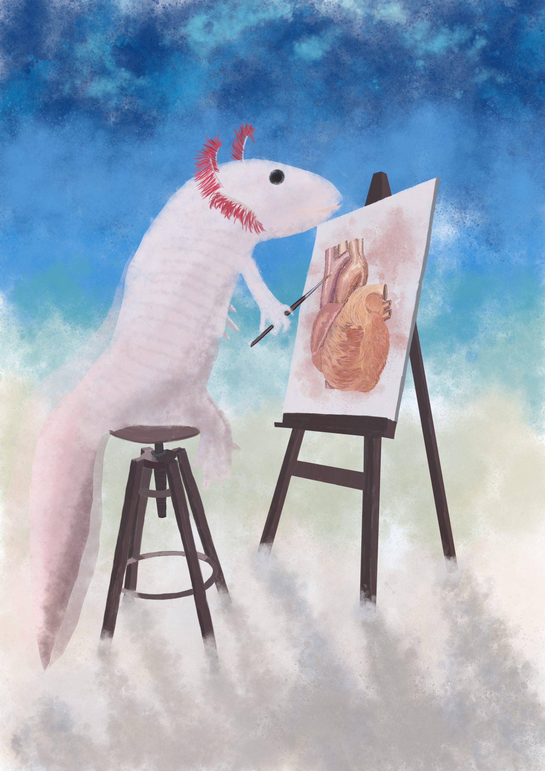
In this SciArt profile, we get to know more about Elad Bassat, the scientist behind the artwork ‘Klimt-olotl’, which was chosen as both the Judges’ choice and People’s choice in the ‘Science-inspired art’ category of the Node-BSDB virtual art exhibition.
Klimt-olotl – The decision of Axolotls to stay in water rather than metamorphose painted in the style of Gustav Klimt.
Can you tell us about your background and what you work on now?
I have been a regeneration enthusiast for the majority of my scientific career. Ever since I started my masters (and later my doctoral) work in the lab of Prof. Eldad Tzahor in the Weizmann Institute of Science, I was always fascinated how some animals can achieve amazing feats of regeneration while we cannot. I continued my regeneration journey moving to Vienna and becoming a post doctoral fellow in the lab of Elly Tanaka (a position I still hold today), essentially moving from working with mice to working with the cutest model organism in existence, the axolotl. Specifically, I am working on two different projects: what is the role of the extracellular matrix in axolotl limb regeneration and how does the axolotl regenerate its heart. I hope that in the near future I will be able to wrap up my projects and look for an independent investigator position in which I could combine my previous mouse experience with the axolotl work and study regeneration across species.
Were you always going to be a scientist?
In short, oh yeah!
In my high school, the Hebrew Gymnasium of Jerusalem, at the 9th grade, the students are tasked with choosing their major subjects. This entails sitting and listening to, sometime boring and dry, descriptions of what will be taught by the teachers and as you can imagine, I also found it boring and tedious. However, one of the teachers, which taught Biology, told us: “Instead of telling you what I will teach you, I will show you what this could be useful for…” and then she told us about treating diseases and improving yields of crops and genetic engineering and… I was hooked.
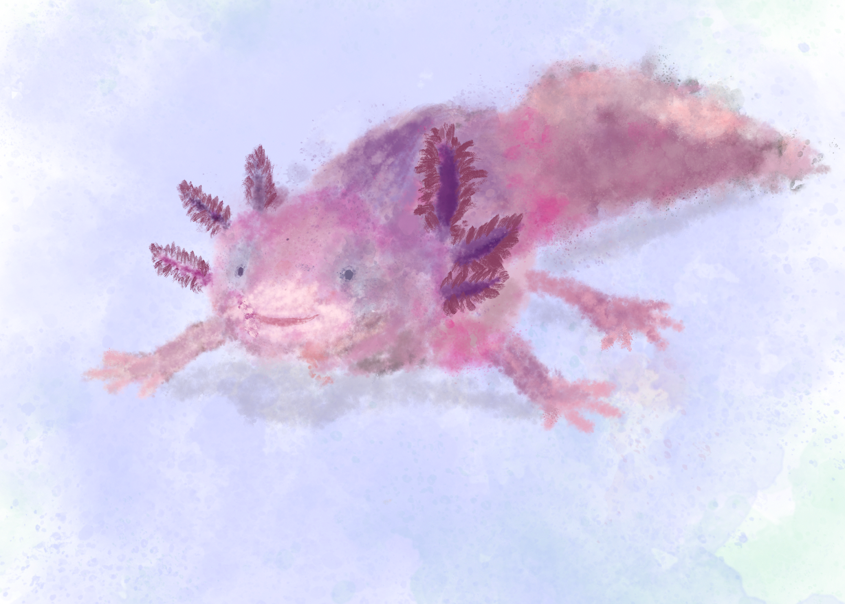
Just a cute axolotl – I was playing around with the different brushes available when I got my new tablet and the water color effect was amazing.
And what about art – have you always enjoyed it?
I want to put an asterisk here – *compared to the amazing artists who participated in the competition, I consider myself a non-artist. I always enjoyed seeing art, but I was and still am very bad at this. My mother is very skilled in painting, jewelry making and other arts and crafts, so I dabbled in painting as a kid, but I was always inpatient and unhappy with myself when the straight line I wanted to draw turned out curved. So, for many years after this, I didn’t attempt to paint or do anything “artsy” again. During my university days, I started to play with digital art, mostly experimenting with photoshop and illustrator and then I identified the one thing I was missing when I was painting as child, an “undo” button. From this point on I started to generate models and illustrations for colleagues and for the labs I was working in. To this day, the majority of the art I generate is aimed to make my presentations more fun or to communicate my science better.
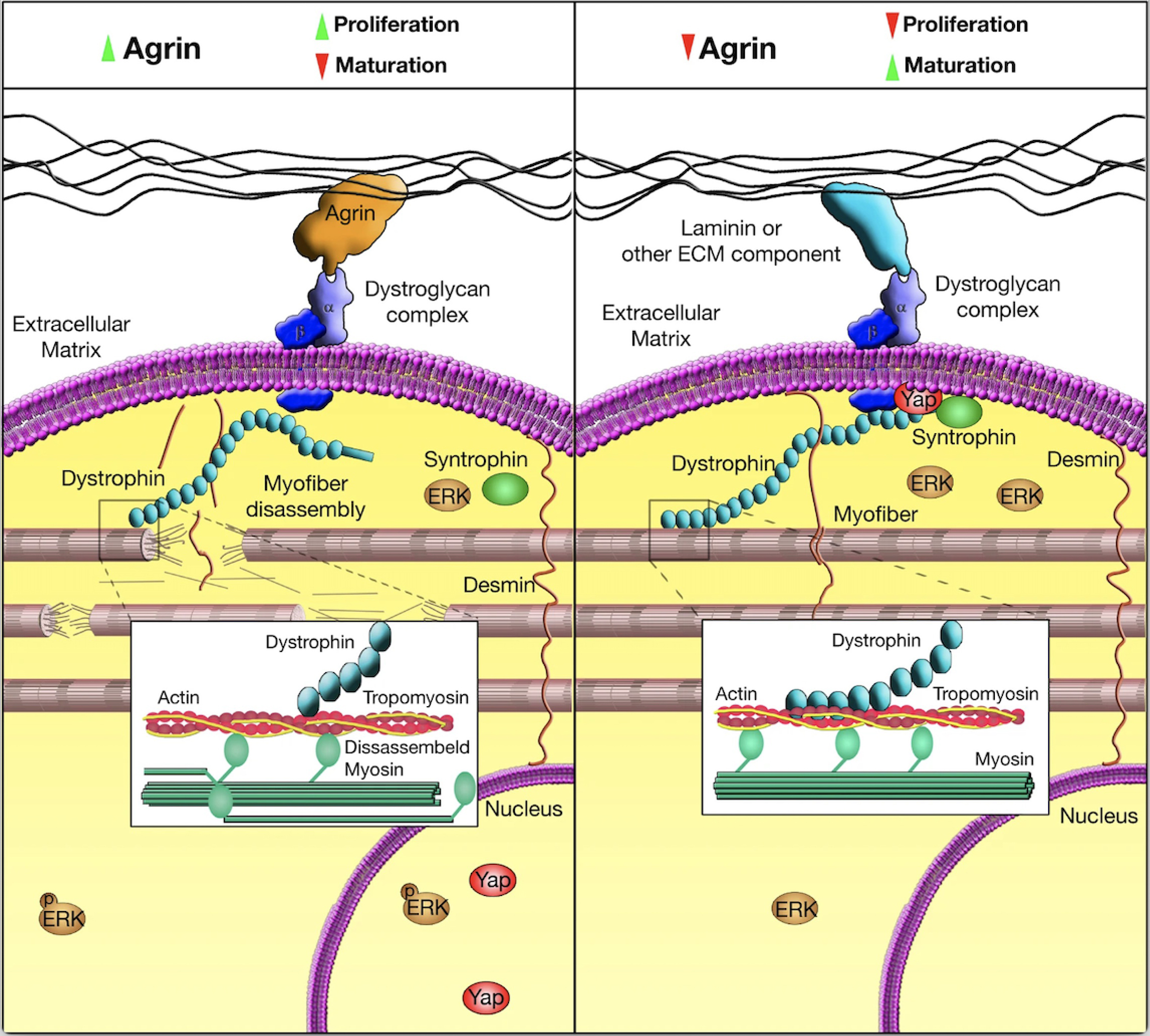
Model from Bassat et al, Nature 2017 publication – A model I drew for my 2017 publication depicting the mechanism of action of Agrin in inducing cardiac regeneration.
What or who are your most important artistic influences?
Honestly, I don’t think I have “most important” artistic influences, I appreciate specific pieces of art in museums and exhibitions, and I admire those who created them. For example, I visited museums in Vienna and saw beautiful works of the Austrian artist, Gustav Klimt. Following this, I went home and tried to copy his style and that is how “klimt-olotl” was born, combining my science model with the art I admired. Similarly, I visited the Albertina museum in Vienna and saw Pointillism paintings by Seurat and Signac which apart from being inspiration for amazing R-packages for bioinformatics, are artists I truly admire. I was so impressed with “the pink cloud” by Signac that I tried to draw with a similar style immediately when I got home only to fail miserably (although I am still practicing this technique).
I think I should also include one additional avenue of inspiration and that is the works of many Sci-Artists on twitter which I follow (such as @DrawImpacts, @maayanvisuals and @Ella_Maru). I often find the beautiful illustrations, models, and graphics they generate, truly inspiring.
Peripheral Neural circuits – an image I made for the lab, not really my expertise but I think it turned out nicely
Your painting ‘Klimt-olotl’ was both the People’s and the Judges’ choice in the BSDB-the Nodevirtual art exhibition. Can you tell us more about the story behind the artwork?
As I mentioned before, I currently live in Vienna and I enjoy visiting the many art museums we have in the city. As Gustav Klimt was an Austrian artist there are many locations in the city you could see his works and stylistic influence. About two years ago there was an interactive exhibition titled the “Klimt: the immersive experience” which we decided to go with my 3-year-old son, and he loved it. After questioning him a bit more he mentioned that he loved, what I would paraphrase, his color palette especially from the golden period. After that we went home and we started drawing together and he asked me to outline spirals and shapes that he would later color with the Klimt palette. After he went to bed, I took my tablet and started playing with the actual color palette and shapes used in “The Kiss”, “The Woman in Gold”, and “Death and Life” and noticed I am seeing a separation of water and land. From this point a few days have passed and my mind was focused on my scientific work and I thought about how axolotls have the same type of separation between water and land as they would normally would not metamorphose in nature, so it just clicked that they should be also represented in the image.
How do you make your art?
Apart from painting with my son (which is currently limited to painting unicorns), I only paint electronically using my tablet and computer. I would generate the image using the Apple pencil and then perform special functions, post-processing and add effects using photoshop and illustrator.
Axolotl drawing a heart – As a fun way to introduce my research topic in presentations I ask the audience: what can the axolotls teach us about mammalian cardiac regeneration, this image is the representation of this question.
Does your art influence your science at all, or are they separate worlds?
I don’t think anything is really separated in my head. I can’t think of a specific example where seeing an art exhibition influenced me to run a specific experiment but when I see or make art my mind wonders and then new ideas are generated. In the end I think we are the sum of all our experiences, they are intermingled, and they shape the next decisions you make.
What are you thinking of working on next?
Honestly I don’t know, I haven’t drawn anything in the last few months and my motivation is down given what is happening in my home country. Since the 7th of October attack on Israel I find it hard to think about what’s next.
Nawseen Tarannum, postdoc, University of Manchester
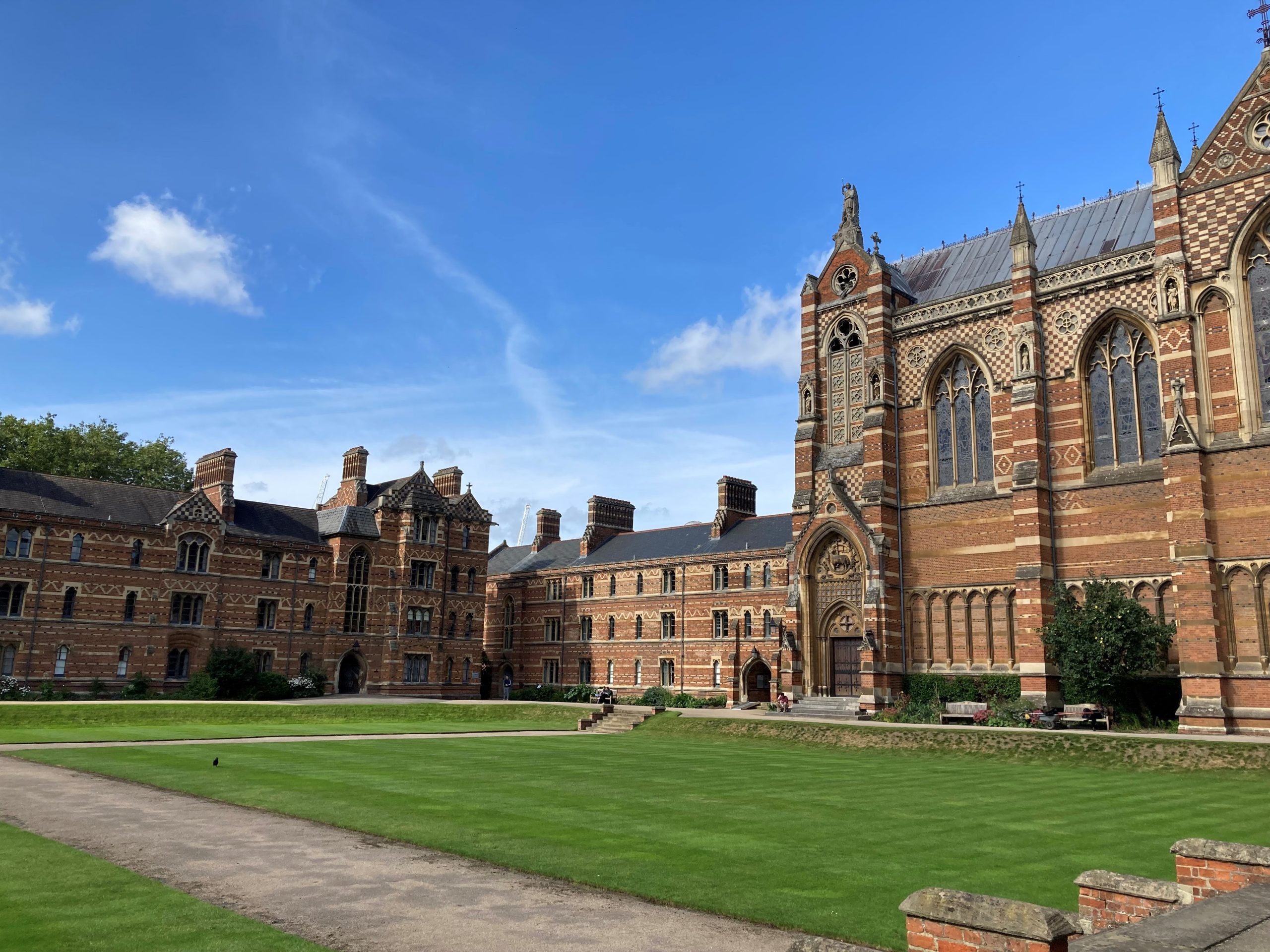
While conference hunting in early 2023, I saw an advertisement for the 2nd EDBC on the BSDB website. Given my love for cell and developmental biology, wanting to attend it was a no-brainer decision. Held from 25th-28th September 2023 in Oxford, the conference attracted developmental biologists from across Europe. Packed with a collection of great talks, posters and social events, this conference was unique for the science and how it was organised. In addition to Oxford, there were two hubs – Paris and Barcelona – from where talks were live-streamed and intertwined with the Oxford schedule. I’m not sure what technology gods the organisers and the AV team prayed to, but they managed to pull off the least technologically disruptive hybrid conference I’ve been to. Truly impressive. Now let’s turn to the real showstopper, the science.
The conference was nestled in beautiful Keble College, University of Oxford
Pilot
Season two of the conference started in the afternoon with an opening address from Paul Martin (BSDB chair). He promised fantastic talks prioritising early-career group leaders and researchers and the conference programme indeed delivered on this promise. He also introduced the winner of the Cheryll Tickle medal, a mid-career female scientist who has made outstanding contributions to the field. This year’s medal was awarded to Madeline Lancaster. She took us on a trip through her PhD and postdoc days before focusing on her lab’s interest in understanding how the human brain develops compared to other primates. Her work has identified that the tissue architecture of the developing brain impacts cell fate, with temporal progression from progenitors to neurons depending on the correct spatial positioning of cells (1).
Madeline’s presentation paved the way for other talks that afternoon that were based on “patterning”. During this session, I listened to Teresa Rayon talk about discrepancies in the developmental timescale of neural tube ventral patterning in mice and humans. She showed that although the process is conserved between both species, increased protein stability in human cells may explain our slower development (2). Jacqueline Tabler discussed skull morphogenesis in the context of a noncanonical form of cell motility. She showed that a collagen gradient drives osteoblast movement, divisions and differentiation towards a softer matrix with feedback between the stiffness gradient and cell fate controlling bone size (3). Markéta Kaucká Petersen elaborated on how her lab outlines the blueprint of cellular heterogeneity underlying craniofacial morphogenesis using single-cell genomics and transcriptomics.
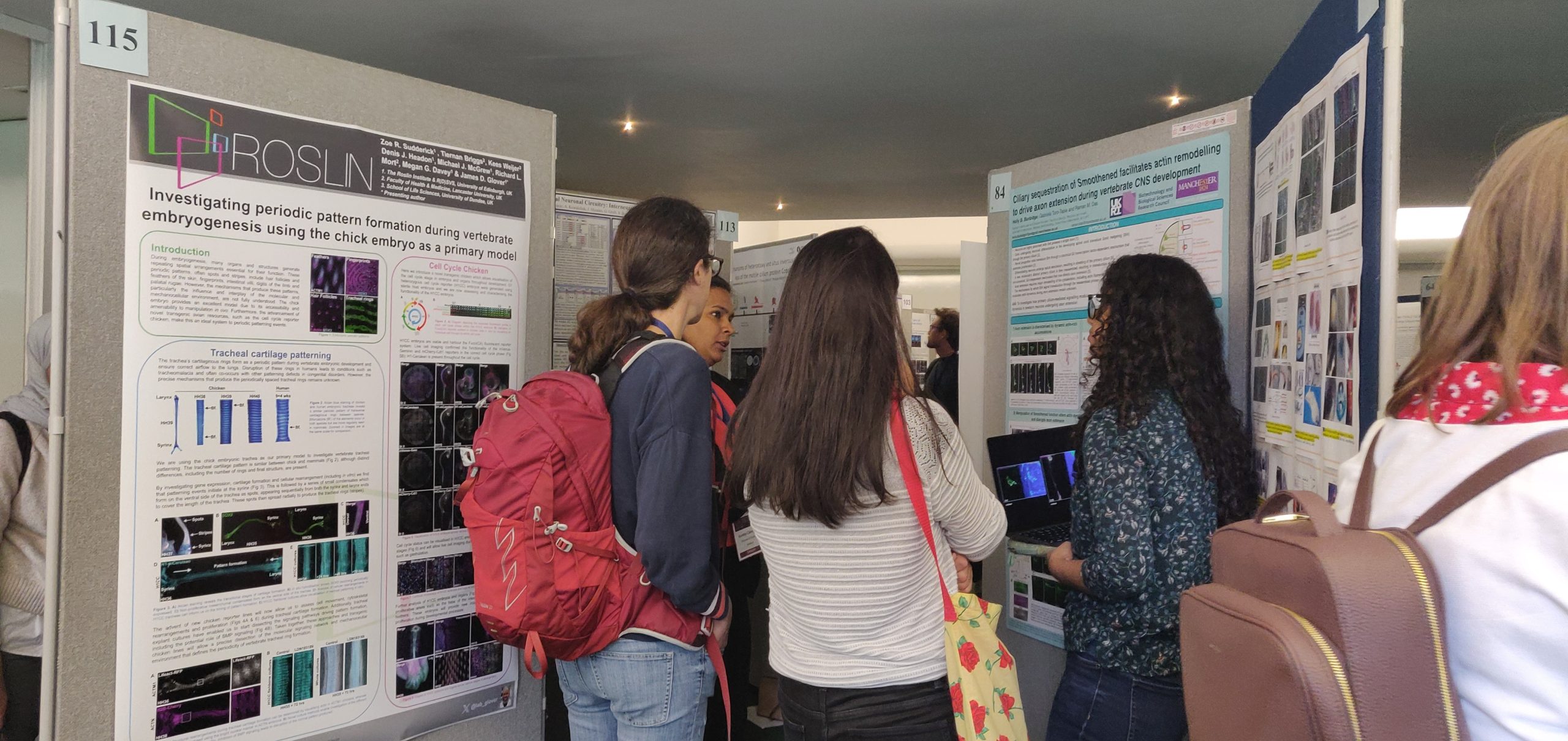
The relatively short first day was brought to a close with a poster session featuring a virtual reality experience, designed by Shaping Destiny, involving dance forms that depict embryonic growth. Unfortunately, I missed this as I got too immersed in the poster session (recruiting people to my poster), but those who experienced it said it was good fun. Hopefully, I’ll be able to try it at the next EDBC (or another BSDB conference)!
Poster presenters and their avid listeners
Episode 2
After a well-needed sleep, day two began with some morning Tai chi led by in-house Shifu Paul Martin. Those of us who braved the drizzly outdoors witnessed the Shifu very meticulously yet hilariously demonstrate how we could potentially defend ourselves. I, however, was embarrassingly hopeless, and then it was time to join the first session of the day to remind myself of biology which I feel I am marginally better at compared to Tai chi.
Day two was all about the hybrid format with interdigitating talks between Oxford and Paris. We started with the “morphogenesis” session, where Magali Suzanne described how apoptotic cells exert apicobasal forces on the neighbouring tissue and actively contribute to neural tube bending (4). Similar forces are also generated during epithelial-to-mesenchymal transition (EMT) in Drosophila (5). I also listened to Diana Pinheiro explain how a gradient of Nodal signalling fractionates the zebrafish mesendoderm into highly protrusive leader cells and less protrusive followers. The leader cells readily undergo internalisation while they pull the followers inwards in a mechanism that preserves mesoderm patterning (6). An interesting talk by Thibaut Brunet introduced the multicellular choanoflagellate, Choanoeca flexa. These remarkable organisms form multicellular sheets of polarised cells reminiscent of an epithelial monolayer (7) and reversibly transition between the multicellular colony state and unicellular dormant cysts depending on environmental conditions. The session ended with a visually stunning talk from Kate McDole who has developed an advanced light sheet microscope to investigate mouse organ development. By applying machine learning to the images, her lab follows the journey of individual cells up to organogenesis (8). Interestingly, this microscope can track the embryo’s position at complex developmental stages and adapt for optimal imaging without requiring manual input. How I wish I could use this fancy smart scope for my experiments!
After a morning of fascinating talks, the afternoon provided the much-awaited opportunity to explore beautiful Oxford. Some of us visited the botanical garden while others went punting. I decided to see some pretty flowers and when I returned in two hours, my phone was brimming with photos of colourful petals while my airways were revved up with pollen. Thankfully, I did not let out one of my signature deafening sneezes to disrupt the upcoming talks.
The afternoon was exclusively dedicated to talks from Paris with the theme “Gene regulation”. The opening talk by Claire Rougeulle showed that X chromosome inactivation and dampening are regulated by the same molecular players during early development in females (9). Nicola Festuccia then highlighted the transcription factor Nr5a2 as a master regulator of gene expression coordinating proliferation and genome stability during preimplantation in mice (10). At the end of the talks, we were treated to a grand dinner at the Hogwarts-esque dining hall of Keble College. With our stomach content, we celebrated the success of some ace researchers. Jonathan Slack was awarded the Wolpert medal recognising his impact through public outreach and teaching especially as he has written multiple books for scientific and non-scientific audiences. The Waddington medal recipient, whose identity was kept top secret until the award, was Marysia Placzek. The medal symbolised her major contributions to developmental biology, which became apparent as she took us through her research journey. The thousands of embryos she has dissected throughout her career were enough grounds to earn her the medal although her research on understanding patterning of the vertebrate nervous system was probably what did the trick. Alongside her career trajectory, it was amazing to see how she balanced her family life, a quality I find inspiring being a woman in science. To top it all off, the evening ended with a Hollywood-style trailer of the upcoming BSDB film that collated the diverse scenes of developmental biology in the UK.
Episode 3
The morning of day three was designed around “Concepts and theories in developmental biology”. Inaugurating the session, Jeremy Green delved into the theories of pattern formation based on the “reaction-diffusion model” and “positional information” theory (11). He also proposed two new ideas – mechanics and an omics approach to morphogenesis – in symmetry breaking. In a related aspect, Berta Verd showed that mathematical models of pattern formation should implement cell movements that are ignored in such models. Her research has established, for the first time, a framework to reverse-engineer gene regulatory networks (GRNs) of pattern formation in tissues under robust morphogenetic cell rearrangements (12). I also listened to James DiFrisco discuss the conundrum of how generalised principles can be extracted from diverse biological systems and applied across phylogenies. He suggested that combining mathematical modelling and our knowledge of evolutionarily conserved mechanisms may help address such complexities. Finally, Ruth Baker demonstrated that calibrating a mathematical model to high throughput experimental data from scratch assays can be used to infer mechanistic details underlying wound closure (13). Fitting right in with the session’s theme, the Beddington medal was awarded to Rasa Elmentaite for her fantastic PhD thesis investigating human intestinal development using single-cell RNA sequencing and spatial transcriptomics (14, 15).
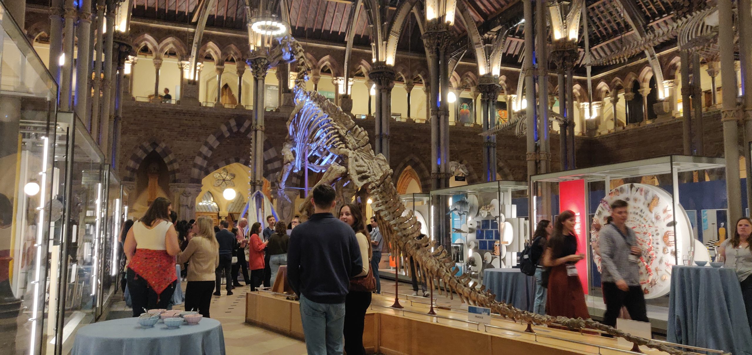
The afternoon was Barcelona’s moment in the sun (quite literally), similar to the format of day two. With quite a vast genre encompassing “Dynamics, mechanics and evolution”, this session was packed with interesting talks. Alejo Rodriguez-Fraticelli outlined how combining single-cell genomics and lineage tracing can be used to understand cellular heterogeneity and its consequences during development, ageing and cancer (16, 17). Yanlan Mao spoke about how tissues maintain their shape during repair and homeostasis. Focusing on wound closure in the Drosophila wing disc, she showed that tissue fluidity at the wound edge helps repair (18) with mechanical force-mediated cell shape changes contributing to the process. Next, Elvan Böke probed the question, “How do oocytes remain healthy for decades?” to which a part of the answer could be due to the ability of dormant oocytes to keep two detrimental factors in check – the production of reactive oxygen species (19) and the accumulation of protein aggregates in the cytosol. In his talk, Cristian Cañestro revealed how the tunicate, Oikopleura dioica, can be used to study gene loss and the deconstruction of GRNs to understand evolutionary diversity (20). Lastly, Antonio Scialdone showed that loser cells possessing mitochondrial defects tend to be eliminated in the mouse embryonic epiblast with cell competition selecting for optimal mitochondrial function before gastrulation (21). After a day of back-to-back talks, the evening was party time with the dinosaurs. Drinks in hand, we explored the quirky Pitts River Museum before ending up at the Museum of Natural History, our final stop for the night. With great food, drinks, music and dancing I unwinded in the revelry. If only the dinosaurs had come alive, it would have been a perfect re-enactment of Night at the Museum…
An evening with the dinosaurs at the Oxford University Museum of Natural History
Season finale
After partying (and one too many drinks), it was nothing short of a miracle that I made it to the first talk the following morning. It was presented by Julien Leclercq who was also awarded the Thesis Prize from the French Society for Developmental Biology for his outstanding PhD research identifying the genetic mechanisms that regulate eye formation in different morphotypes of the fish Astyanax mexicanus (22). Julien’s presentation was followed by the final instalment of talks revolving around “Regeneration, Disease and Ageing”, where Mathilda Mommersteeg challenged the current notion that oxidative phosphorylation inhibits regeneration. She highlighted that oxidative phosphorylation is required for the re-differentiation of cardiomyocytes and the long-term regeneration of the zebrafish heart. Stephanie Ellis then focused on how cell competition maintains tissue structure during mouse skin development. She outlined two sequential mechanisms – (a) the elimination and engulfment of loser cells by winners and (b) the expulsion of losers from the basal layer by differentiation (23). In the penultimate talk of the conference, Daria Siekhaus demonstrated that BMP signalling specifies the fate of leader macrophages that then infiltrate the Drosophila embryo to regulate homeostasis. Lastly, the closing keynote was delivered by Angela Nieto who talked about different programmes of EMT concerning three scenarios – development, fibrosis, and cancer. She discussed the commonalities and differences between these EMT forms and how they can maintain or breach epithelial homeostasis.
After four days of excellent talks, fruitful discussions and making new science buddies, it was time to return to life as usual. While on my train to Manchester, I felt grateful to have attended the EDBC. It not only provided important feedback for my work but also instilled in me new ideas that I could explore. I learned about aspects of developmental biology that I wasn’t familiar with before, helping broaden my outlook on the field and making me realise the scale of the versatile things that are indeed possible. While writing this report, I eagerly look forward to the next one. Here’s to EDBC 2027!
1. Chiaradia I, Imaz-Rosshandler I, Nilges BS, Boulanger J, Pellegrini L, Das R, et al. Tissue morphology influences the temporal program of human brain organoid development. Cell Stem Cell. 2023;30(10):1351-67.e10.
2. Rayon T, Stamataki D, Perez-Carrasco R, Garcia-Perez L, Barrington C, Melchionda M, et al. Species-specific pace of development is associated with differences in protein stability. Science. 2020;369(6510).
3. Dang Y, Lattner J, Lahola-Chomiak AA, Afonso DA, Taubenberger A, Ulbricht E, et al. Self-propagating wave drives noncanonical antidurotaxis of skull bones in vivo. bioRxiv. 2023:2023.07.10.547677.
4. Roellig D, Theis S, Proag A, Allio G, Bénazéraf B, Gros J, Suzanne M. Force-generating apoptotic cells orchestrate avian neural tube bending. Dev Cell. 2022;57(6):707-18.e6.
5. Gracia M, Theis S, Proag A, Gay G, Benassayag C, Suzanne M. Mechanical impact of epithelial-mesenchymal transition on epithelial morphogenesis in Drosophila. Nat Commun. 2019;10(1):2951.
7. Brunet T, Larson BT, Linden TA, Vermeij MJA, McDonald K, King N. Light-regulated collective contractility in a multicellular choanoflagellate. Science. 2019;366(6463):326-34.
8. McDole K, Guignard L, Amat F, Berger A, Malandain G, Royer LA, et al. In Toto Imaging and Reconstruction of Post-Implantation Mouse Development at the Single-Cell Level. Cell. 2018;175(3):859-76.e33.
9. Alfeghaly C, Castel G, Cazottes E, Moscatelli M, Moinard E, Casanova M, et al. XIST dampens X chromosome activity in a SPEN-dependent manner during early human development. bioRxiv. 2023:2023.10.19.563078.
10. Festuccia N, Vandormael-Pournin S, Chervova A, Geiselman A, Langa-Vives F, Coux R-X, et al. Nr5a2 is essential for morula development. bioRxiv. 2023:2023.01.16.524255.
11. Green JB, Sharpe J. Positional information and reaction-diffusion: two big ideas in developmental biology combine. Development. 2015;142(7):1203-11.
12. Spiess K, Fulton T, Hwang S, Toh K, Saunders D, Paige B, et al. Approximated Gene Expression Trajectories (AGETs) for Gene Regulatory Network Inference on Cell Tracks. bioRxiv. 2022:2022.01.12.476060.
13. Martina Perez S, Sailem H, Baker RE. Efficient Bayesian inference for mechanistic modelling with high-throughput data. PLoS Comput Biol. 2022;18(6):e1010191.
14. Elmentaite R, Kumasaka N, Roberts K, Fleming A, Dann E, King HW, et al. Cells of the human intestinal tract mapped across space and time. Nature. 2021;597(7875):250-5.
15. Elmentaite R, Ross ADB, Roberts K, James KR, Ortmann D, Gomes T, et al. Single-Cell Sequencing of Developing Human Gut Reveals Transcriptional Links to Childhood Crohn’s Disease. Dev Cell. 2020;55(6):771-83.e5.
16. Rodriguez-Fraticelli AE, Wolock SL, Weinreb CS, Panero R, Patel SH, Jankovic M, et al. Clonal analysis of lineage fate in native haematopoiesis. Nature. 2018;553(7687):212-6.
17. Weinreb C, Rodriguez-Fraticelli A, Camargo FD, Klein AM. Lineage tracing on transcriptional landscapes links state to fate during differentiation. Science. 2020;367(6479).
18. Tetley RJ, Staddon MF, Heller D, Hoppe A, Banerjee S, Mao Y. Tissue Fluidity Promotes Epithelial Wound Healing. Nat Phys. 2019;15(11):1195-203.
19. Rodríguez-Nuevo A, Torres-Sanchez A, Duran JM, De Guirior C, Martínez-Zamora MA, Böke E. Oocytes maintain ROS-free mitochondrial metabolism by suppressing complex I. Nature. 2022;607(7920):756-61.
20. Ferrández-Roldán A, Fabregà-Torrus M, Sánchez-Serna G, Duran-Bello E, Joaquín-Lluís M, Bujosa P, et al. Cardiopharyngeal deconstruction and ancestral tunicate sessility. Nature. 2021;599(7885):431-5.
21. Lima A, Lubatti G, Burgstaller J, Hu D, Green AP, Di Gregorio A, et al. Cell competition acts as a purifying selection to eliminate cells with mitochondrial defects during early mouse development. Nat Metab. 2021;3(8):1091-108.
22. Leclercq J, Torres-Paz J, Policarpo M, Agnès F, Rétaux S. Evolution of the regulation of developmental gene expression in blind Mexican cavefish. bioRxiv. 2022:2022.07.12.499770.
23. Ellis SJ, Gomez NC, Levorse J, Mertz AF, Ge Y, Fuchs E. Distinct modes of cell competition shape mammalian tissue morphogenesis. Nature. 2019;569(7757):497-502.
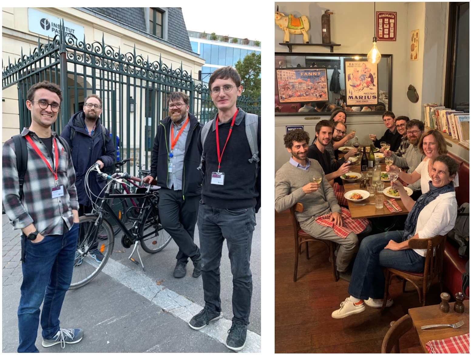
This year, the European Developmental Biology Congress experimented with an innovative conference format. A main meeting at Keble College in Oxford, UK, was complemented by two mini-hub meetings across continental Europe: one at Institut Pasteur in Paris, France, and the other at the Institute for Research in Biomedicine (IRB) in Barcelona, Spain. This format, proposed by Sally Lowell and the British Society of Developmental Biology, was conceived to maintain the benefits of in-person scientific interaction, a valuable component of conference attendance, while decreasing the environmental impact and cost associated with travel.
As part of the Paris hub organizing team, alongside Sigolène Meilhac, Nicola Festuccia, Tom Cumming, and Guillaume Frasca, I was excited to be part of this ambitious project. The Paris hub hosted 68 participants from around the world, most of whom had travelled from either within France or continental Europe, thereby achieving the main goal of the meeting.
The morning of September 26 began with a Morphogenesis session shared between Paris and Oxford. This series of talks opened with Magali Suzanne from Université Paul Sabatier in Toulouse. Magali spoke from Paris, sharing her work on mechanical forces exerted by apoptotic cells that contribute to tissue remodeling.
Following this, a series of fascinating talks seamlessly alternated between the Oxford and Paris locations. Those who spoke from Paris included Thibaut Brunet, who shared work from his lab at Institut Pasteur investigating the influence of environmental factors on multicellularity in choanoflagellates. He was joined by Adriano Bolondi from the Max Planck Institut for Molecular Genetics in Berlin who shared work from his PhD thesis exploring the mechanisms by which transient progenitors undergo coordinated changes during embryonic development, and by Amélie Elouin, a PhD student from École Polytechnique in Paris, who presented her work on non-cell autonomous functions of myosin in cell migration during gastrulation. The diversity of research topics and model organisms represented within this session, shared by speakers from all career stages, made for an exciting start to the integrated portion of the meeting.
After a poster session at the Paris hub, a second session on Gene Regulation was hosted exclusively from Paris and streamed in Oxford. During this session, we were delighted by talks covering diverse mechanisms of gene regulation in embryonic stem cells by both Claire Rougeulle, from Université Paris Cité, who shared work from her group on the role long noncoding RNAs in X chromosome inactivation in primates, and a talk by Nicola Festuccia who presented his work on the essential role of orphan nuclear receptors during the transition from genome activation to lineage specification. Postdoctoral fellow Cara Piciotto from Institut Pasteur shared work on the effect of cell-to-cell heterogeneity in binary fate decisions mediated by Notch signaling, and Robin Rondon, a PhD student at Institut Jacques Monod, spoke about the molecular mechanisms by which BMP signaling regulates patterning in the developing spinal cord.
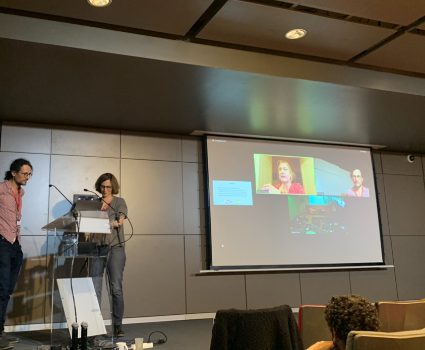
Romain Levayer (left) and Sigolène Meilhac (right) at the Paris hub interacting with Daria Siekhaus (UCLA) and the Oxford site over Zoom after Daria’s talk, streamed from California, on the role of BMP signaling in regulating immune cell infiltration during development in Drosophila.
During these integrated sessions, highly engaged participants asked many questions from both locations, with the interconnected format both promoting scientific curiosity and establishing a synergy between the two sites that was maintained throughout the week.
During the rest of the conference, sessions were streamed from both Oxford and Barcelona on campus in the Francois Jacob amphitheater, maintaining the community atmosphere that was initiated earlier in the week. Paris delegates listened to the talks from Oxford and Barcelona as a group, discussing new and exciting concepts over coffee during breaks between each session. The collaborative spirit of the conference became particularly strong on the afternoon of September 27, when the Barcelona hub had their shared session with Oxford and all three sites were connected online at once.
Right: Paris hub participants Tom Cumming, Thibaut Brunet, Guillaume Frasca, and Julian Leclercq outside of Institut Pasteur on a sunny afternoon. Left: Meeting participants enjoying an evening out for dinner in Paris.
In addition to the shared program integrated with Oxford and Barcelona, the Paris site hosted a poster session and evening cocktail social to promote further interaction among those at the hub. During the social event, Liza Sarde and Nisha Veits were each awarded a poster prize, and travel grants were given to Zeinab AlKobra AlHajj Hassan, Charlene Guillot, Joseph Leger, Xiaohui Liu, and Marcia Peixoto who travelled to Paris for the meeting. Congratulations to the awardees, as well as everyone who participated for their impressive achievements!
On September 28, the final day of the meeting, the French Society of Developmental Biology (Société Française de Biologie du Développement) Thesis Prize lecture was given by Julian Leclercq from Institut des Neurosciences, Paris-Saclay, who shared his PhD work on the evolution of gene regulation in the Astyanax mexicanus embryo with those present both in Paris and Oxford. This was followed by a final series of talks on Regeneration, Disease, and Aging, streamed from Oxford for those in Paris. Closing words from Paul Martin, Sally Lowell, and Shankar Srinivas from the Oxford organizing team marked the end of this first experiment in sustainable conferencing, which was a great success!
Within our collective of zebrafish labs at UCL, the sheer volume of single-use plastic Petri dishes we breeze through is staggering – a staggering 130 kg per year, to be exact. To put this into perspective, it’s a whopping 8.4% more plastic waste than the average person in the UK generates annually, and that’s solely from our Petri dish used for housing larvae. And petri dishes are not the only problem, we use thousands of plastic tips, tubes, PCR plates…and on and on. It’s a dilemma that resonates deeply with many of us in the scientific community. As sustainability takes centre stage across institutions, our host, UCL, has set commendable goals: aiming to eliminate non-essential single-use plastic on campus by 2024 and achieve net-zero carbon status by 2030. While sweeping institutional initiatives mark significant progress, the reality remains that not all these strides trickle down to our individual laboratories. In the scientific community, single-use plastic consumables are gold-standard, and ultracold storage is indispensable. Finding ourselves in this situation, all the fish groups sharing the communal labs got together and sat down to brainstorm ways to make our work more sustainable. We came up with ideas to reduce our environmental impact while continuing our research, in three main areas:
Plastics
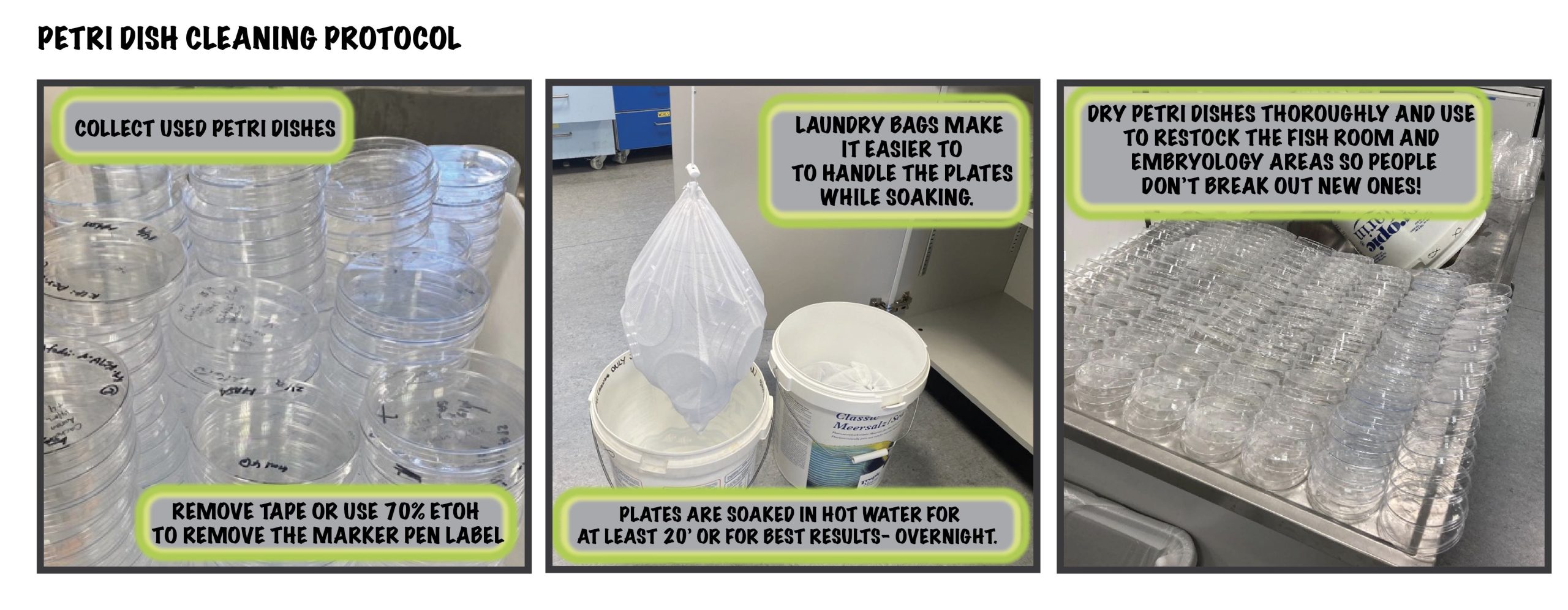
Even though single-use plastic tips and Petri dishes aren’t yet on UCL’s sustainability radar for 2024, we’re already taking steps to reduce our use across the groups. Where possible, we are swapping plastic for glassware and when we can’t use glassware, we are finding ways to reuse plastic tips and petri dishes whenever we can. For instance, to reuse Petri-dishes for housing zebrafish embryos we remove labels with ethanol, soak them in hot water for at least 20’ and then dry, ready to be re-used! We’ve also tracked our plastic usage by weighing our Petri dish bins weekly. This not only gauges our sustainability efforts but also aids in estimating equipment stock levels.
One lab protocol that significantly contributes to plastic usage is genotyping, a routine procedure consuming lots of plastic tips and PCR plates. Not commonly known is the practice of reusing tips for gel loading, a neat plastic-saving tip resurrected by the more mature lab members involved in large genetic screens and manual mapping of mutants. These mapping projects required running copious amounts of PCRs on lots of embryos and we did it reusing the same tips again and again. Credit originally to William Talbot’s lab in Stanford University (the master mapper!) for this thrifty and sustainable tip!
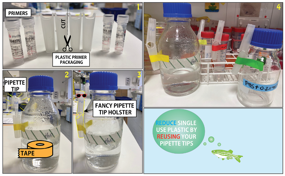
Not quite sure how to re-use pipette tips? Check out our video at http://zebrafishucl.org/plastic for simple tips. Reusing tips can serve various purposes, from simple DNA digestion controls to PCR products and RNA checks – any application where gel band extraction or subsequent analysis isn’t necessary. To reduce plastic use further, we also keep partially-used PCR plates for future runs. Start now by collecting used tips in a dedicated box for hassle-free diagnostic gel loading today!
And it’s not only for loading gels, pipette tips can have multiple lives. From water and buffers to alcohol and various solutions that are often used, tips can be reused! Everyone has their unique and fun way to store pipette and Pastette tips – discover yours or explore some ideas here.
Now that we can re-use some tips, what about plastic tubes and falcons? We all use lots of Eppendorf tubes and there isn’t much we can do about this. At a recent sustainability event at the Royal Society, Eppendorf’s life cycle assessment delivered a sobering result – their biobased tubes only show an underwhelming reduction of 16% in CO2 compared to their standard fossil-based counterpart. While there’s a projected 27% decrease in this, the current findings are disappointing. Not to mention the biobased tips are unfortunately more expensive.
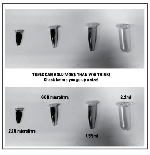
As we left the conference, we felt there was room for improvement. How often do we use a classic 1.5 mL Eppendorf for a miniscule 50 µL sample or for storing small aliquots of enzymes and antibodies? Turns out, the comfortably-stored volume for:
PCR tube – 0.220 mL
0.5 mL Eppendorf – 0.6 mL
1.5 mL Eppendorf – 1.6 mL
2 mL Eppendorf – 2.2 mL
Therefore, by just considering the right tube size for an experiment, instead of defaulting to the larger more commonly used 1.5 mL could drastically reduce our plastic usage and associated CO2 footprint. It seems we have the power to make a bigger impact than even Eppendorf can achieve currently, with minimum effort.
The principles of “reduce and reuse” can also be applied to other areas of the lab such as reagents. For diagnostics requiring only a few lanes, consider using a smaller gel. Here, we laser-cut a miniature electrophoresis gel mould for running extra small gels by repurposing our old/broken electrophoresis chamber plastics lids that uses only a third of agarose compare to our smallest commercially bought mould. You can download the laser cutting template from our website here.
Energy
We reduce energy consumption by turning off equipment when not in use, utilizing energy-efficient appliances, and optimizing equipment settings. This can be as simple as switching freezers from -80°C to -70°C (and reduce 30-40% energy consumption!) or switching off PC monitors after work, to deleting any useless data accumulating in the cloud! We also find that installing plug timers set to working hours on appliances such as water baths and heat blocks works really well.
Certain labs are still hesitant to switch ultra-low temperature freezer to -70°C because of sample safety concerns. We store our plasmid and antibodies stocks as well as most of our RNA at -20°C while total RNA, tissues, and competent cells are stored at -70°C. And we’re not the only ones, check out this list of samples successfully stored at higher temperatures by universities in the US. Increasing number of labs have now made the switch. Start 2024 right by joining a sustainability framework like the Freezer Challenge.
It’s always useful to regularly rethink ingrained lab practice. Some procedures appear to be engrained without clear origins. Take, for instance, the ‘cold hold’ – the infinite hold step at the end of a PCR protocol at the 4˚ C. Considering that our PCR products are double-stranded DNA, a fairly stable molecule, the need for this step becomes questionable. Don’t believe us? Here’s the evidence. Now, we’ve opted to terminate the cycle after the final extension, not only conserving energy but also prolonging the machine’s lifespan!
Culture of change
To maintain a consistent and concerted effort toward sustainability, we initiated a floor-wide green committee comprising volunteers from each of our labs. This committee also serves as a bridge between labs and core facilities, such as the UCL Fish Facility. Expanding on this initiative, we’ve established similar committees at the Departmental level, ensuring sustainability remains a priority. As part of this commitment, every new member, including students, postdocs, and PIs, undergoes sustainability training, and we’ve mandated plastic-free events throughout the department (take your own glass to socials, for instance). Our efforts are bolstered by frameworks like the laboratory efficient assessment framework (LEAF) which standardises sustainable practices. We joined LEAF in 2018 and thanks to contributions from many participants, the framework has evolved over the years and will keep improving and expanding. Being part of a framework that is endorsed by leading scientists and that is recognised by funding bodies helps with the acquisition, reinforcement and spreading of good, ideally, GOLD practices. Our entire department has set its sights on having all laboratories taking part of the LEAF and achieving Gold awards by 2025, a testament to our commitment to sustainability.
Despite having come a long way, there is still lots to do including in the way we think about how we conduct and publish research. Open research practices and open access publishing is indeed helping scientists to have better access to data, methods and resources and to minimise unnecessary duplication (while acknowledging that validating results is of course critical!). Sharing protocols and reagents pre-publication as well as publishing negative results should become the norm and we would love to see sustainability approaches within the methods section of papers (including positive as well as the negative outcomes).
While lots of science will remain competitive and sometimes secretive, the balance is shifting to more cooperative, collaborative, sustainable research with funders recognising the importance of community projects and resources that bring widespread benefits to the global research endeavour.
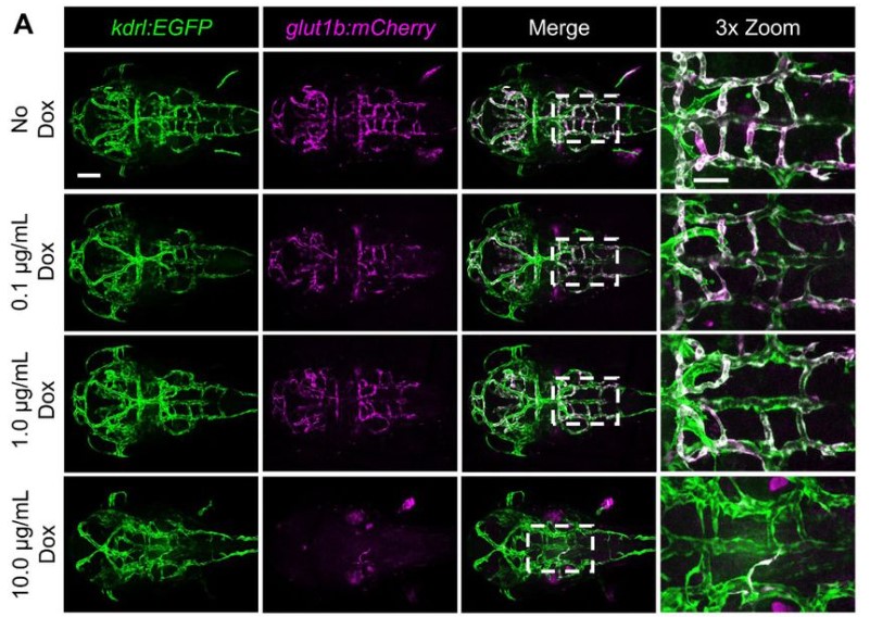
Alexandros Sountoulidis, Alexandra Firsova, Andreas Liontos, Jonas Theelke, Janine Koepke, Pamela Millar-Büchner, Louise Mannerås-Holm, Åsa Björklund, Athanasios Fysikopoulos, Konstantin Gaengel, Fredrik Bäckhed, Christer Betsholtz, Werner Seeger, Saverio Bellusci, Christos Samakovlis
Kieran M. Short, Giovane G. Tortelote, Lynelle K. Jones, Fabiola Diniz, Francesca Edgington-Giordano, Luise A. Cullen-McEwen, Jan Schröder, Ashley Spencer, Andrew Keniry, Jose M. Polo, John F. Bertram, Marnie E. Blewitt, Ian M. Smyth, Samir S. El-Dahr
Pedro Vallecillo-García, Mira Nicola Kühnlein, Mickael Orgeur, Nils Rouven Hansmeier, Georgios Kotsaris, Bernd Timmermann, Claudia Giesecke-Thiel, René Hägerling, Sigmar Stricker
Silvia Carvalho, Luna Zea-Redondo, Tsz Ching Chloe Tang, Philipp Stachel-Braum, Duncan Miller, Paulo Caldas, Alexander Kukalev, Sebastian Diecke, Stefanie Grosswendt, Ana Rita Grosso, Ana Pombo
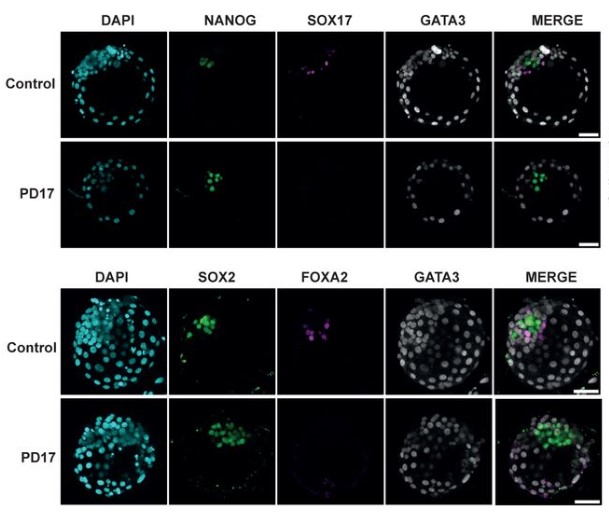
Anish Dattani, Elena Corujo-Simon, Arthur Radley, Tiam Heydari, Yasaman Taheriabkenar, Francesca Carlisle, Simeng Lin, Jonathan Mill, Peter Zandstra, Jennifer Nichols, Ge Guo
Valerio E.C. Piscopo, Alexandra Chapleau, Gabriela J. Blaszczyk, Julien Sirois, Zhipeng You, Vincent Soubannier, Geneviève Bernard, Jack P. Antel, Thomas M. Durcan
Stephanie Farhat, Bahaeddine Tilouche, Spencer Short, Medjie Piron, T. Mark Campbell, Alex Fernandes, Mariya Somyk, Hina Bandukwala, Eric Arezza, Quentin Sastourne-Arrey, Katherine Reilly, Maria Abou Chakra, Gary Bader, Leo Kunz, Timm Schroeder, Sasha Carsen, Pierre Mattar, Jeffrey Dilworth, Daniel L. Coutu
Karina O. Brandão, Viviana Meraviglia, Daniela Salvatori, Xu Cao, Luca Sala, Loukia Yiangou, Mervyn P.H. Mol, Milena Bellin, Christine L. Mummery, Richard P. Davis
Kuan-Hung Lin, Jamie E Hibbert, Jake L Lemens, Melissa M. Torbey, Nathaniel D. Steinert, Philip M. Flejsierowicz, Kiley M. Melka, Marcos Lares, Vijayasaradhi Setaluri, Troy A. Hornberger
Ioannis Oikonomakos, Melina Tedesco, Fariba Jian Motamedi, Mirko Peitzsch, Serge Nef, Stefan Bornstein, Andreas Schedl, Charlotte Steenblock, Yasmine Neirijnck
Anthony E. Postiglione, Allison M. Delange, Mohammad Foteh Ali, Maarten Houben, Eric Y. Wang, Stacy L. Hahn, Colleen M. Roark, Molly Davis, Robert W. Reid, James B. Pease, Ann E. Loraine, Gloria K. Muday
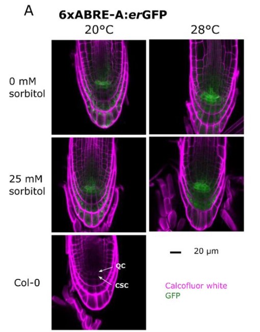
Nataliya E. Yelina, Eftychios Frangedakis, Zhemin Wang, Tina B. Schreier, Jenna Rever, Marta Tomaselli, Edith Forestier, Kumari Billakurthi, Sibo Ren, Yahui Bai, Julia Stewart-Wood, Jim Haseloff, Silin Zhong, Julian M. Hibberd
Yuan Zhang, Deepak Sharma, Yan Liang, Nick Downs, Fleur Dolman, Kristen Thorne, Ian Black, Jose Henrique Pereira, Paul D. Adams, Henrik Scheller, Malcolm O’Neill, Breeannna R Urbanowicz, Jenny C Mortimer
Anastasia I. Kurtova, Alexander D. Finoshin, Margarita S. Aparina, Guzel R. Gazizova, Olga S. Kozlova, Svetlana N. Voronova, Elena I. Shagimardanova, Evgeny G. Ivashkin, Elena E. Voronezhskaya
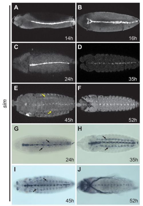
Luis Baudouin Gonzalez, Anna Schönauer, Amber Harper, Saad Arif, Daniel J. Leite, Philip O. M. Steinhoff, Matthias Pechmann, Valeriia Telizhenko, Atal Pande, Carolin Kosiol, Alistair P. McGregor, Lauren Sumner-Rooney
Matthew T Biegler, Kirubel Belay, Wei Wang, Christina Szi, Paul G Collier, Ji-Dung Luo, Bettina Haase, Gregory L. Gedman, Asha V. Sidhu, Elijah Harter, Carlos Rivera-Lopez, Kwame Amoako-Boadu, Olivier Fedrigo, Hagen U. Tilgner, Thomas T Carroll, Erich D. Jarvis, Anna L. Keyte
Sona Relovska, Huafeng Wang, Xinbo Zhang, Pablo Fernández-Tussy, Kyung Jo Jeong, Jungmin Choi, Yajaira Suárez, Jeffrey G. McDonald, Carlos Fernández-Hernando, Jean-Ju Chung
Matthew Lefebvre, Jonathan Colen, Nikolas Claussen, Fridtjof Brauns, Marion Raich, Noah Mitchell, Michel Fruchart, Vincenzo Vitelli, Sebastian J Streichan
Quan Xu, Lennard Halle, Soroor Hediyeh-zadeh, Merel Kuijs, Umut Kilik, Qianhui Yu, Tristan Frum, Lukas Adam, Shrey Parikh, Manuel Gander, Raphael Kfuri-Rubens, Dominik Klein, Zhisong He, Jonas Simon Fleck, Koen Oost, Maurice Kahnwald, Silvia Barbiero, Olga Mitrofanova, Grzegorz Maciag, Kim B. Jensen, Matthias Lutolf, Prisca Liberali, Joep Beumer, Jason R. Spence, Barbara Treutlein, Fabian J. Theis, J. Gray Camp
William Dalleywater, Alexander V. Predeus, Batuhan Cakir, Pavel Mazin, Jayakumar Vadakekolathu, Sergio Rutella, Marian L. Meakin, Alison A. Ritchie, Shamir Montazid, Sara Cuevas Ocaña, Nadine Holmes, Victoria Wright, Fei Sang, Adam Bills, Declan Sculthorpe, Rasa Elmentaite, Sarah A. Teichmann, Shazia Irshad, Ian Tomlinson, Andrew Silver, Ricky D. Wildman, Nicholas R.F Hannan, Felicity R.A.J. Rose, Mohammad Ilyas
Mirazul Islam, Yilin Yang, Alan J. Simmons, Vishal M. Shah, Musale Krushna Pavan, Yanwen Xu, Naila Tasneem, Zhengyi Chen, Linh T. Trinh, Paola Molina, Marisol A. Ramirez-Solano, Iannish Sadien, Jinzhuang Dou, Ken Chen, Mark A Magnuson, Jeffrey Rathmell, Ian G Macara, Douglas J Winton, Qi Liu, Hamim Zafar, Reza Kalhor, George M. Church, Martha J. Shrubsole, Robert J. Coffey Jr., Ken Lau
C. Lapoujade, M. Blanco, M. Givelet, A.S Gille, I. Allemand, L. Lenez, N. Thiounn, S. Roux, J.P. Wolf, C. Patrat, L. Riou, V. Barraud-Lange, P. Fouchet
Malgorzata Lagisz, Joanna Rutkowska, Upama Aich, Robert M. Ross, Manuela S. Santana, Joshua Wang, Nina Trubanová, Matthew J. Page, Andrew Adrian Yu Pua, Yefeng Yang, Bawan Amin, April Robin Martinig, Adrian Barnett, Aswathi Surendran, Ju Zhang, David N. Borg, Jafsia Elisee, James G. Wrightson, Shinichi Nakagawa
Melanie M Cooper, Marcos D. Caballero, Justin H. Carmel, Erin M. Duffy, Diane Ebert-May, Cori L. Fata-Hartley, Deborah G. Herrington, James T. Laverty, Paul C. Nelson, Lynmarie A. Posey, Jon R. Stoltzfus, Ryan L. Stowe, Ryan D. Sweeder, Stuart Tessmer, Sonia M. Underwood
To provide more visual content on the Node, we are starting a new series called ‘Show and Tell’. The aim of these short posts is to act as a hook for people to find out more about a paper, a technique or a location that is of interest to the developmental and stem cell biology community.
Write a ‘Show and tell’ post yourself!
Do you have an incredible image or video from one of your recent papers? Are you optimising a technique and want to showcase a piece of equipment you are using? Or perhaps your research involves going to a field site to collect samples?
Post an image, photo, or video of your choice, and answer the questions below. Keep the answers short and snappy, and always include a link at the end for people to find out more. Be as creative as you want with what you show people, as long as it is relevant to #devbio!
What is this?
Where can this be found?
How was this taken?
What does it do?
Why should people care about this?
How would you explain this to an 8-year-old?
Where can people find more about it?
(Note: you can choose to answer the questions that apply to you, and feel free to adapt the questions to fit your answers)
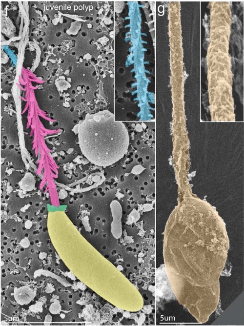
Scanning Electron Micrographs (SEM) of a sea anemone stinging cell (F) and change in cell identity following the knockout of a gene called NvSox2 (G). Images are pseudo-colored to highlight characteristics of these cells, collectively called cnidocytes. Taken from Babonis et al. (2023).
What is this?
A sea anemone stinging cell (F; left panel) and change in cell identity following the knockout of a gene called NvSox2 (G; right panel).
How was this taken?
With a Scanning Electron Microscope. Images are pseudo-colored to highlight characteristics of these cells.
What does it do?
Sea anemones have various stinging cells, called cnidocytes, with different functions such as piercing prey for feeding or sticking to surfaces so they don’t get swept away by ocean currents.
Why should people care about this?
Piercing cells come in over 30 different sizes and shapes, all of which were thought to share a single “piercing” ancestor. But at some point in the starlet sea anemone’s evolution, a gene called NvSox2 silenced the molecular decisions that led stinging cells to become “sticky” and instead instructed them to become a specific type of “piercing” cell (F), effectively inventing a new cell type.
When NvSox2 is disabled, these “piercing” cells are restored to their ancestral “sticky” identities (G) that have been forgotten in this particular anemone’s long evolutionary history.
These results suggest that single-gene switches driving cell fate have been around for a long, long time and provide a flexible molecular mechanism for animals to adapt to new and changing environments as needed to survive. It’s plausible — maybe even likely — that other types of “piercing” cells have a similar gene-induced amnesia blocking cellular memories of a distant past.
How would you explain this to an 8-year-old?
Sea anemones (pronounced “uh-neh-moh-nees”) don’t look like you and me. They’re related to jellyfish and have special stinging cells. If you’ve ever been stung by a jellyfish, you know that can hurt!
These are pictures of two cells that come from a sea anemone. Someone has colored the cells for us so we can see them better. They’re very different from each other. Can you see how they’re different?
The smooth yellow cell on the left shoots pointy darts into the sea anemone’s food so it can eat. The dart is colored pink and blue in the picture.
The tan cell on the right has large, sticky strings that keep the sea anemone from being washed away.
When these cells are young, they’re not sure if they’re supposed to shoot small pointy darts or large sticky strings when they grow up. Do you know what you want to be when you grow up? How do you know what to choose?
These cells are told what to do with an on-off switch. When the switch is flipped on, the cell forgets how to be large and sticky and looks like the small pointy cell on the left. That’s what this sea anemone normally does. But when scientists turn the switch off, the cell suddenly remembers it should be large and sticky like the cell on the right.
Wouldn’t you like to have a switch to help you remember something that you forgot?
Switches like this can teach us how animals invent new cells. It also shows us how cells store old memories that might help them survive through hard times. When something works, you don’t want to forget it!
Where can people find more about it?
This paper was featured on Nature Communications Editors’ Highlights page. You can find the full-length article here.
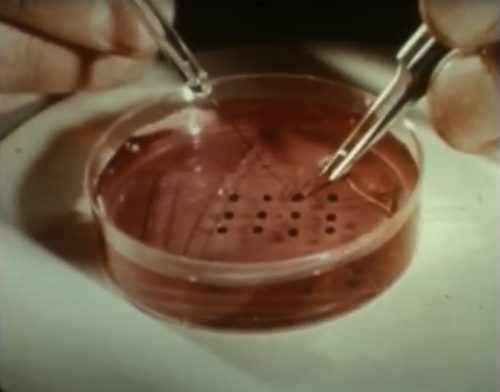
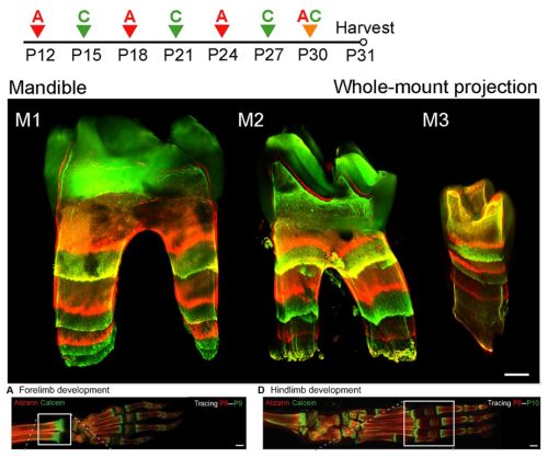
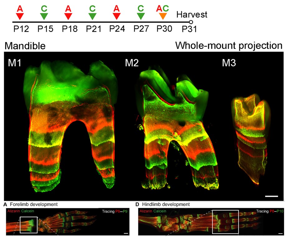
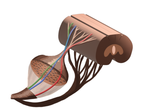
BEE-ST (Bone and tEEth Spatio-Temporal growth monitoring) is a method to monitor mineralization dynamics across species. It is based on the intraperitoneal (abdominal cavity) administration of fluorescent calcium-binding dyes in an animal of interest at the specific time you want to observe newly-forming mineralized tissue. Sequential administration of dyes that fluoresce at different wavelengths (alizarin, red; and calcein, green) enables the observation of bone and tooth mineralization dynamics across time.
Where can this be found?
The technique can be applied to all tissues that undergo mineralization in various species, so the fluorescent dyes will be found in parts of bones and teeth that have developed since the administration of the dyes. The technique was developed by graduate student Marcos González López and his colleagues in Jan Křivánek’s lab at the University of Masaryk in Brno, Czechia.
How was this image taken?
The image was taken by high-resolution confocal laser-scanning microscope. Confocal microscopes use fluorescent lasers to illuminate different depth spots in your sample. By using confocal microscopy, you can obtain several images in different planes that can be put together to recreate a 3D model of your sample, in this case, the mouse molar.
What does it do?
The BEE-ST technique enables researchers to investigate how mineralized tissues (bones and teeth) develop or repair themselves after injury. This is achieved through the binding of dyes to the newly-synthesized or newly-repaired regions of bone or teeth. Once harvested, bones and teeth can be subjected to an optical-clearing protocol, enabling light to pass through them, and a couple of days later the tissue is ready to be imaged by fluorescent laser-scanning microscopy.
Why should people care about this?
The BEE-ST method is a powerful to understand how hard tissues become mineralized.
In the developmental biology and tissue engineering research communities this tool can be utilized to:
Gain a better understanding about how bone and teeth obtain their shape.
Evaluate how changes in the expression of genes, proteins or signalling pathways influence bone or tooth regeneration.
Assess the effectiveness of small molecules or drugs in promoting repair and healing of bones.
How would you explain this to an 8-year-old?
Teeth and bone have a lot of calcium in them. When they grow, or when they break and repair themselves, they add more calcium to themselves. We can use special, coloured dyes that “grab onto” growing teeth and bones to see how fast they grow or repair themselves. This way, when we look at a tooth or bone from an animal that we gave these dyes to, we can see which parts grew recently. Like you can see in the picture, when we switch between red and green dyes, we can find out how long ago each part of the tooth or bone grew, or how quickly. Look at the bones from the front and back leg of the mouse: the red parts grew before the green parts. This way, we can study how teeth and bones grow, and how they repair themselves after they break. We can also test new medicines to see if they can help bones heal more quickly after they break.
Where can people find more about it?
If people want further information on how this method works and its versatile use in all mineralized tissues across species, they can read the recent paper from the Křivánek lab at the Department of Histology and Embryology at Masaryk University in Czechia.
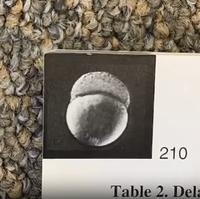
Flipping through the pages of Development Volume 123 Issue 1 (1996) shows the dynamics of zebrafish embryogenesis from the 2-cell stage to the 16-somite stage.
What is this?
A physical copy of Development Volume 123 Issue 1 (1996), with a flipbook at the upper corner of the issue showing zebrafish embryogenesis over 17 hours from the 2-cell stage to the 16-somite stage.
Where can this be found?
The Company of Biologists office in Cambridge, UK
Why should people care about this?
Consisting of 37 papers, this zebrafish special issue presented the results of two large screens for zebrafish mutants. The papers describe about 1500 mutations in more than 400 new genes involved in a wide range of processes that govern development and organogenesis. The mutants described in this issue provide a rich resource for many zebrafish laboratories to study embryogenesis, neuronal networks, regeneration and disease.
How would you explain this to an 8-year-old?
What happens to the first 17 hours of the life of a little zebrafish? In this science book, when you flip through the pages, you can see how the zebrafish grows from just two cells to 16 cells by the end of the book, where you can start to see the shapes of the eyes and brain of the zebrafish.
These zebrafish may look very different from us humans, but they are actually very useful for scientists to learn more about the general rules of how we grow, and what happens to us if something inside us is not working properly.
Where can people find more about it?
For more details about the making of the ‘flipbook’, read this article.
 (6 votes)
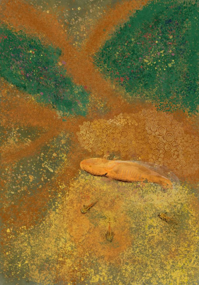
 (No Ratings Yet)