The FocalPlane Network is an international directory of researchers with expertise in microscopy. It is designed to promote networking, as well as a resource for those looking for collaborators, reviewers, speakers and committee members.
Like the Node Network, the FocalPlane Network is an inclusive site and we hope that it will help promote diversity in the microscopy community. Members provide information on their scientific field, microscopy area of expertise, place of work and career stage. They can also voluntarily provide details on aspects of diversity such as gender, race/ethnicity, LGBTQ+ identity and disability status.
You can find out why our members think that the Network is important on a new post FocalPlane. If you use microscopy as part of your research, we would like to encourage you to join and use the FocalPlane Network.
To showcase the variety of interests and artistic talents among the developmental biology community, the Node and the British Society for Developmental Biology (BSDB) is jointly hosting a virtual art exhibition, to accompany the European Developmental Biology Congress(EDBC).
Voting is now open to the public to pick their favourite artwork from the three categories:
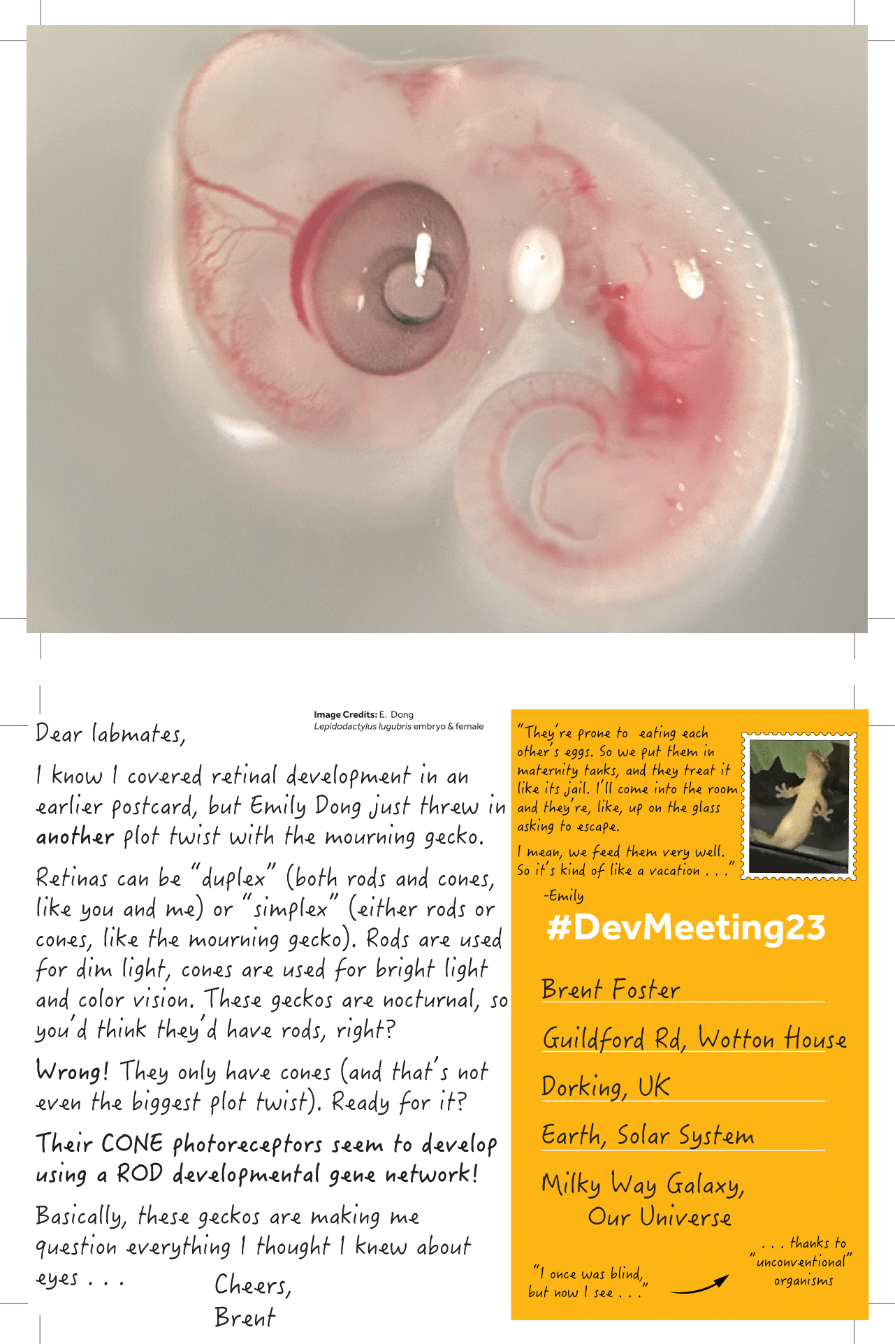
1) Scientific images
2) Science-inspired art
3) Art by scientists
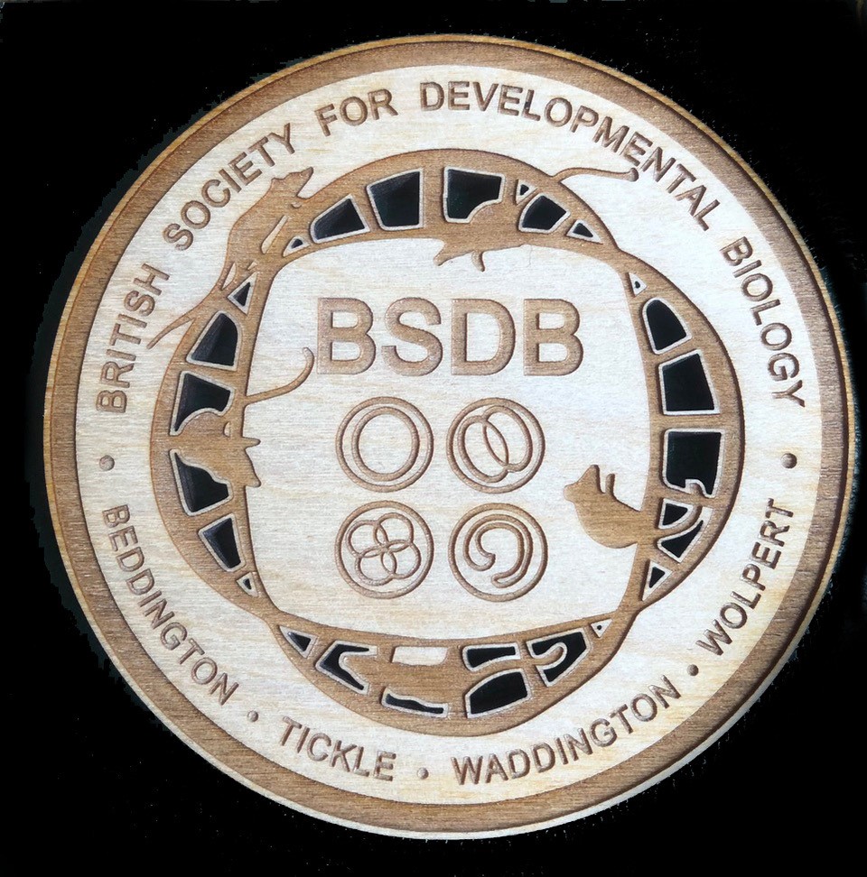
There will be a ‘People’s Choice’ from each category. A panel of judges will also pick a winner from each category, who will receive a laser-cut wooden coaster designed by Helen Weavers.
Voting closed on 25 October 2023 23:59 GMT.
Visit the different rooms of the exhibition
The ‘Science-inspired art’ and ‘Art by scientists’ room
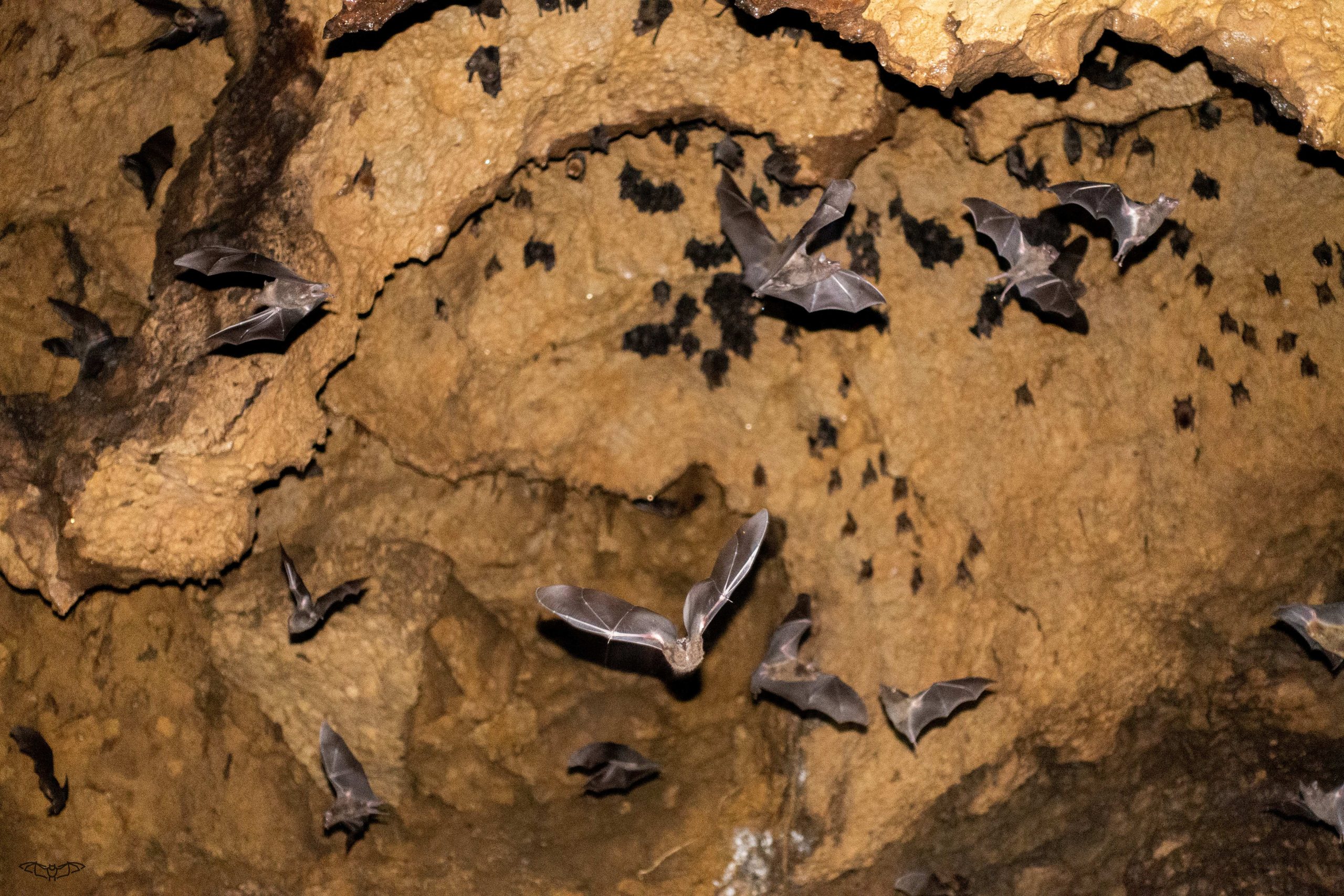
Dr. Alexa Sadier is a Research Scientist in Karen Sears’ lab at UCLA. She is using bats as a model system to understand the origin and diversification of a key mammalian innovation, the tooth classes (e.g. incisors, canine, premolars and molars). From the µCT scans and two-photon microscope in the lab to the jungles and caves of Trinidad, find out more about the story behind the team’s recent paper!
What brought you to join Karen’s lab? I joined Karen Sears’ lab in 2015 to study the evolution of sensory systems in noctilionoid bats, the family I am focusing in. As an evolutionary developmental biologist, I quickly fell in love with both the animals and the system.
How did the project get started? What was known about the origin of mammalian teeth before your work? I did my PhD on molar evolution in mice, in which we demonstrated that the particular shape of the mouse first molar can be explain by the complex evolution of dental patterning in this species. This training gave me a deep knowledge of tooth development and the potential of this system. After working on bat vision evolution, I decided to launch my own area of research in Karen Sears lab (I thank her SO MUCH for giving me this opportunity) because I realized that bat teeth are so diverse that they can be a good model system to study phenotypic diversification given our deep knowledge of tooth development in mice. On the contrary to mice, bats possess all tooth classes so we can study not only molars and very derived incisors, but all of them. We can investigate mechanisms that are still not known such as what makes an incisor an incisor, a canine a canine, a molar a molar, etc. Indeed, most of the developmental studies have been done in mice. We know that tooth class identity seems to be determined early during development, through a prepatterning of the jaw, but our understanding of what happens after and their respective morphogenesis is extremely limited.
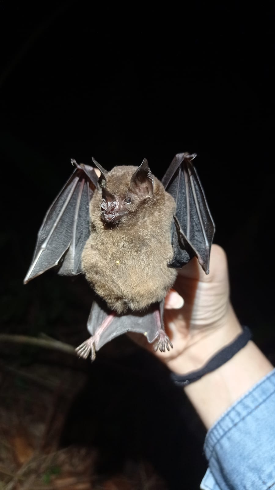
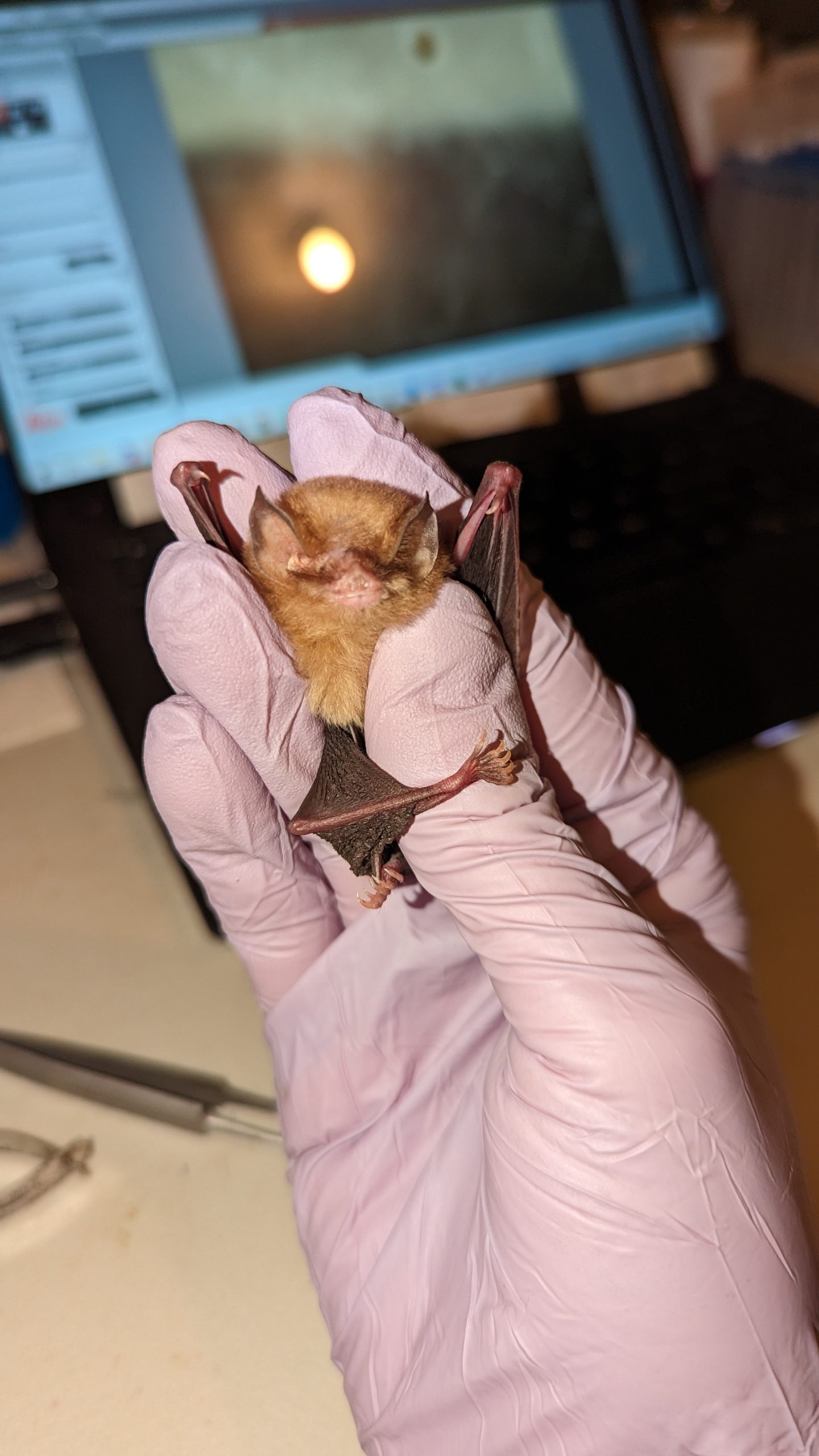
What made you choose noctilionoid bats as your model organism? For an evolutionary biologist, noctilionoid bats are a fantastic model since phyllostomids (a group of noctilionoids) underwent an adaptive radiation (like Darwin finches) and have evolved various diets. You probably know the vampire bats which eat blood but other species specialize on fruit, nectar, insects, vertebrates, fishes or even pollen. This adaptation to various diets had shaped their evolution at all levels: the shapes of their skull and teeth are adapted to their main diet, so are other systems such as vision, echolocation, etc. In the Sears’ lab, I realized that what I thought was only possible in model species, such as mice, was also doable in bats (up to a certain point). Before working with them, I would have been skeptical about the ability to perform developmental biology or even functional experiments on non-model species. Now that the genomes are available through Bat1K, and developmental material thanks to field expedition and museum specimens, we have all the tools we need to study them from genotype to phenotype. It’s a kind of an eco-evo-devo researchers’ dream.
Can you summarise your key findings? Noctilionoid bats exhibit a huge variation in their tooth number, size and shape due to the colonization of various dietary niches in only 25 million years, making them a fantastic model to study the developmental basis of rapid morphological evolution. We used integrative approaches (morphological measurement on adults and embryos, cell proliferation labeling and modeling) to investigate the development and evolution of two tooth classes, premolars and molars.
We found that premolars and molars develop and evolve independently by two different Turing-like rules in bats, and probably other mammals, that deviate from previous models (the Inhibitory Cascade (IC) model). This important result brings new insights regarding the developmental and evolutionary differences between tooth classes – a major mammalian innovation – that remain relatively obscure and limited in their taxonomic scope. Then, by linking this variation with the variation in jaw length, we show that the interaction between Turing-like mechanisms and growth rate is sufficient to generate the observed variation. Our work demonstrates how new morphologies are reached by modulating the interaction between multiple developmental constraints during the burst of diversity that accompanies adaptive radiations. While the idea that growth rate variation is important for Turing mechanisms is not novel, our work proposes that it can facilitated the apparition of new phenotypes in teeth and potentially other ectodermal appendages that develop like teeth.
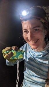
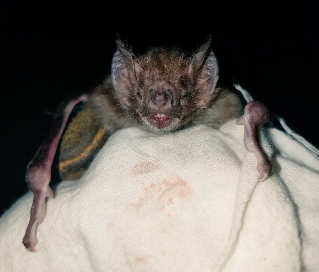
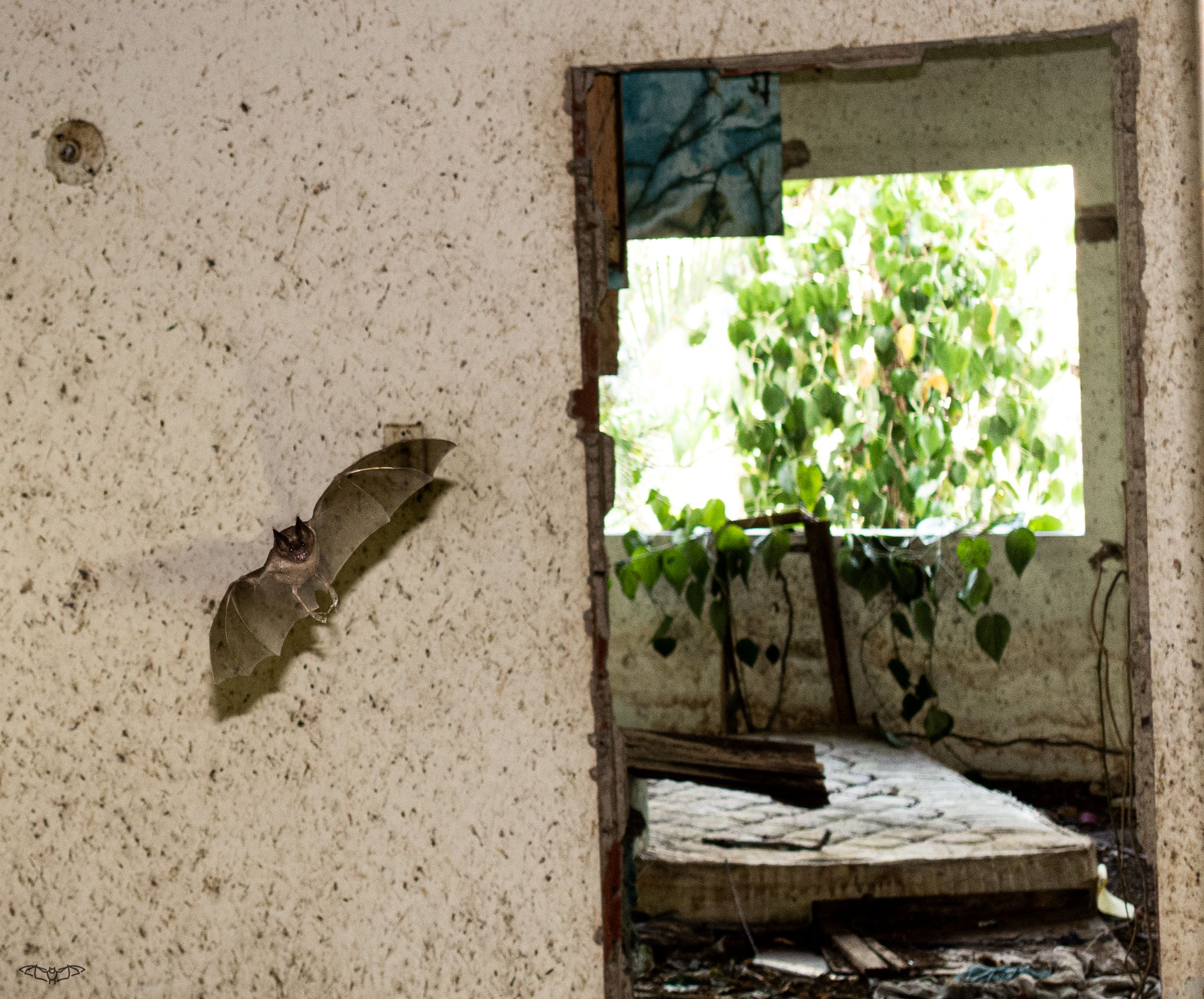
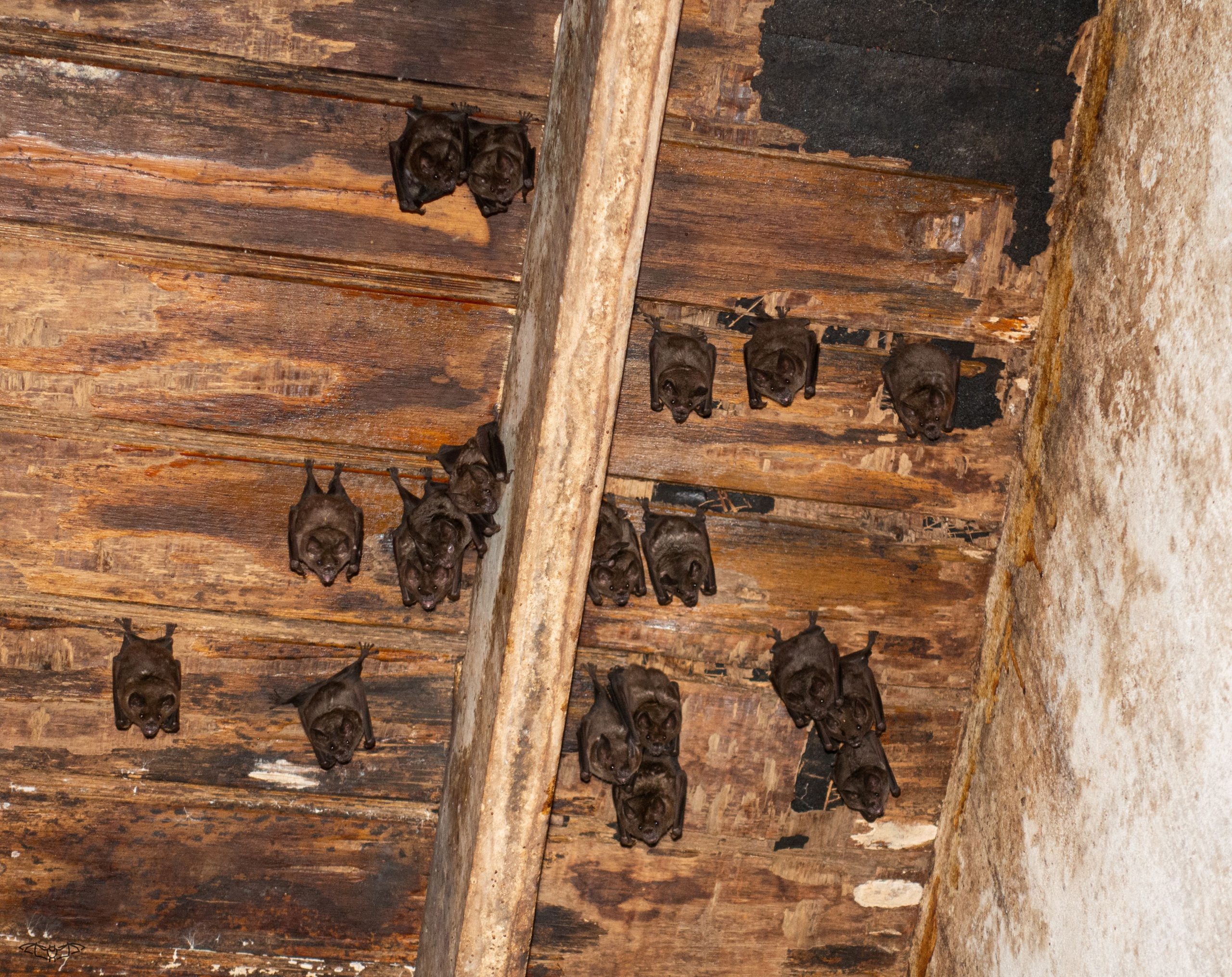
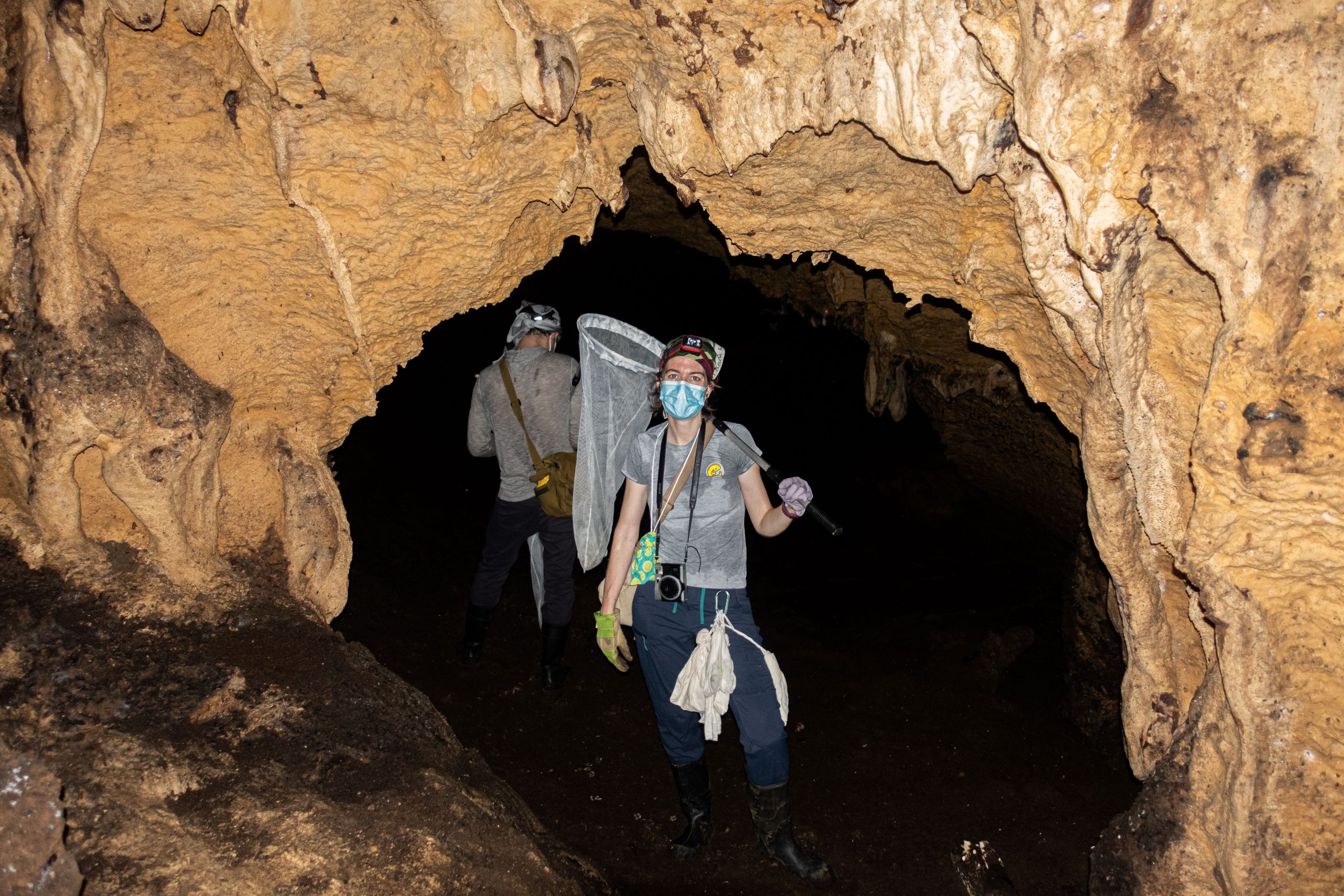
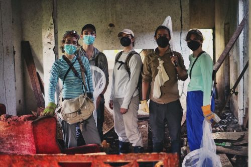
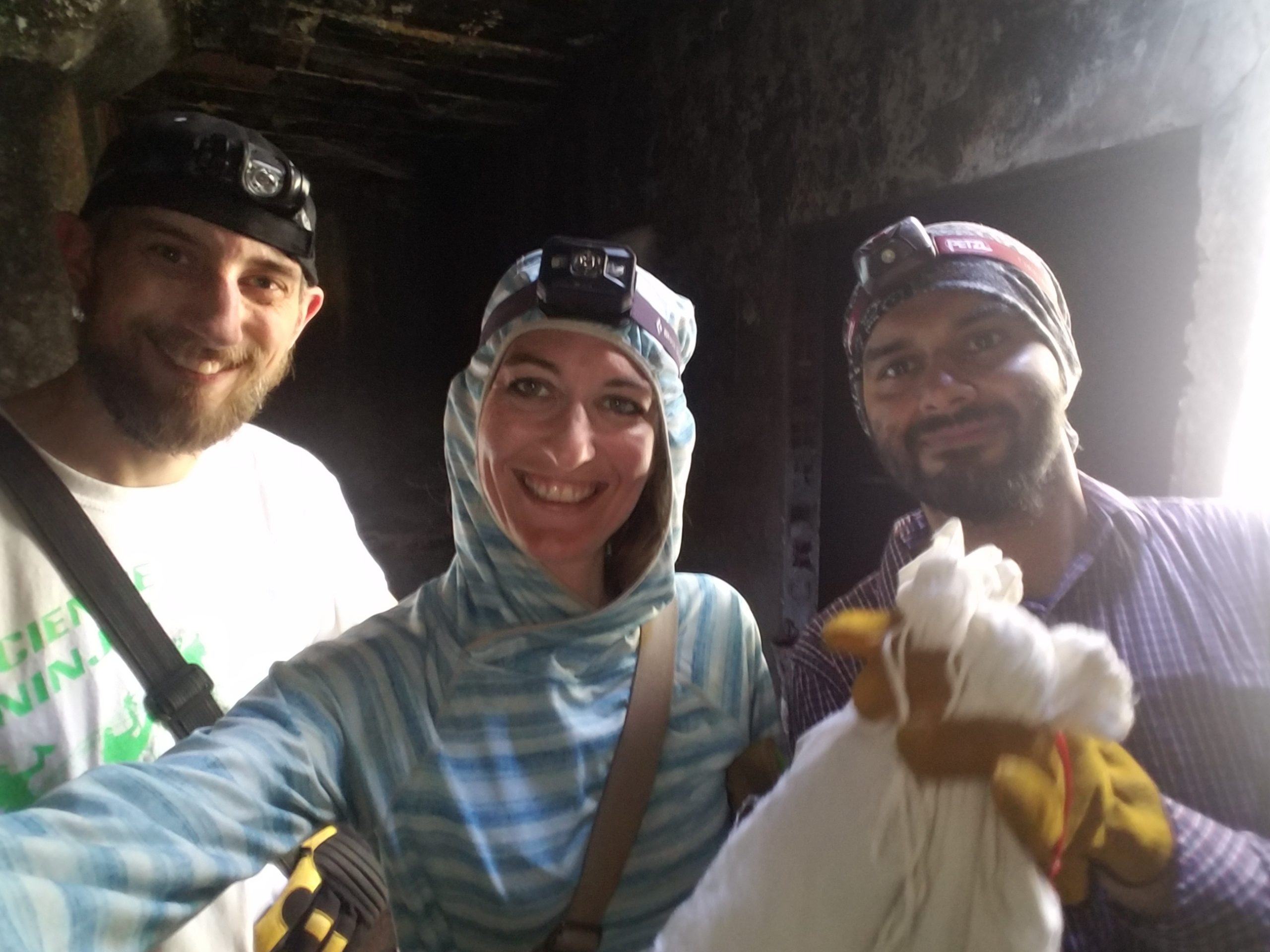
How was it like working with bats? Any memorable stories about the fieldwork? It’s probably my favorite moment of the year even if it implies being sleep deprived, long nights, a lot of work and administrative tasks but all of this disappear when we hold a bat with its little personality. Typically, we travel to Trinidad, Dominican Republic or Puerto Rico for 2 to 3 weeks at the time. Our days and nights are organized around bats. At 2-3 pm, we generally drive to a field site to be ready with our traps at 5-6pm when the bats come out. We then catch them until 11pm and put up a triage station to decide which one we release (95% of them) before driving back with them in cotton bags. Then, the long processing night starts, sometimes often until 5-6 am. We then sleep for a few hours, eat, and repeat. Some people think we go to the beach and enjoy the Caribbean life with rum every night but the reality is that we barely have time to complete our tasks. And when we do, we try to catch some sleep. As though as it could be for the body, there is something magical when we go down into a cave with all the bats flying around us. Forests are also special places, with so many species, so are abandoned houses, sometime frozen in time. I have so many fieldwork memories, but one is particularly fun. When looking for fishing bats, we had, one day, to swim into a bat cave from a boat with our butterfly nets. As we were swimming into the cave, which was like a narrow tunnel, we started to smell them. Then the cave became wider and all the bats were there, with pup, looking at us. We couldn’t believe it was true. Finally, the best of all is probably sharing this with our local collaborators, year after year, triaging bats sitting in the back of the truck, laughing, sharing, nerding, eating the delicious Trinidad doubles, working hard but in such a special atmosphere.
Do you think that doing fieldwork change the way you perform your research? Yes! In eco-evo-devo, it’s a new way to think about the species you work on. Seeing them in their environments can really make a difference in your research. I remember this conversation with a researcher who solely study the genomic aspects of bat evolution. From his dataset, he thought that one bat species was blind although it’s clear, when seen it in the field looking at you, following your finger, looking around, that it is not the case. Fieldwork adds another dimension to our work as evolutionary biologists. From a more personal point of view, as an outdoor person who grew up in the French Alps, I have always been skiing, climbing, hiking, etc. I love being outside and I always think of science as a way to explore, exactly like explorers who discover new territories. Fieldwork represents the ultimate fusion between geographical and intellectual exploration: we are looking for new species, new specimens, new results while we explore new areas and make link between everything.
How was your experience collaborating with people with different expertise for the paper? It was really great, especially regarding the modeling aspect of it. Being able to find a mathematician interested in biology and vice versa was really one of these fun moments in research. Interdisciplinary research is not always easy and it’s always a special moment when everything is finally getting together. It’s also a way to push the boundaries when it works well like this.
Did you have any particular result or eureka moment that has stuck with you? Yes! And it was so good. I was segmenting teeth at the computer and my colleague and friend Neal, who is a co-author, was in the lab. I realized that, in some bats, the premolar that disappears is the middle one (on the contrary to molars) and that it happens gradually during evolution. I was like WTF! and asked him to come next to me to tell him what was his conclusion (without telling him first). We looked at each other being like: that’s AMAZING! It really changed the study and the way I then thought about these results.
And the flipside: were there any moments of frustration or despair? Of course, when the two-photon images were too big (1.6 Tb) to be opened and we had to start imaging everything again not knowing if the dyes would have survived in our precious samples. Or during COVID, when fieldwork was not possible despite our need to get more developmental stages or more museum specimens and thought it would delay the paper. And more than anything else, being a postdoc and then a research scientist with all the uncertainty that comes with it. While it’s exhilarating in so many ways, it’s still a temporary situation that implies a lot of sacrifices, long distance relationships, and living on grants, sometimes not knowing if everything will stop after 6 months. Delaying results can have devastating consequences on an application cycle. I knew I couldn’t abandon this project because I deeply believed in it (its significance, the science behind it and where we can go from it), but I had some close calls because I was so sick of sacrificing many things I love (including my personal life) for my career. I made these sacrifices but not anyone can do it (or even want). For me, this is the most challenging part of developing new risky research at this career stage.
Where will this story take the lab? This paper really showed the potential of this model to study the origin of tooth classes and is the foundation of my future research program. Next research will investigate how the variation of the dental gene network drives tooth class diversification. We will still use bats as a model system and plan to extend to other mammals, and long term, ectodermal appendages that develop the same way!
What is next for you personally after this paper? Hopefully, the best is yet to come! I’m about to start my group using this model system to study the evolution of tooth classes, so this paper constitutes the foundation of the future, it’s a beginning. Developing this model and program (along with the other paper that came out of it) helped me to gain confidence in my science and research and establish my area of research. It will always have a special place.
Reference Sadier, A., Anthwal, N., Krause, A.L. et al. Bat teeth illuminate the diversification of mammalian tooth classes. Nat Commun14, 4687 (2023). https://doi.org/10.1038/s41467-023-40158-4
Pictures: Alexa Sadier, group picture: Marie Treibert
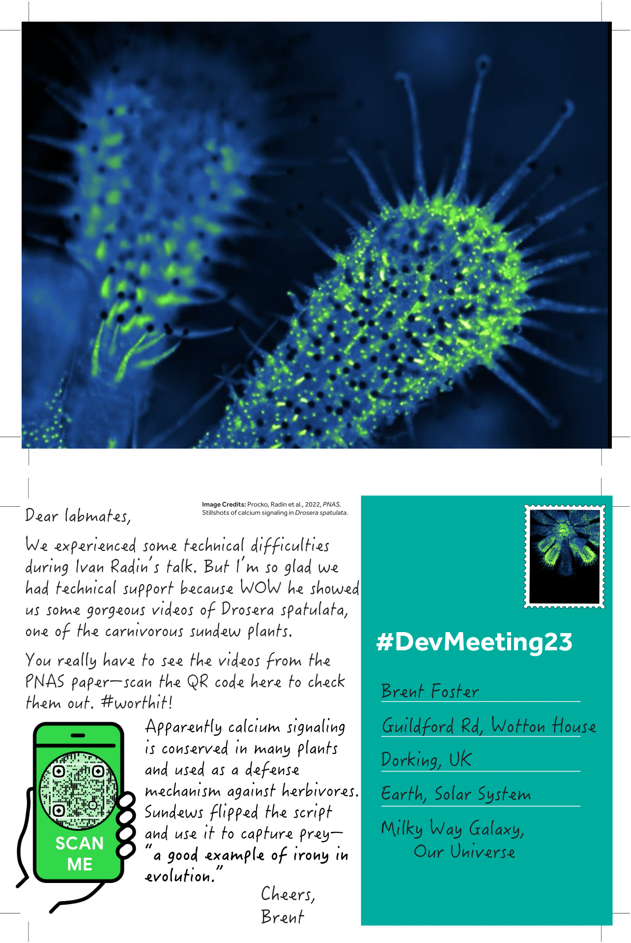
From Procko, Radin et al., 2022. “Dynamic calcium signals mediate the feeding response of the carnivorous sundew plant.” PNAS.
More plant representatives
For this postcard, I’ve selected Ivan’s talk about calcium signaling in the sundew, but I wanted to give a shoutout to some of the conference’s other plant representatives. We’ve learned about
how the brown algae Ectocarpus can help us answer questions about the origin and evolution of multicellular development (FUN FACT: this algae’s female gametes emit a pheromone that smells like gin)
how the moss Physcomitrella patens teaches us about single-cell perspectives of tissue patterning
how the wild grass Brachypodium distachyon addresses developmental questions of how form impacts function in plant stomata (FUN QUOTES: “Stomata are cellular bouncers” and “”Plants are tunneling experts, much like the Swiss”).
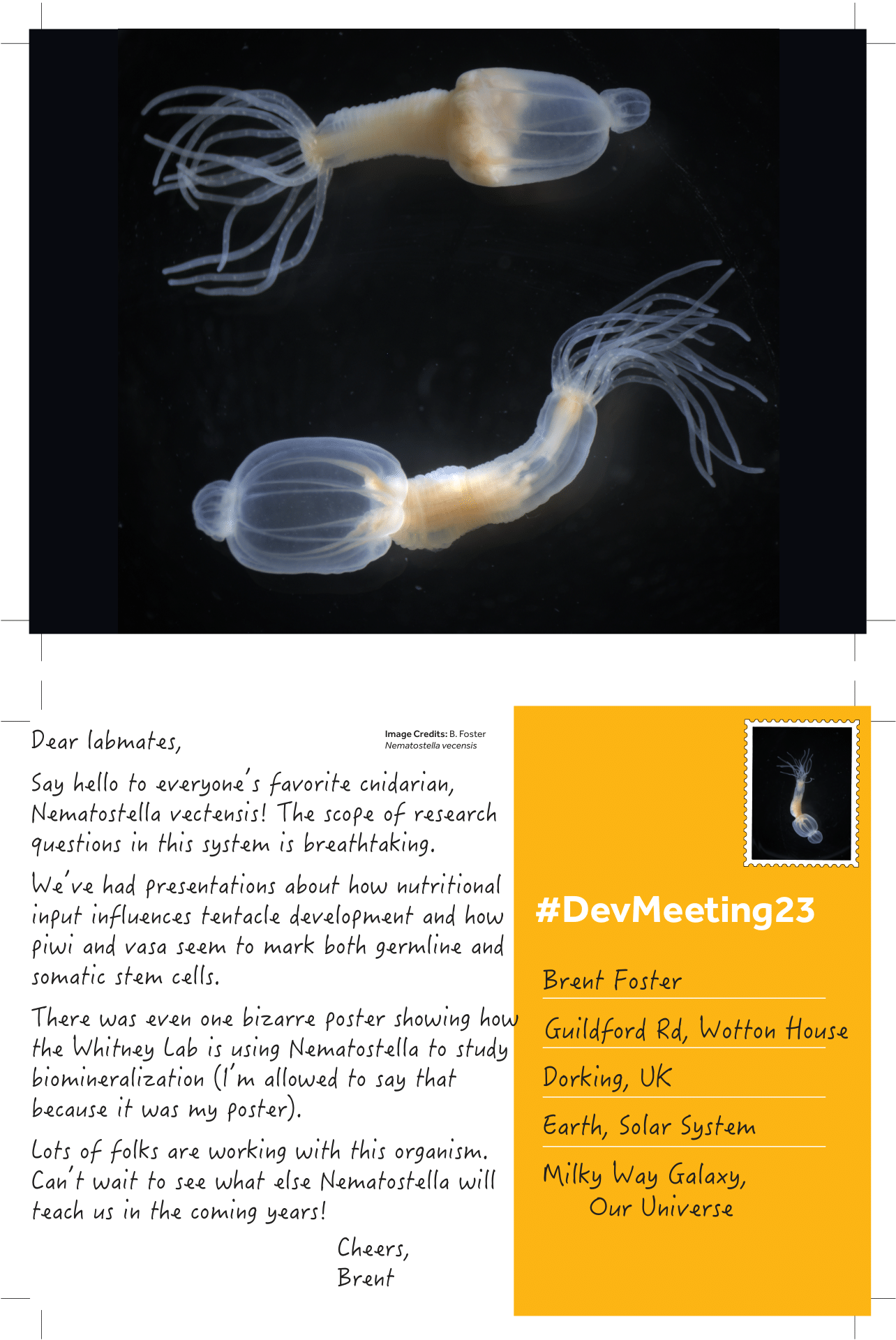
On Monday we learned a bit about early branching animals, especially the cnidarians. One poster mentioned ongoing work in ctenophores.
Note: I should point out that Nematostella wasn’t the only cnidarian mentioned at the conference. We’ve had representation from Hydra showing us how cell types may have evolved, as well as from Aiptasia teaching us about intracellular host-algal symbiosis.
Have you ever had to interview someone for an article? What are the considerations before, during and after the interview?
A group of active members of the three community sites (the Node, FocalPlane and preLights) got together to discuss their experiences of interviewing people and shared a few tips and good practices.
 (No Ratings Yet)
 (10 votes)