March 5-10, 2023, Cold Spring Harbor Asia will host a human development meeting in Awaji (Japan). Join us in this beautiful island and meet like-minded, human-centric dev bio and stem biol researchers. Register early for opportunity to present your work orally (twenty slots to be filled), and/or if you need assistance for entering Japan. Check out how to register here: https://www.csh-asia.org/?content/505
In our first SciArt profile of 2023, we hear from Mol Mir, a Science Visualization specialist in Alejandro Sánchez Alvarado’s lab at the Stowers Institute for Medical Research. Mol shares how a tour of an EM facility turn into a job, and how science and art are deeply intertwined for them.
Where are you originally from and what do you work on now?
I grew up in North Carolina in the United States. I moved to Kansas City, Missouri in 2015, where I attended Kansas City Art Institute and received my bachelor’s degree in Interactive Arts in 2019. I’m currently working at Stowers Institute for Medical Research in Kansas City as a Science Visualization Specialist in Alejandro Sánchez Alvarado’s lab.
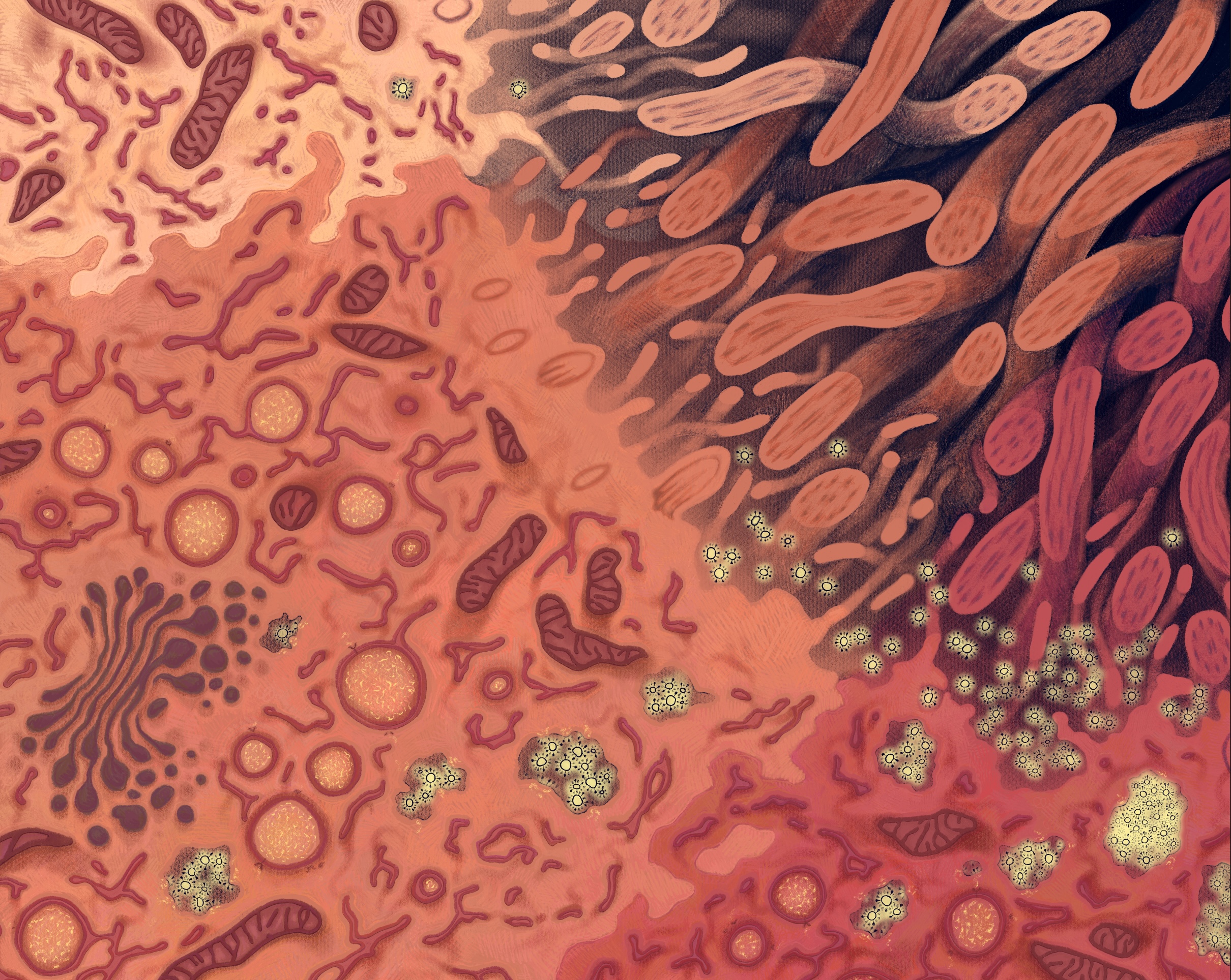
As a Science Visualization specialist, my responsibilities are to learn and to share. With a background in art and user-centered design, I get to spend my time solving puzzles about how to present data to both scientists and non-scientists. I mostly work with planarians, using electron microscopy (EM) data to see their cells in 3D, at a high resolution, and within the context of the animal. Planarians are flatworms with an incredible ability to regenerate due to their stem cells, which happen to be the only cells in this animal that divide. I spend a lot of time creating 3D models of dividing cells or differentiated cell types. The amount of time I’ve spent with my eyes on this data makes me an expert resource for other lab members as their research leads them to a particular type of cell or region of the animal.
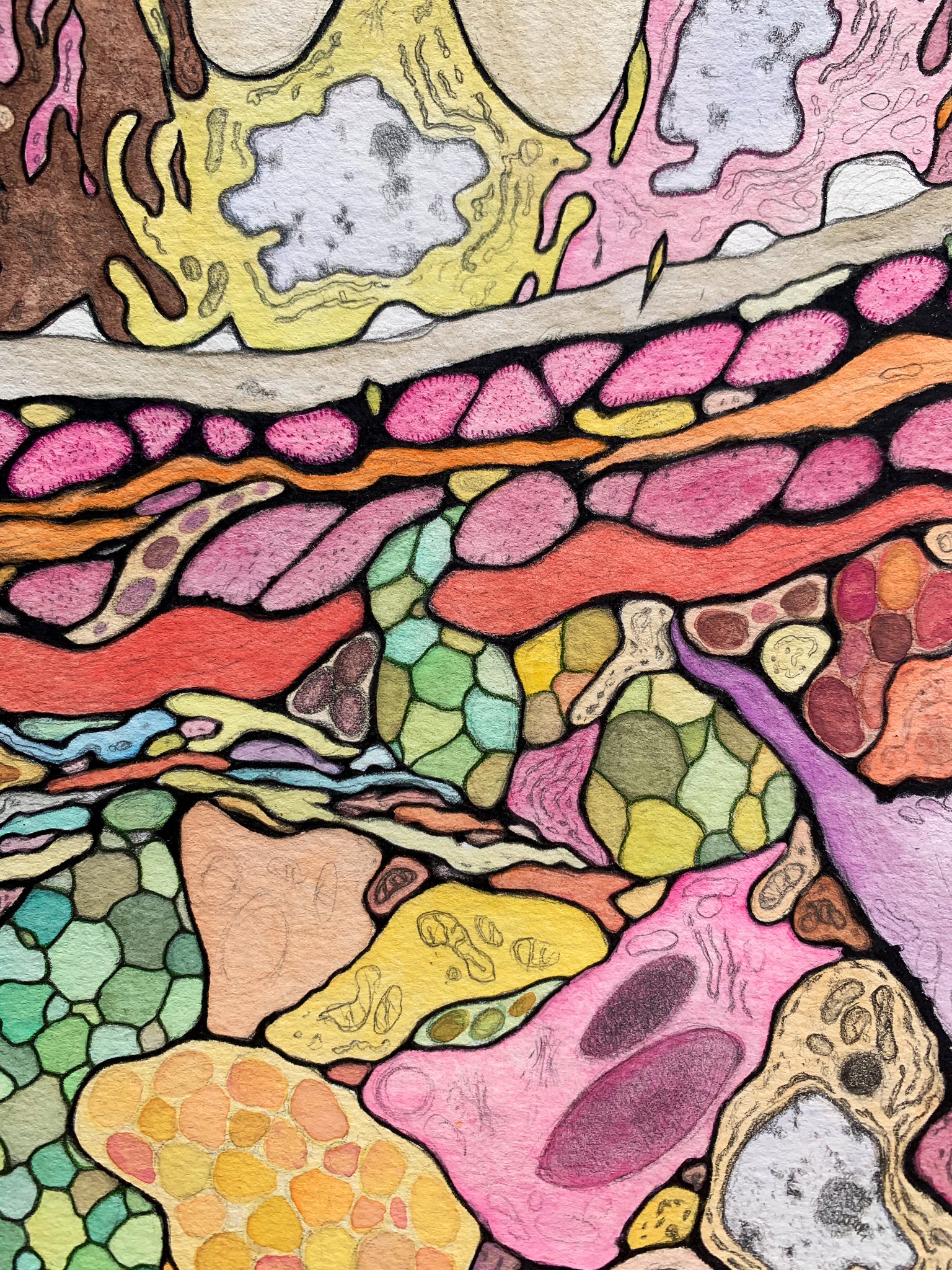
‘Middle Juice’ – Watercolor, graphite, and colored pencil on paper
Were you always going to be an artist?
Yes, I was always going to be a creative of some sort. The first thing I ever said I wanted to be when I grew up is a ‘coloring dentist’ – don’t ask me what I meant by that, because I don’t really know! Although, I imagine it’s actually pretty similar to the work I’m doing now!
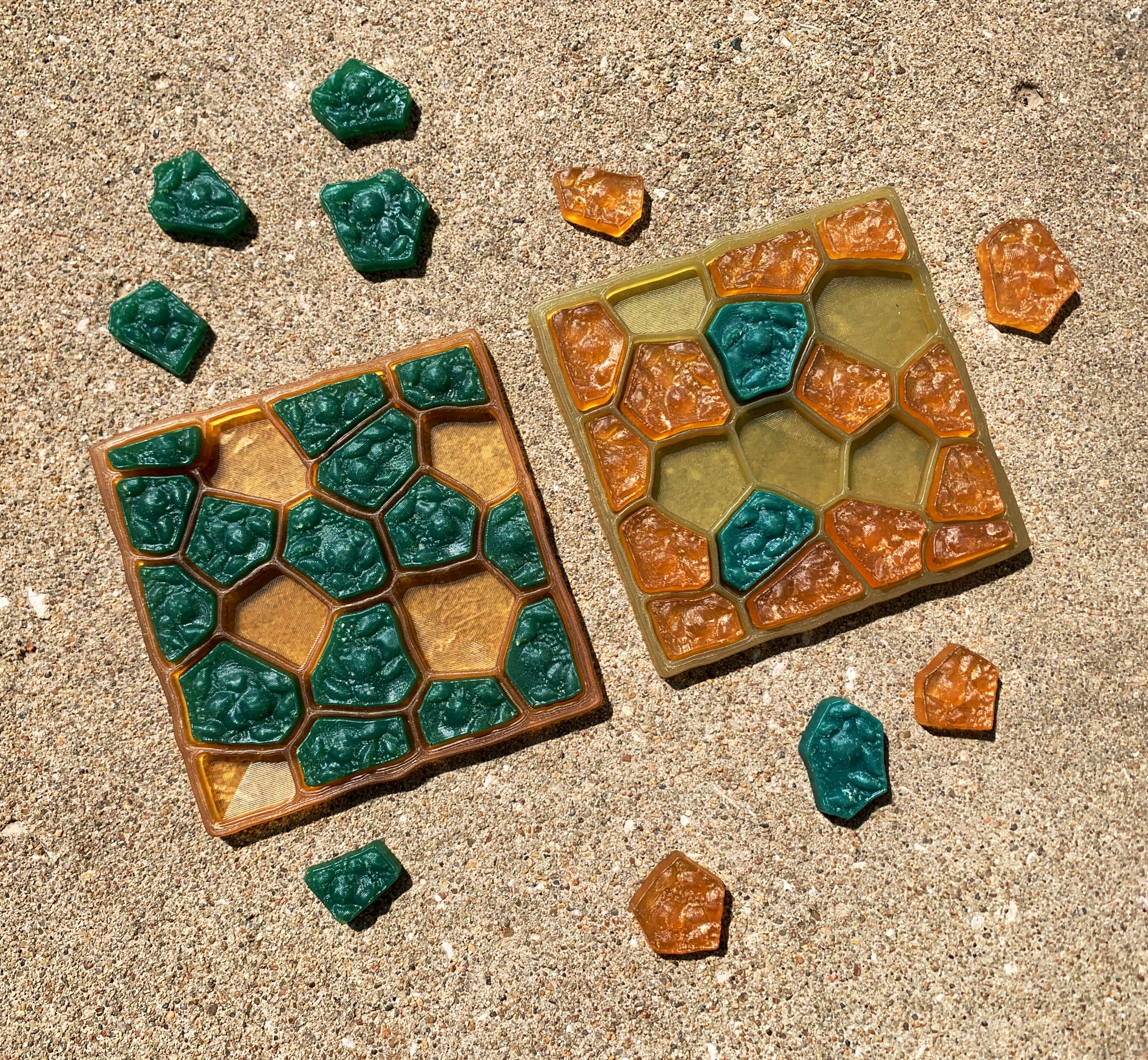
‘Rubber Cell Puzzles’ – Urethane rubber cast from a CNC milled HDPE mold
And what about science – have you always enjoyed it, and how did you begin working in the lab?
Yes, I’ve had a love for biology all my life. I thought I missed my chance to be involved in science because I had a difficult time in school (I’m neurodivergent). I headed in the direction of art because I thought that was the only thing I could do. But while I was in school – searching for the reason why I want to make things – I fell in love with the cell! I began making artwork of cells, including a 3D printed cell model, a quilt, and several puzzles. At the end of my third year at KCAI I met my mentor, Steph Nowotarski, during a tour of Stowers in the electron microscopy department. What started as a wonderful internship (where I traded 3D printing knowledge for learning about cell & developmental biology) later turned into my full-time job. I’ve learned so much about science, biology, planarians, and electron microscopy since then.
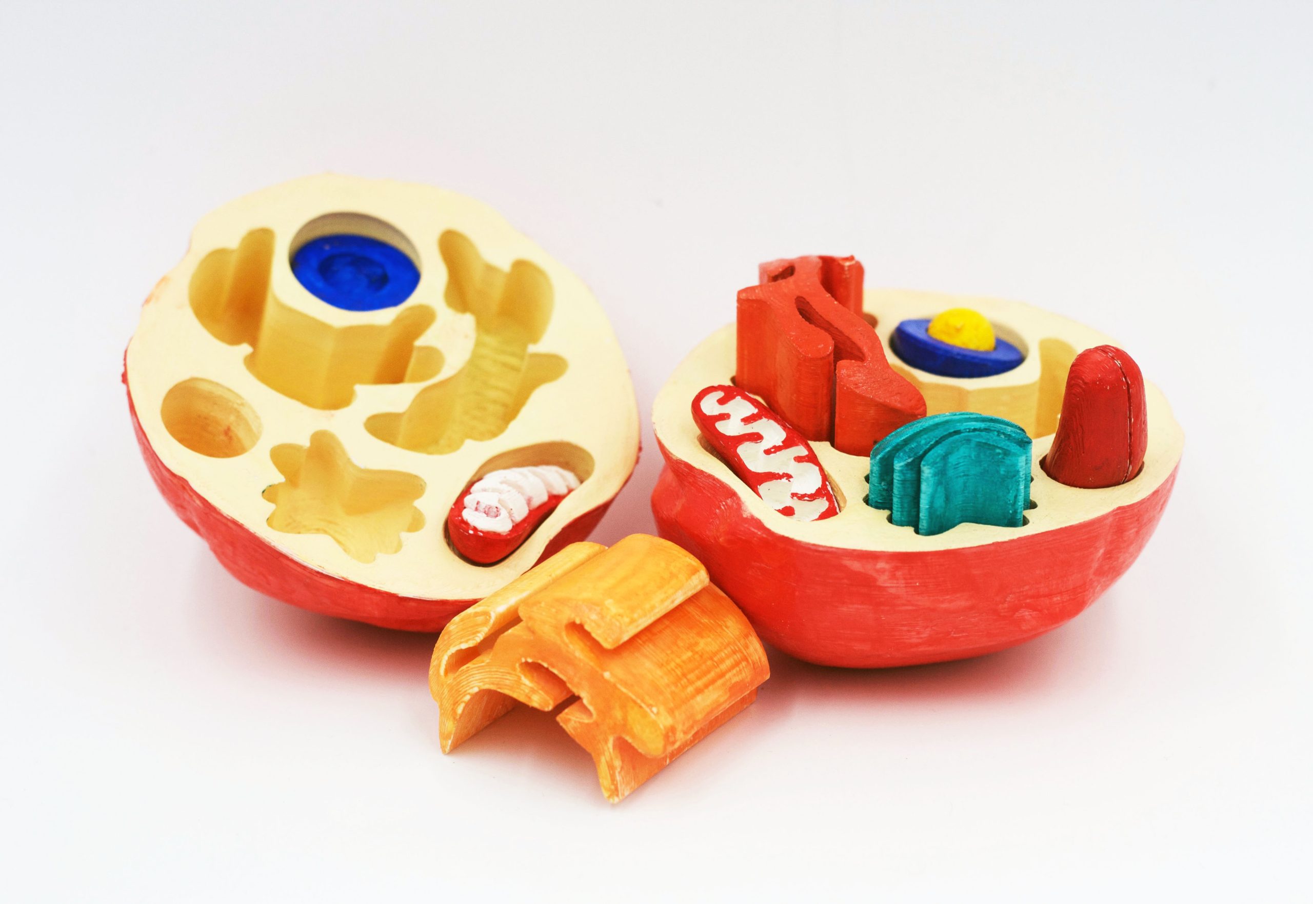
‘Cell Model’ – 3D printed ABS
What or who are your most important artistic influences?
Growing up, I had this amazing set of books: the Childcraft collection. These books and science museums for children have been huge influences on my work. Recently, I’ve also been inspired by the work of Agnes Pelton, Gemma Anderson, and Ipsa Jain.
‘Infection Illuminated’ – Digital illustration
How do you make your art?
I really have two categories of art-making: art for communication, and art for expression. My communication artwork centers around a topic I want to explore or share with others. Here, accuracy is very important, and so is ‘testing’. It’s important to test my work with a smaller audience before I set it free into the world, to make sure it’s communicating what it is supposed to communicate! My artwork for expression is something else. It is about making for the sake of making and most of this happens in my sketchbook.
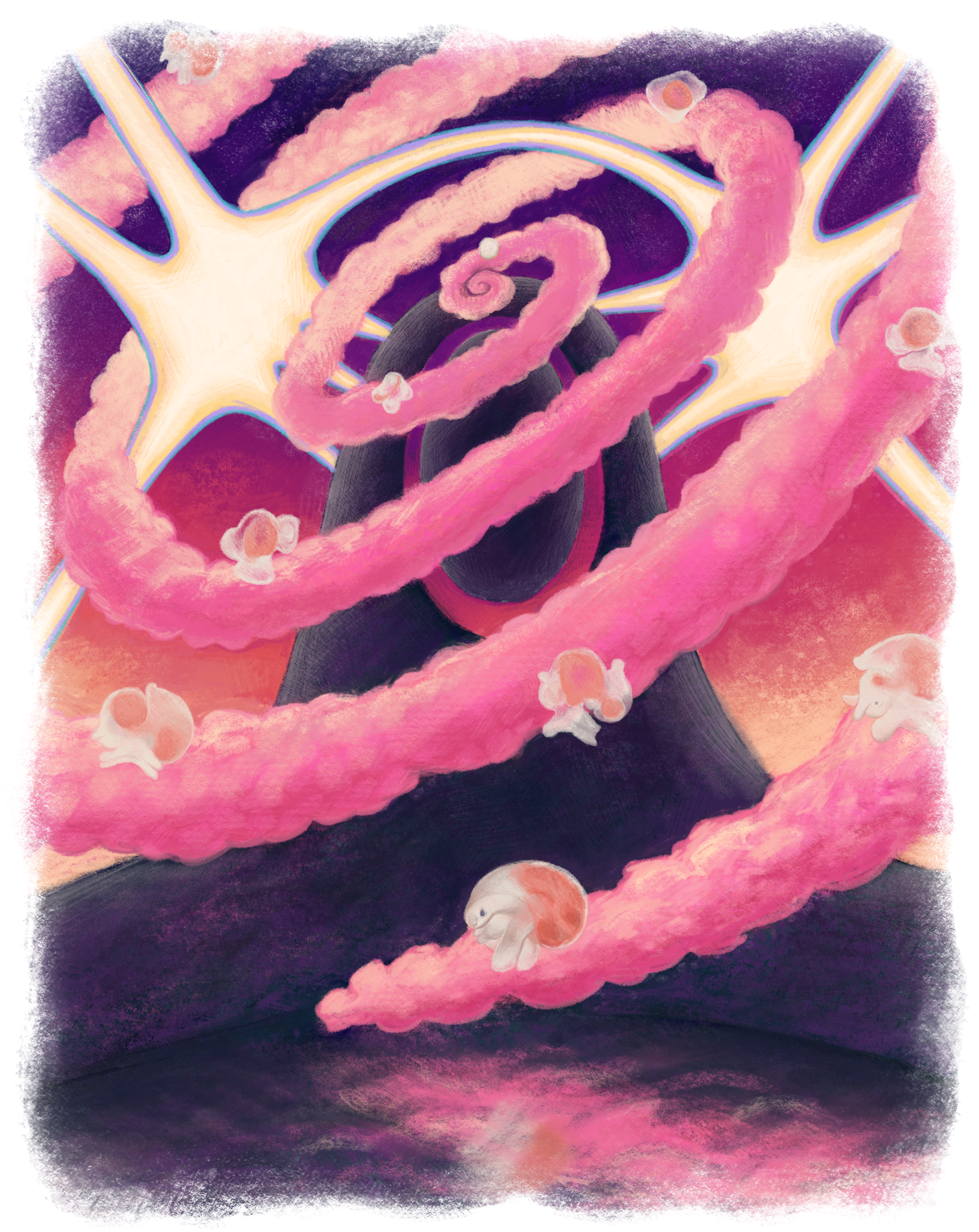
‘Snail Mountain’ – Digital illustration
Does your art influence your science at all, or are they separate worlds?
My art and science are deeply intertwined. Most of my work involves looking at electron microscopy images of cells. This very directly inspires my artwork, as cells are a common subject for me. Sometimes my sketchbook explorations will give me new ideas for data visualization and communication.
So, my art for communication is inspired by science and my science is inspired by my art for expression!
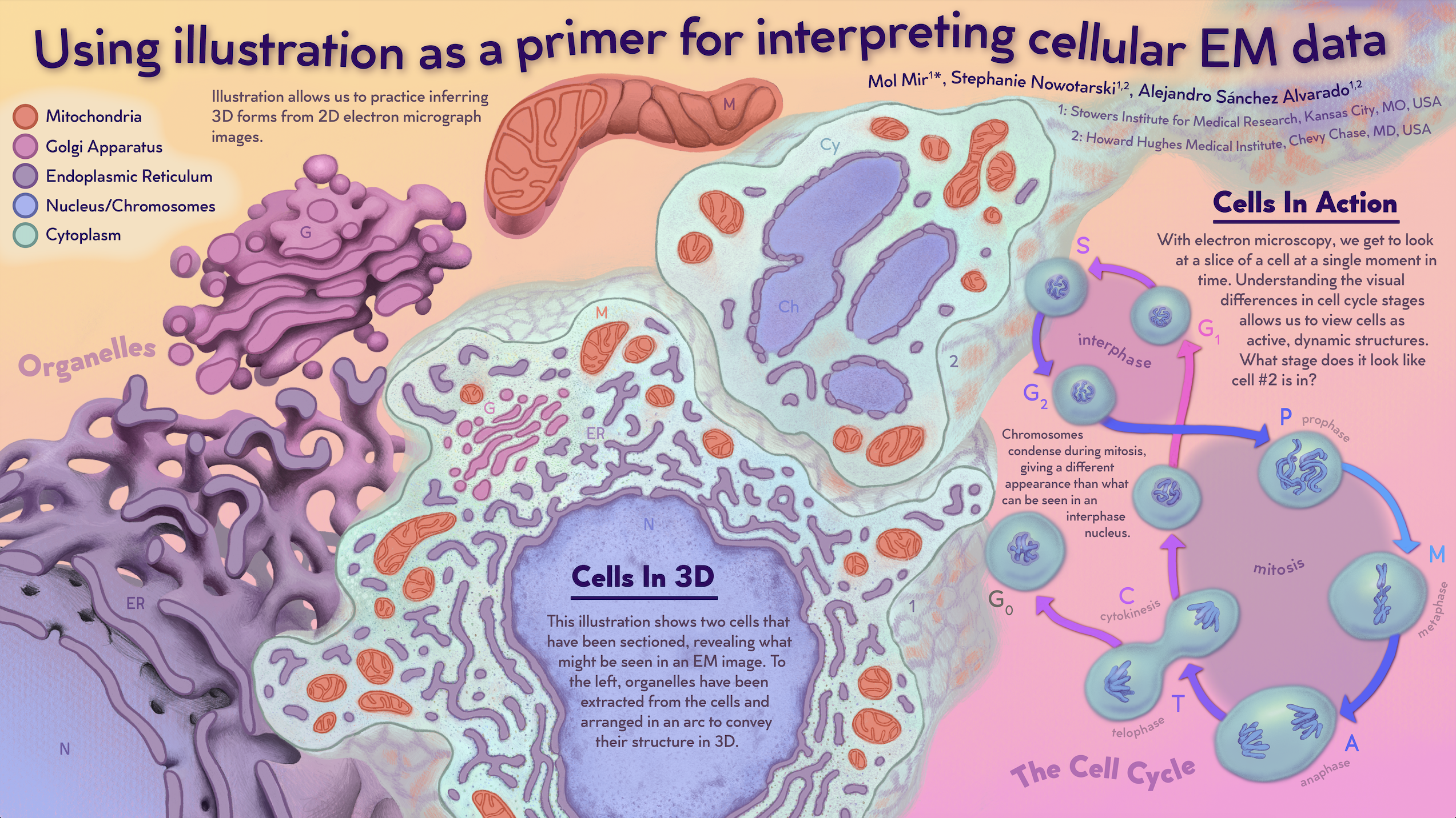
‘Using illustration as a primer for interpreting cellular EM data’ – Scientific poster for VIZBI 2022
What are you thinking of working on next?
My next big project is a book! I’m working on a book that will serve as a collection of planarian cell types through electron microscopy that also helps people to understand EM images and how they are created. This is a project I’ve been thinking about for a long time, so it’s very satisfying to be working towards it now.
Thanks to Mol and all the other SciArtists we have featured so far.We’re looking for new people to feature in this series – whatever kind of art you do, from sculpture to embroidery to music to drawing, if you want to share it with the community just email thenode@biologists.com (nominations are also welcome!)
Exploring how germ cells are metabolically supported in Drosophila testes
During the summer I had the privilege to work in Dr. Amoyel’s lab at UCL, to study the mechanisms providing metabolic support to germ cells in Drosophila testes. As my first lab project, it was thrilling to realize how much I enjoy academic research and feel invested in shining light on the unknown.
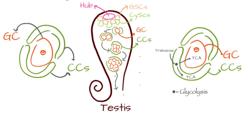
The lab seeks to understand how stem cell interact with their environment and with each other, to influence their fate and maintain tissue homeostasis. In fact, stem cells can either undergo self-renewal or differentiation, to fulfil their functions in tissue maintenance and regeneration. The balance between these two fates is determined by a micro-environment called a niche, which delivers signals promoting proliferation to the adjacent stem cells, while cells that are displaced from the niche differentiate. In particular, the Drosophila testis niche, called the hub, supports two stem cell lineages, namely germline stem cells (GSCs), which differentiate into germ cells, and somatic stem cells (CySCs), differentiating into post-mitotic cyst cells. When differentiating, two somatic cyst cells (CCs) envelop one germ cell (GC), forming a complex called a cyst (See Fig. 1). The enclosed GC then undergoes several mitotic events, increasing its number within the cyst from 1 to 16 cells (See Fig. 2).
Fig. 1 A cyst is composed of two somatic cyst cells (CCs) surrounding one germ cell (GC). Fig. 2 Drosophila testis, containing the hub and two cell lineages. A differentiated germ cell undergoes mitotic events within a cyst. Fig. 3 In a cyst, our theory is that CCs provide glycolysis products to support TCA in both cell lineages.
Since the enclosed GCs are sealed off from the environment by the two somatic CCs, the way they receive the metabolites necessary for the glycolytic and TCA pathways to survive was still unknown. In carbon metabolism, sugars – Trehalose molecules in flies – are broken down through a process called glycolysis, which produces pyruvate and ATP. Pyruvate is then either converted to lactate, or transported into mitochondria to fuel the TCA cycle, which provides most of a cell’s energetic needs. Hence, my project’s objective was to understand whether the two stem cell lineages interact metabolically, exploring the hypothesis that CCs provide glycolysis products to support germ cell metabolism (See Fig. 3). This shuttling of metabolites is already known to happen in the brain, where glia conduct glycolysis and provide lactate to neurons to feed their mitochondria. To test this theory in Drosophila testes, we knocked out glycolytic and TCA enzymes to downregulate the respective metabolic processes. A prediction of our hypothesis is that different metabolic pathways should be required in different cell types. In particular, knocking down glycolysis only in CCs should affect GCs, while knocking it down in GCs should have no effect, while both cell types should rely on TCA cycle enzymes.
During the first week, I learnt about Drosophila handling and husbandry: the basics about fly development and genetics, how to set up crosses, and how to identify markers to select the correct flies for analysis. I was also trained in immunostaining, to identify cell types in the tissue and whether their distribution changed as a result of my genetic manipulations. With plenty of practice and patience, I was soon comfortable enough with the staining procedure to start building my own experiments, and that’s how slowly the puzzle finally started coming together and I found myself in charge of boxes of crosses, and creating my own routine in order to keep crosses and experiments going.
This project was also an opportunity for me to discover and apply the Gal4/UAS-RNAi system, one of the main techniques used to conduct large-scale gene disruption in flies. Gal4 encodes a transcription factor which specifically binds to an enhancer called UAS, and activates expression of target genes downstream of the UAS sequence. Spatial control of expression is achieved by placing Gal4 expression under the control of tissue-specific promoters. When RNA interference (RNAi) sequences are placed downstream of UAS, their expression inhibits the expression of targeted proteins, disrupting gene expression. Therefore, when crossing a female fly carrying a Gal4 transgene with a male carrying a UAS-RNAi for a specific metabolic enzyme, the enzyme will be knocked down in the progeny carrying both transgenes.
To test these hypotheses, I used specific Gal4 drivers, including Traffic jam (Tj-Gal4), which targets CCs, Nanos-Gal4, which is only expressed in GCs, and Tubulin (Tub)-Gal4, which is expressed in all cells. These metabolic genes should all be essential for viability during development, so knocking them down in all cells using the UAS-RNAi system should result in lethality and a failure to obtain offspring carrying both the Gal4 and UAS-RNAi transgene.
My results showed that knocking down all glycolytic and TCA enzymes with Tub-Gal4 led to lethality, except for knockdown of Trehalose transporters (Tret), which did not affect viability. This might indicate either that the many versions of Tret proteins overlap each other in their functions, or that there are other pathways that might lead to the same downstream outcome without the use of Tret enzymes. Together with Holly Jefferson, we went on to show that knocking down glycolysis genes in CCs led to decreased GC survival, while knockdown in GCs had no effect. These results altogether support the hypothesis that cyst cells produce metabolites through glycolysis to support germ cell metabolism.
The sense of responsibility I felt, and the way other lab colleagues, with many more years of experience than me, relied on me to complete the necessary experiments, was in a sense enlightening for me to understand dynamics in a laboratory and in a work environment. A dynamic in which colleagues confide in each other’s’ abilities and strengths, in which every question is a good question, and in which a wrongly dissected testis or a failed experiment is an opportunity to learn and fully comprehend mechanisms without blindly following protocols. This wholesome experience has shown me how working in research, contributing to the scientific community, and entering the world of scientific discovery, is a life full of suspense and serendipity, struggle and competition, failure and accomplishment. It is a selfless life to which my future career aspirations lean fully, allowing me to explore the mechanisms through which life is possible.
Sonia Paoli – UCL, BSc Biomedical Sciences, Cells and Molecules
Supervisors: Marc Amoyel, Diego Sainz de la Maza, Holly Jefferson
The role of canonical Wnt signalling in embryogenesis of the invertebrate chordate Ciona intestinalis.
Ascidians, as the closest invertebrate sister group of vertebrates, are important to study the development and evolution of our own species. Their gene networks are closely related to those of humans, but without the complexities that were introduced via the whole genome duplications that occurred at the origin of vertebrates. A particular conserved mechanism, canonical Wnt (cWnt) signalling is essential to various development processes, especially in patterning along the anterior-posterior axis. Understanding the role of the cWnt pathway in ascidians would be helpful to elucidate the emergence of chordates and their evolution.
The summer project was complementary to ongoing work in the lab of David Ferrier (in the Scottish Ocean Institute, University of St. Andrews), focused on understanding the mechanisms of cWnt controlling their potential target genes in Ciona, such as the ParaHox genes (Gsx, Xlox and Cdx) that are the evolutionary sisters to the Hox genes, with roles in patterning the anterior-posterior axis in the central nervous system and gut. I had the opportunity to try both experiments with my two lab mates (Dr Nuria Torres-Aguila and Anastasia Ellis, see Figure 3) who use different ways to disrupt the cWnt signalling pathway.
Background
The cWnt pathway features the activation of the β-catenin transcription factor and modulation of specific target gene expression. T-cell factor/lymphoid enhancer (TCF/LEF) is the main transcription factor mediating the cWnt pathway. In the absence of Wnt ligands, responsive genes are generally repressed by TCF/LEF due to the transcriptional cofactor β-catenin constantly being degraded by the proteosome. When Wnt ligands interact with the transmembrane receptor proteins, several proteins (Frizzled, LRP5/6 and Disheveled) are brought to form a multimeric complex attached to the membrane, inhibiting the phosphorylation of β-catenin and its degradation. The stabilised β-catenin then converts the former repressor TCF/LEF into a transcriptional activator of the Wnt-target genes (Gilbert and Barresi, 2022).
Heat shock experiment
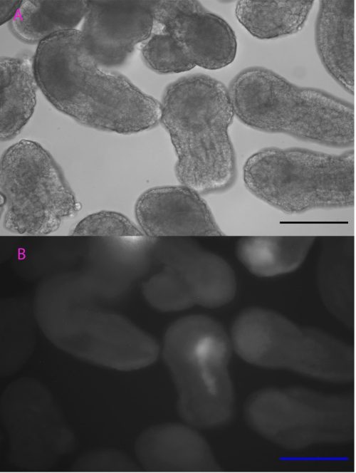
Controlling when and where TCF is expressed is useful to help analyse the function of TCF in cWnt signalling. Based on the heat-inducible cis-regulatory element initially characterized by Kawaguchi’s team (Kawaguchi et al., 2014), we tested the efficiency of a DNA construct with heat-inducible gene Ci-HSPA1/2/6-like and mCherry gene in Ciona embryogenesis to adapt this versatile technique to our species and population (i.e. a temperate Scottish population versus a more tropical Japanese population). Heat shock was initiated at different stages of development with different lengths of time and temperatures before being observed under an epifluorescence microscope.
Although the precise heat-shock conditions need to be further refined, it was encouraging to see the induction of mCherry in embryos with this technique (Figure 1), raising the prospects of using the heat-shock approach to over-express genes like TCF in the near future. In this experiment, the microscope was probably the most exciting but also challenging piece of equipment, the microscope was probably the most exciting but also challenging piece of equipment I used. It took me some time to patiently check every detail like light intensity, exposure time, and magnification to ensure comparability between my images. The experience has been valuable for me to be familiar with this essential equipment for studying developmental biology.
Figure 1. Heat shock-induced mCherry fluorescence under the microscope. 25 degrees 30 minutes heat shock treated embryos. Photos were taken 3 hours after the treatment A: Embryos under white light B: Embryos showed mCherry fluorescence under fluorescent light. Scale bar: 100µm
Chemical treatment
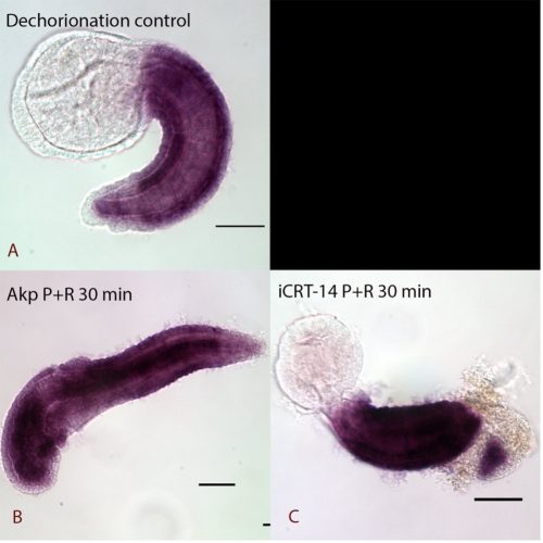
Chemical treatment investigated whether the TCF-dependent ParaHox gene Cdx is potentially directly controlled by the cWnt pathway. Pharmacological agents, iCRT-14 and azakenpaullone (Akp) were used to downregulate and upregulate the level of the cWnt pathway. Embryos were left to develop to the desired stage for recovery as a post treatment to distinguish rapid (potentially direct) responses from slower (possibly secondary) changes. The samples were processed for in-situ hybridization with a probe against the Cdx gene and images were captured by Nomarski microscopy.
The results showed that the expression of Cdx is possibly regulated by a secondary effect instead of directly disrupted by cWnt cascade, as the samples fixed immediately after treatment showed no obvious changes in expression (data not shown) whereas dramatic anterior extension of Cdx expression was seen with a recovery period in cWnt activator treatment (Figure 2). The lack of apparent response to iCRT14-treated samples may be due to TCF not normally being expressed in tail epidermal cells (Garstang et al., 2016). Further work aiming to check the Cdx expression in deeper cells where TCF is expressed will be conducted in the future.
All of this research helped me to appreciate the importance of time management in the lab. I had the opportunity to repeat the in-situ hybridization several times, and each time I got more skilled in the procedure. My first two times of in-situ hybridization turned out with either the embryos being accidentally lost because of I rushed during wash steps, or they were left in the solution for too long, so I stayed in the lab until very late. It is a relief that eventually they were successfully mounted to be observed, but wisely making use of time during short waits and multitasking largely improved the efficiency in my later experiments.
Figure 2. Expression of Cdx in Ciona intestinalis at late tailbud stage I A: Normal expression of Cdx at the late tailbud stage of dechorionated control samples. Expression is observed in the epidermis of the tail except at the very posterior end. B: 30 minutes pulse and recovery samples be treated by cWnt activator (Akp). It had dramatic anterior extension of Cdx expression and lost some of the anterior features. C: cWnt inhibitor (iCRT-14) pulse and recovery treated embryos with extension of Cdx expression to the posterior tip of the tail. Pulse and immediately fixed samples did not show obvious changes in expression. (data not shown) All the embryos are lateral views scale bar: 50 µm
Personal experience
I appreciate that I have experienced the systematic research process as a whole. From collecting sea squirts in the harbour and manipulating the embryos, to finally assaying their responses to various treatments. I have tried many essential techniques in developmental biology and see how the experiments proceed. I learned how the protocols are designed and improved based on the techniques developed from previous research. Even finding the potential mistakes I made by recalling the steps with my supervisors was a valuable and rewarding experience. Thanks to Dave, Nuria, Anastasia and the friendly people around SOI, all of who created a warm and supportive environment. I am more comfortable working in the laboratory and more determined to pursue further studies now. Thanks to the BSDB and Gurdon scholarship for making this opportunity possible. It has been one of my best memories doing scientific exploration with such a great team, besides the beautiful East Sands beach at St. Andrews.
Figure 3. From left to right. Dave, Anastasia, me and Nuria on the seafront in front of Scottish Ocean Institute.
Reference list:
-Garstang M.G., Osborne P.W. and Ferrier D.E.K. (2016) TCF/Lef regulates the Gsx ParaHox gene in central nervous system development in chordates. BMC Evolutionary Biology 16:57.
-Gilbert S.F. and Baresi M.J (2022) Developmental Biology 12th edition Chapter 4 Cell to cell communication. Oxford University Press 109-111
-Kawaguchi A, Utsumi N, Morita M, Ohya A, Wada S. (2014) Application of the cis-regulatory region of a heat-shock protein 70 gene to heat-inducible gene expression in the ascidian Ciona intestinalis. Genesis. ;53(1):170-82.
Role of PAX6 in human cerebral organoid development
The cerebral cortex contains two major classes of neurons: excitatory and inhibitory. The imbalance between them is postulated to underlie some autism spectrum disorders (ASDs) (Rubenstein, 2010; Uzunova et al., 2016). The transcription factor PAX6 is an important regulator of embryonic forebrain development and mutations in PAX6 have been associated with ASDs (Kikkawa et al., 2019; Manuel et al., 2015). It is therefore possible that PAX6 could be involved in the regulation of the inhibitory-excitatory balance, hence playing a role in the development of ASDs. Supporting this hypothesis, recent findings indicate that PAX6 deletion in mice leads to the appearance of ectopic GABAergic (inhibitory) cells (Manuel et al., 2022). While animal data are useful, the mechanisms of this effect, and whether it is present in humans remains unclear, as human neurodevelopment is difficult to study directly. However, human cerebral organoids offer an exciting, new way to directly investigate early human embryonic neurodevelopment, including the effects of PAX6 mutations (Mason & Price, 2016). Hence, as a first step towards uncovering the mechanisms of the possible PAX6-dependent regulation of the inhibitory-excitatory balance in humans, this project aimed to examine PAX6-/- organoids at early developmental stages to investigate the effects of PAX6 mutations.
Methods
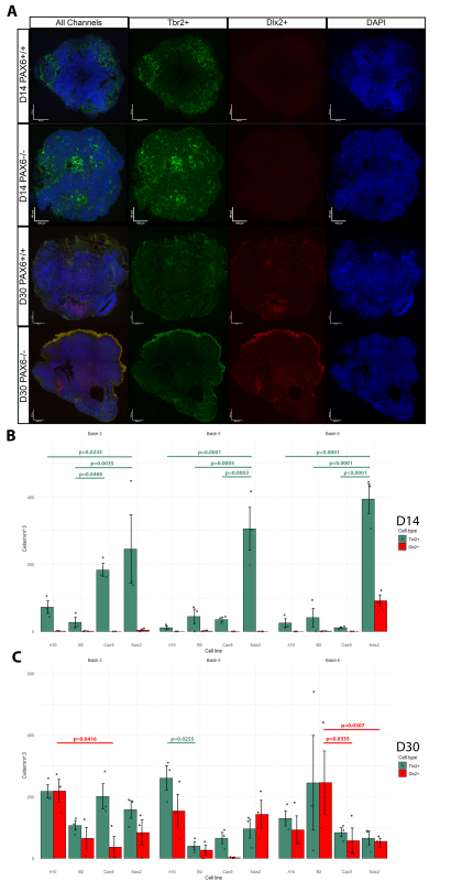
The Mason Lab has previously grown cerebral organoids from PAX6-/- induced pluripotent stem cells and PAX6+/+ controls. At culture day (D)60, the cellular phenotypes of these organoids were tested using scRNA-seq analysis. Indeed, ectopic GABAergic populations were observed in the mutants and not in controls. To examine the earlier stages of PAX6-/- organoid development, I analysed three batches of organoids originating from four cell lines (Controls: Nas2,Cas9; Mutants: A10,B2) at D14 and D30. Organoids were cut into 10μm sections. Inhibitory/excitatory neurons were visualized by tagging Tbr2 (marker of excitatory glutamatergic neurons/progenitors) and Dlx2 (marker of inhibitory GABAergic neurons/progenitors) with fluorescent antibodies. Hence, two distinct cell types could be visualized using fluorescent microscopy (Fig.1A). The number of Tbr2+ and Dlx2+ cells was counted in three randomly selected sections from each organoid using ImageJ software. The cell count was normalized by area of each section and averaged per organoid (Fig.1B-C).
A) Representative immunofluorescent images of control (PAX+/+) and mutant (PAX6-/-) organoids at D14 and D30, counterstained with DAPI. B-C) Counts of Tbr2+ and Dlx2+ cells at B) D14 and C) D30. Each point represents the average cell count from three sections of an organoid, normalised per area (n=3/group, 72 total). 2-Factor (Batch and cell line) ANOVA, evidence for interaction between variables for D14 Tbr2+ counts (F=3.0612, p=0.02278), D30 Dlx2+ counts (F=3.1609, p=0.01984), D30 Tbr2+ counts (F=2.5636, p=0.04615). Significant results of post-hoc pairwise Turkey comparisons presented as lines with p-values above relevant bars.
Results
No D14 organoid (apart from an anomalous batch 6 Nas2 organoids) featured Dlx2+ cells. Hence, ectopic GABAergic cells likely arise after D14, which narrows the window of investigation in potential future research. In many cases, the number of Tbr2+ cells was significantly higher in the Nas2 organoids when compared to other cell lines, including the second control line, Cas9 (Fig.1B). This may suggest the presence of unknown variables which affected organoid development, or possibly that PAX6 has an effect on the speed/timing of Tbr2+ neuron emergence.
Both mutant and control organoids at D30 exhibited comparable counts of Tbr2+ and Dlx2+ cells, with significant differences in cell counts present between only a few groups (Fig.1C). This is surprising, as prior literature suggests that ectopic GABAergic cells (in PAX6-/- mice) appear at some point during development due to environmental signals such as Shh found in cerebrospinal fluid (CSF) and/or immigrating interneurons (Manuel et al., 2022). It seems that in human cerebral organoids (in the absence of CSF or immigrating interneurons) GABAergic cells emerge as a normal part of development whether PAX6 is present or not, but disappear by D60 in controls.
PAX6 could possibly be triggering the death of Dlx2+ cells between D30 and D60. PAX6 could also be causing Dlx2+ cells to switch to an excitatory fate by D60. It would be interesting to expand upon these findings by conducting lineage tracing experiments: this would allow for the tracking of the changes in cellular phenotypes to reveal whether the presence of PAX6 affects cell fate decision making.
My experience at Mason Lab
I found this project very exciting. The problem-solving aspect of research following inevitable experimental failures was genuinely great. It was very interesting to brainstorm the next steps when it becomes apparent that the current experimental procedure is not yielding any results. For instance, originally the experiment involved tagging dividing cells with EdU at D13.5, to see whether these cells switch between Tbr2+ and Dlx2+ identities between D14 and D30. However, the experiment had to be modified as the EdU fluorescent signal became too faint at D30 due to dilution during cell division.
The project also showed me the more labour-intensive side of research as I spent many days cutting organoid sections at the cryostat. After the baptism of fire by cutting my finger, I eventually found it a compelling task. As one of the Mason Lab members said, “it builds character”. I was also introduced to fluorescent microscopy and image analysis using ImageJ. The former forced me to become organised and efficient, as one must tame the unruly microscope and take the hundreds of pictures before the booked time runs out. The latter I found challenging as it was my first time using image analysis software, but it will undoubtedly be useful in my future work.
Every part of this project, no matter how menial or monotonous it might have seemed to an observer, I found thrilling. I have obtained experience in numerous laboratory techniques, and again reaffirmed research as my chosen career path. I am very grateful to Mason Lab and BSDB for this opportunity, and I am looking forward to being able to conduct projects of larger scales in the future.
Figure 2. Right Panel: Me, together with my supervisors: Prof. John Mason and Dr. Calvin Chan! Left Panel: me with my best friend, the cryostat. (2 votes) Loading...
A new article on embryos and the beginning of independent human life:
In her January 1 New York Times article, “When does life start? A post-Roe conundrum,” reporter Elizabeth Dias cites some material from a recent paper that I wrote. The article, “Pseudo-embryology and personhood: How embryological pseudoscience helps structure the American abortion debate. Natural Sciences 2022: e20220041″ is open access and can be found at DOI: 10.1002/ntls.20220041 . The paper reviews scientific opinions as to where independent human life begins, and contends:
There is no consensus among biologists as to when independent human life begins
What passes for science is actually a set of outdated myths that are no longer considered valid
This set of myths denigrates birth and promotes fertilization as the site where personhood begins.
I hope the community of biologists will read and discuss the data and conclusions of this paper.
In response to the front page article in The New York Times of 2 January 2023, “When Does Life Begin,” I am posting a model for the formation of the earliest stages of the embryo to shed light on the question.
A more complete treatise and graphic presentation is available on www.embryogeometry.com.
I look forward to relevant comments.
Stuart Pivar ___________________________________________________________
The heart of biological science is the search for the way the embryo is formed. The rest is known.
This proposed model is an account of embryogenesis, tantamount to the blueprint for the assembly of the embryo.
Included are the phenomena of morphogenesis, organogenesis, segmentation, limb development, and nerve cord development, all the demonstrable consequence of the singular event of gastrulation. The model is illustrated by hundreds of mechanical animation drawings.
The model describes the instantaneous catastrophic embryological event called gastrulation as the bursting of the first embryonic structure, the blastula, and its elastic recoil as demonstrably the universal origin of animal form.
The embryo is the deflated form of the tense, balloon-like blastula membrane upon bursting and elastic recoil.
Organogenesis is the deformation by elastic recoil of the pattern of self-organized circumferential girdles that encompass the blastula from pole to pole.
The radial, vermiform, and bilateral body forms result, respectively, from a spherical, cylindrical, or ovoidal blastula. Morphogenesis is the anterior-dorsally directed migration upon bursting failure of the separate layers of the membrane bilayer, governed by the mechanical exigencies of surface wrinkling pattens. Embryogenesis is predictable by the mathematics of topological surface wrinkling patterns.
Taxonomic differentiation is the result of mechanical error in the repetition of the universal anterio-dorsal elastic reaction. Evolution occurs when the modification is recorded and expressed in the genes generationally.
The genes provide proteins in precisely timed doses that maintain and occasionally change the proportions of the otherwise immutable phyletic form (see S.J. Gould, Ontogeny and Phylogeny, 1977).
The Origin of the Blastula: Incomplete cell division leaves cells connected by a capillary which itself is divided axially in each cell division. At the eight-cell stage the capillaries intersect and fuse centrally to form the blastocoel, a spherical plenum that enlarges–paved with blastocytes, constituting the blastula, and which eventually bursts and recoils as the embryo.
The Cause of Segmentation: The blastula as a water-filled balloon subtends standing oscillatory waves of energy that delineate subdivisions by serial harmonic bisections of the axis that condense chemically at wave intersections and nodes.
Limb Development: The separation and dorsal recoil of the pectoral and pelvic girdles at the ventral midline forms the limb buds in vertebrates comparable to the imaginal discs in insects. Development is the reversal of the action.
I have become passionate about the field of stem cell biology during my undergraduate degree in Molecular Genetics at the University of Edinburgh. Therefore, I was grateful to be awarded the Gurdon Studentship Award from The British Society for Developmental Biology to conduct a summer research project in Dr Soufi lab in the Institute for Regeneration and Repair (IRR). In this report, I will not only describe the aim of my project and how I proceeded to accomplish it, but also how valuable this experience was for my future studies and career.
In 2006, Yamanaka and Takahashi made a breakthrough in stem cell biology by showing that adult cells can be converted to become induced pluripotent stem cells (iPSCs) using the ectopic expression of four transcription factors (TFs) Oct4, Klf4, Sox2 and c-Myc (Takahashia & Yamanaka, 2006). These iPSCs are very similar to embryonic stem cells (ESCs) and have the unique ability to generate all cell types of the body. Therefore, iPSCs have enormous potential in the area of regenerative medicine. However, reprogramming adult cells into iPSCs is highly inefficient which limits the application of this technology for drug discovery and disease treatment. To improve reprogramming efficiency, further research focusing on fundamental mechanisms by which these TFs facilitate and maintain pluripotency is needed.
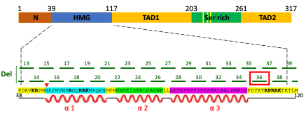
In my project I focused on TF Sox2, which is responsible for both somatic reprogramming and the maintenance of pluripotency in ESCs. Previous work, conducted in the Soufi lab, identified critical regions of Sox2 that are responsible for iPSC reprogramming. This was done by carrying out a systematic mutation screen of the Sox2 DNA-binding domain and mapping which regions of this domain are important for reprogramming fibroblasts to iPSCs. This led to the identification of several deletion mutants who were unable to reprogramme. My aim was to test if one of these deletions, called D36, also abolished the ability of ESCs to maintain pluripotency, which would indicate that this region of Sox2 gene is responsible for both reprogramming and pluripotency maintenance (Figure 1).
Figure 1.Functional domain structure of mouse Sox2 gene. Mouse Sox2 gene is composed of functional domains including high mobility group-box (HMG) domain (i.e. DNA-binding domain), transactivation domain 1 (TAD1), serine-rich domain (Ser rich) and transactivation domain 2 (TAD2). Systematic 5 nucleotide deletions in the HMG domain led to the discovery that the D36 region (among others) is important for the function of Sox2 in somatic reprogramming. The impact of D36 deletion on pluripotency maintenance was investigated in this research project.
I used CRISPR-Cas9 technology to generate mouse ESC (mESC) lines expressing D36 Sox2. The first step in this process was to design Sox2-specific guide RNAs (gRNAs) and test their efficiency to target Sox2 gene in mESCs. This was performed by introducing a designed gRNA together with Cas9 nuclease into mESCs by electroporation, followed by alkaline phosphatase (AP) staining of mESCs. AP is one of the best markers of pluripotency (Stefkova et al., 2015) and therefore it was used determine whether these cells differentiated due to Sox2 knock-out (KO). During this phase of my project, I had to design, order, and test multiple gRNAs to find out which one was the most efficient in targeting Sox2. Ultimately, I managed to design a suitable gRNA able to mediate Sox2 KO as was demonstrated by the decreased number of AP-positive colonies after AP-staining.
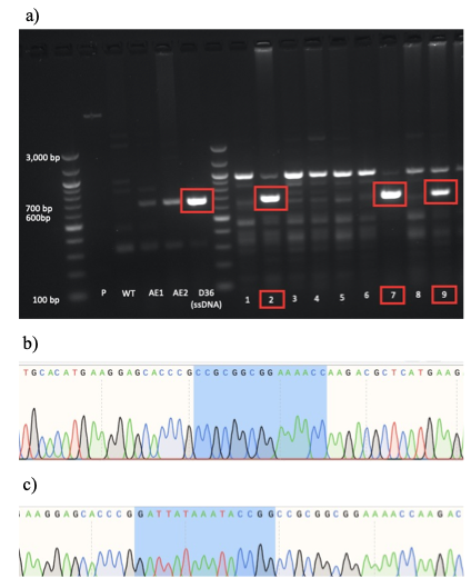
Next, I performed CRISPR-Cas9 knock-in (KI) to generate mESC cell lines containing D36 deletion mutation by homology-directed repair (HDR). For that, I designed a single stranded DNA (ssDNA) template containing D36 and introduced it into mESCs together with gRNA and Cas9 complex by electroporation. Due to the low efficiency of CRISPR-Cas9 technology, single cell sorting had to be performed to identified mESCs with the mutation of interest. After that, the presence of D36 KI in the picked mESC colonies was evaluated by polymerase chain reaction (PCR) screening using D36-specific primers and confirmed by Sanger sequencing (Figure 2). Out of 38 screened colonies, one colony (colony number 27) was identified as a clear D36 homozygote, together with 6 potential heterozygotes. D36 heterozygosity arose because of low CRISPR-Cas9 efficiency, which was only able to mediate KI of only one Sox2 allele.
Figure 2. Identification of D36 mESC colonies after CRISPR-Cas9. a) PCR screening followed by gel electrophoresis was used to identify D36-positive mESC colonies which produced an amplicon size of 654bp (red box). This approach led to the identification of 7 positive colonies numbered 2, 7, 9, 19, 27, 28 and 32 (note: colonies 19, 27, 28 and 32 not shown). b) Sanger sequencing results of colony number 27, showing the clear presence of Sox2 D36 deletion (note: the highlighted region corresponds to the sequence after the D36 locus as the 15bp of the D36 deletion region are missing). c) Sanger sequencing of WT mESCs serving as a negative control (note: the highlighted region corresponds to the locus of prospective D36 deletion, showing the presence of the D36 region).
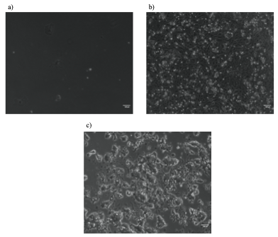
Subsequently, the heterozygosity of the potential heterozygous colonies was validated by TopoA cloning. This approach involved the amplification of the Sox2 D36 region by PCR, its ligation into plasmid vectors and their amplification in E.coli. This allowed me to validate the zygosity of D36 mutation by sequencing individual alleles of Sox2 gene. Unfortunately, the homozygosity status of colony 27 with clear evidence of D36 mutation on both Sox2 alleles could not be validated by TopoA cloning due to the lack of time. Interestingly, there were obvious phenotypic differences between this homozygous clone and WT mESC lines (Figure 3). These differences included slow growth, smaller cell size, and greater adherence to the culturing plates. This suggests that D36 deletion most likely does have an impact on maintaining pluripotency of mESCs. Nevertheless, this important conclusion should further be validated, for example by performing embryo contribution assay to assess the ability of D36 mESCs to differentiate and generate tissues and organs.
Figure 3. Light microscopic images of D36 and WT mESCs. Potential D36 homozygous mESC colony 27 on 6-well plate (a) and 12-well plate (b) had a decreased proliferative ability and smaller cell size compared to WT mESCs E14 IC passage 30 on 10cm plates (c).
During my internship, I have not only learnt different laboratory techniques, but also developed various skills and attributes that are essential for a scientific career. Most importantly, this experience helped me enhance my independent critical thinking when designing and planning experiments. Also, I was required to adjust existing protocols to suit my experiments which sometimes took multiple attempts to make it work and is therefore something I have not experienced during my undergraduate studies. Over time, I also became more confident in trusting my own judgments and making independent decisions, but also asking for help and advice when needed. In this respect, I was lucky to be a part of a stimulating scientific environment, surrounded by experienced scientists always willing to help and share their knowledge and skills.
Overall, being a part of Dr Soufi group in IRR was a very useful experience for me which also significantly influenced my future career plans. This internship cemented my aspirations to dedicate my life towards scientific research. Although sometimes difficult if things do not go as planned, it is nevertheless very rewarding and purposeful for me. Therefore, I aim to continue my research in the stem cell field or related areas that I am passionate about by doing a PhD.
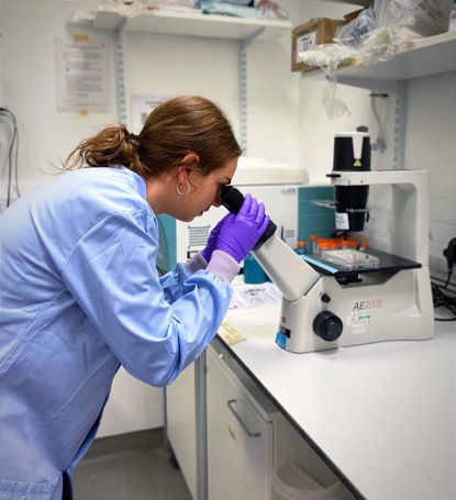
Figure 4. Me looking at mESC colonies using a light microscope.
References
Takahashi, K. and Yamanaka, S., (2006). Induction of pluripotent stem cells from mouse embryonic and adult fibroblast cultures by defined factors. Cell, 126(4), pp.663-676. Available at: https://doi.org/10.1016/j.cell.2006.07.024
Štefková, K., Procházková, J. and Pacherník, J. (2015). Alkaline phosphatase in stem cells. Stem cells international, 2015:628368. Available at: doi: 10.1155/2015/628368
At Development, we know that going on the job market and then setting up a lab is a real crunch time in an academic’s career, and not everyone receives the support they need to manage this transition. As a not-for-profit journal whose aim is to support the research community, we therefore wanted to do something to support individuals through this period. With this aim in mind, we’re excited to launch a new scheme – our Pathway to Independence (PI) programme. This competitive programme will select a small number of researchers (‘PI fellows’) planning to apply for Principal Investigator positions in the coming year, and will provide support, mentorship and networking opportunities.
So if you are a postdoc working in the developmental biology or stem cell field and starting to look for your first independent position, we’d encourage you to apply to this programme. You can find out more information in this Editorial, and also on this webpage. And while we will only be able to select a handful of individuals to become PI fellows, we are also thinking about ways we can support the wider cohort of applicants – we’re hoping to host one or two workshops, and will also give at least some applicants the opportunity to showcase their work through our ‘Development presents…’ webinar series.
This is a pilot scheme, and we’ll be refining the programme with our first cohort of PI fellows. We’re also keen to hear feedback from the wider community, so if you have any thoughts on these plans, do feel free to get in touch.
It’s musical chairs at The Company of Biologists at the moment (though fortunately without any chairs actually being removed!) – after Seema Grewal left Development last month to become the new Executive Editor of Journal of Cell Science, we’re delighted to have appointed Laura Hankins, currently the Company’s Science Communication Officer, as the new Reviews Editor on Development. Laura started working with us a little over a year ago following a PhD with Jordan Raff in Oxford, and I’m really pleased that she’ll be joining the journal in the near future – having worked with her in her current role, I know she’s going to be a great member of the team. And Helen Zenner – who you’ll know as the Node’s Community Manager – is going to be moving over to our sister site, FocalPlane. Helen’s got a very strong microscopy research background, and has some great ideas for developing FocalPlane.
All this means that we now have two openings for scicomm-type roles! Both these positions are suitable either for current researchers looking to move away from the lab, or for people who already have some scicomm experience. They provide a great opportunity to learn about the scientific publishing world, and to contribute to a not-for-profit organisation that really believes in supporting the biological community. To find out more, take a look at the job adverts for the Node Community Manager and the Science Communications Officer positions, and please feel free to reach out if you want to find out more about either role.
 (1 votes)
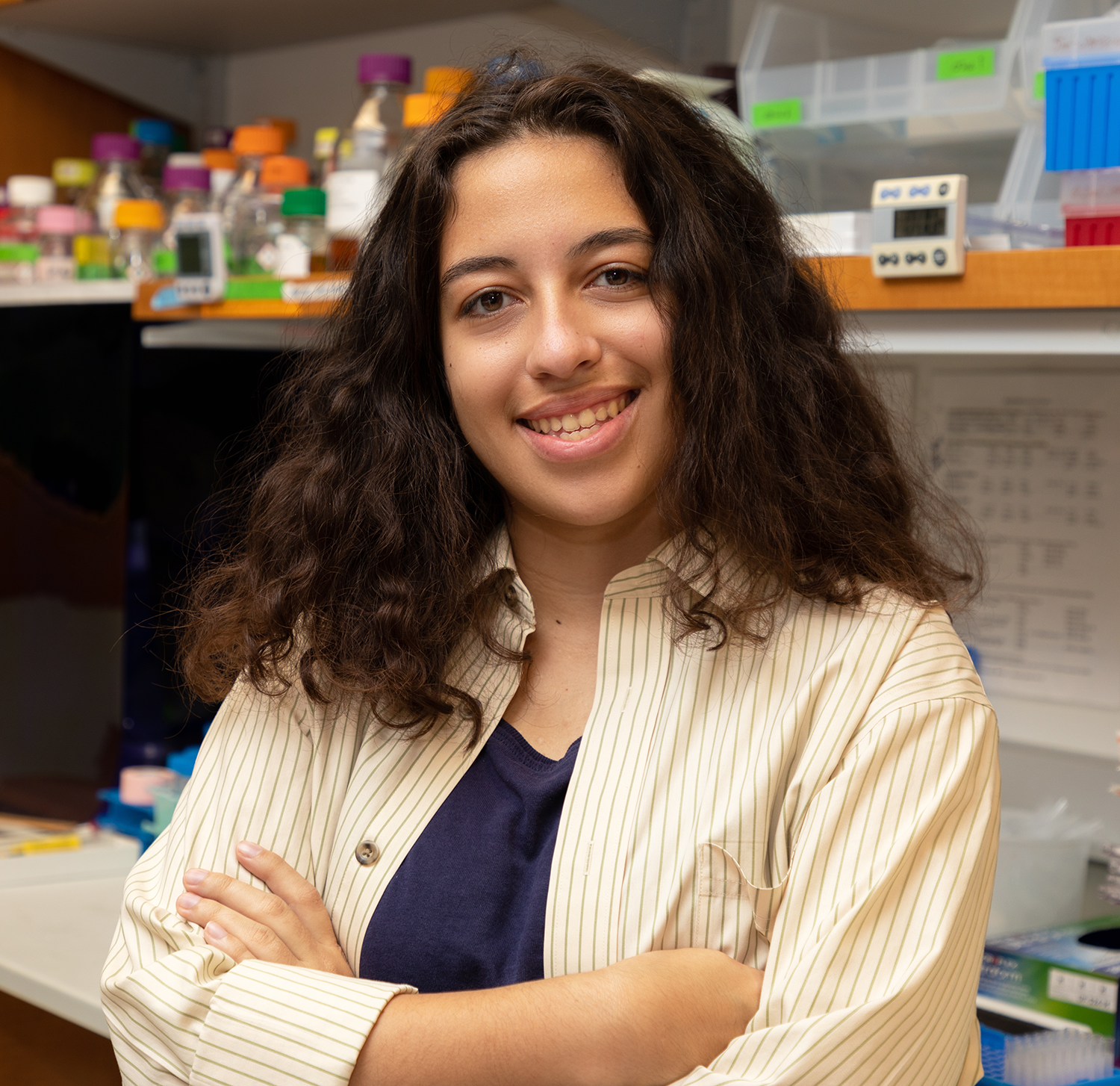

 (No Ratings Yet)