With almost 200 posts published on the Node in 2024, below are just a few of our highlights.
Thank you to everyone who has contributed to the Node in the past year.
Have you read a Node post that you really enjoyed this year? Let us know in the comment section!
Behind the paper stories
Every paper has a story behind it. In these posts, we discover the highs and lows, the unexpected turns, and the fascinating discoveries from the breadth of developmental and stem cell biology.
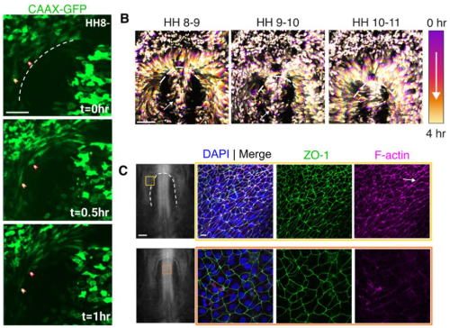
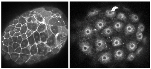
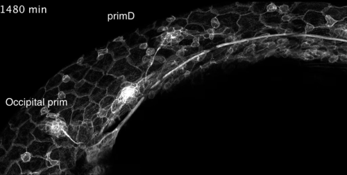
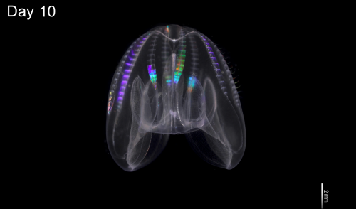
Using an image or a video as a hook, these short posts bring people’s attention to a paper, a technique or a location that is of interest to the developmental and stem cell biology community.
“No such thing as a standard career path” interview series
In this new series, we chatted to several developmental biologists who have had vastly different career trajectories. Check out all the interviews in this series so far.
The Node correspondents
Correspondents are researchers who are also interested in science communication. They work with the Node team to develop and create content on a broad range of topics. Here are a few highlights of posts produced by the correspondents:
Do you want to broaden your science communication experience alongside your research? We are looking for new correspondents for the Node. Find out more and apply by 20 January 2025!
Even though we have grouped posts into different series, we always welcome posts that don’t necessarily fit into any of our existing blog series.
Remember, the Node is your site: once you’ve registered, you can freely share your blog post, job advert or event notice with the community. If you have any questions, just get in touch.
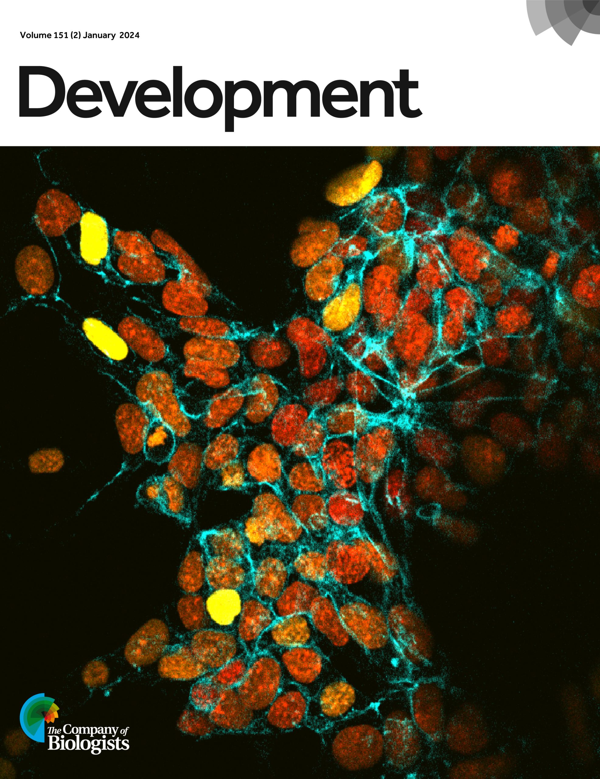
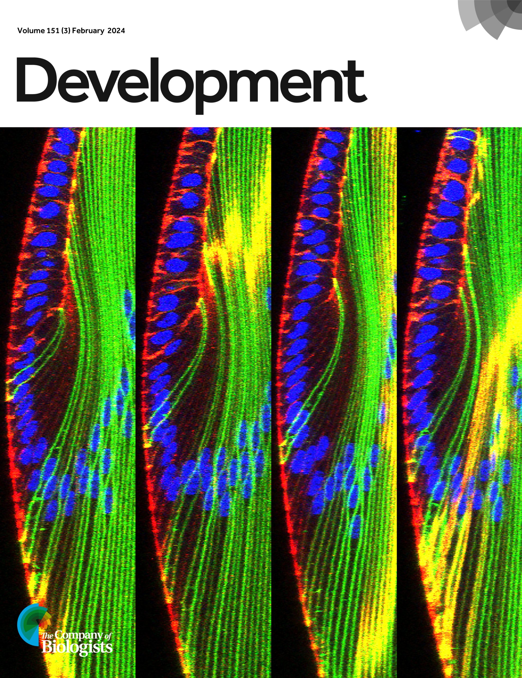
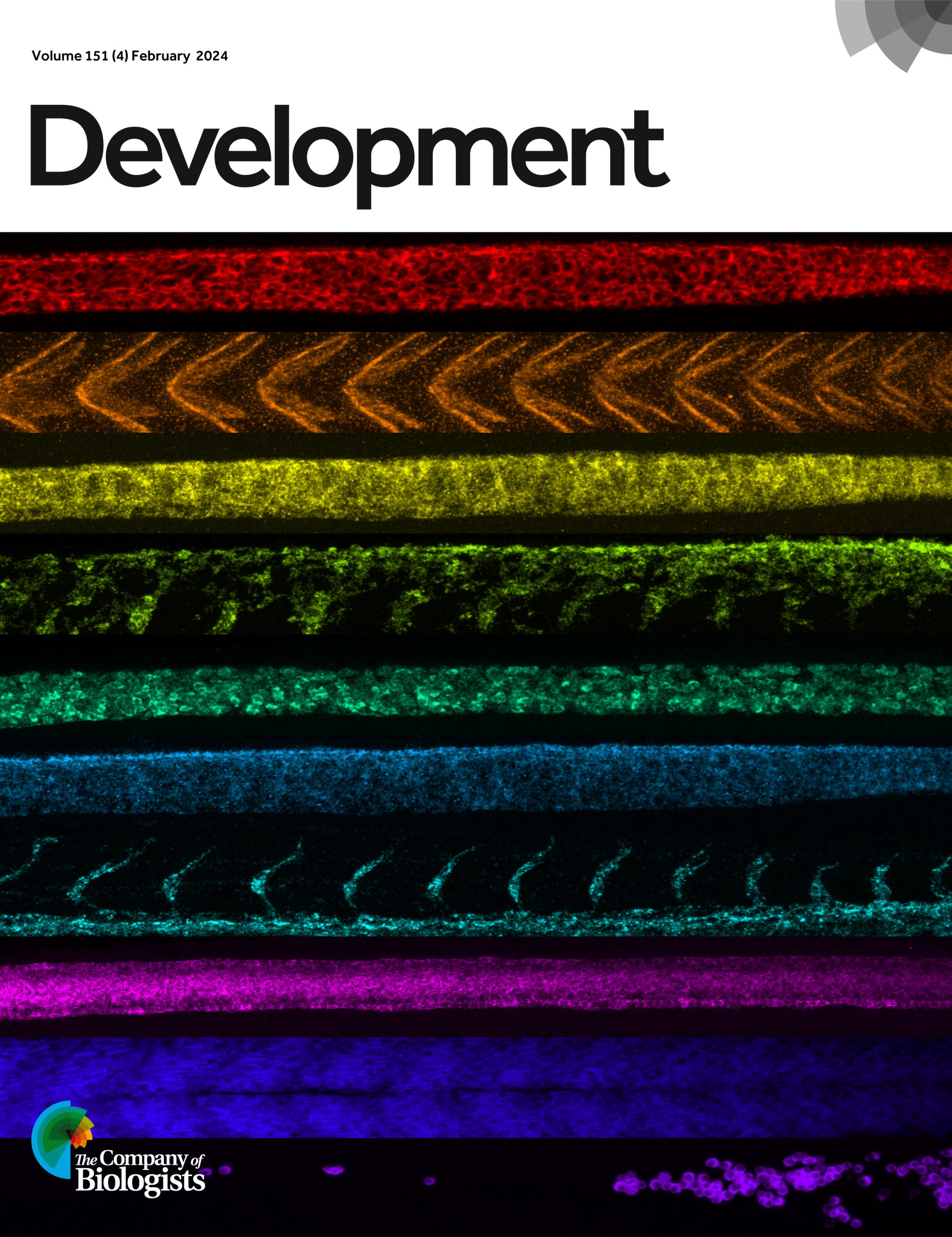
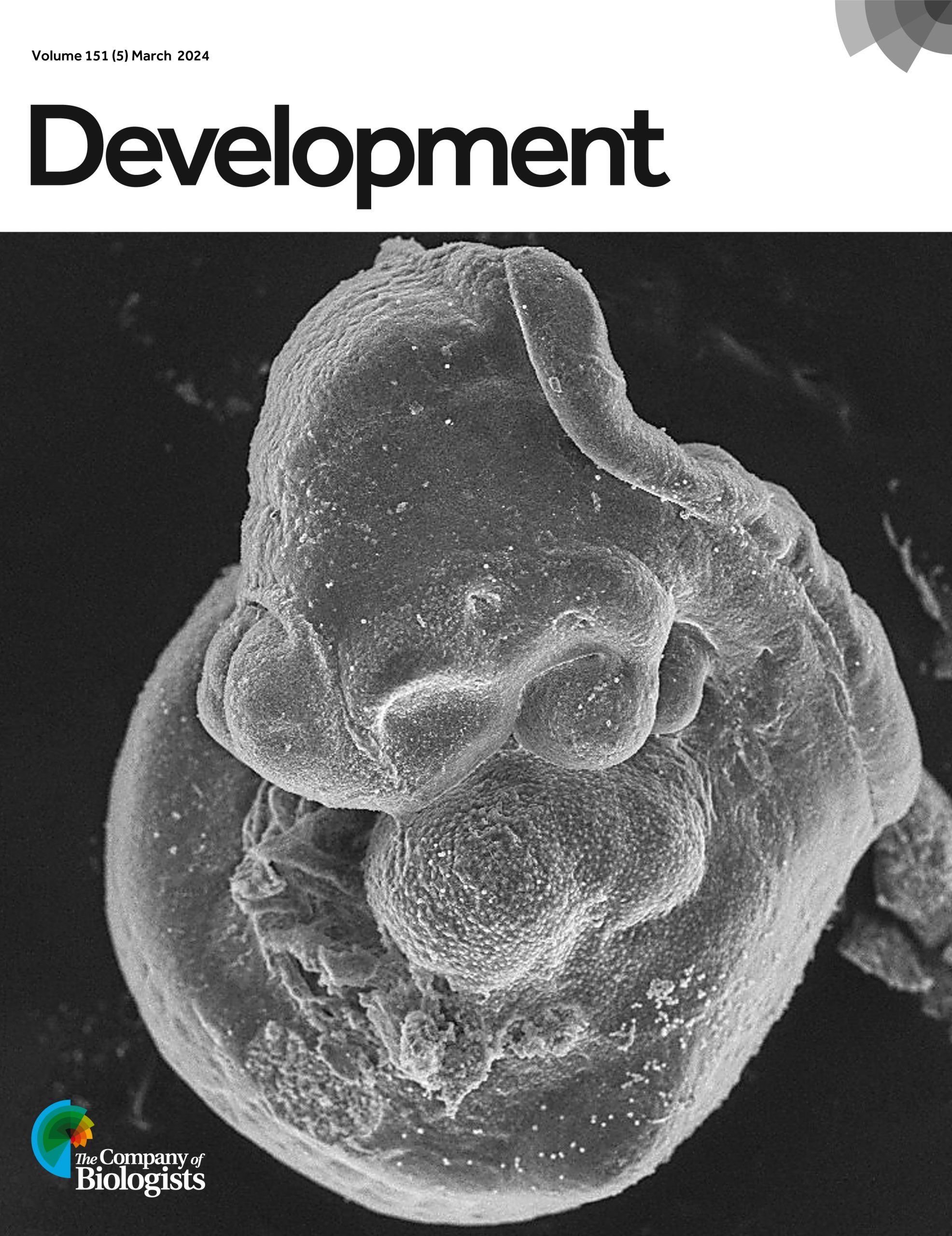
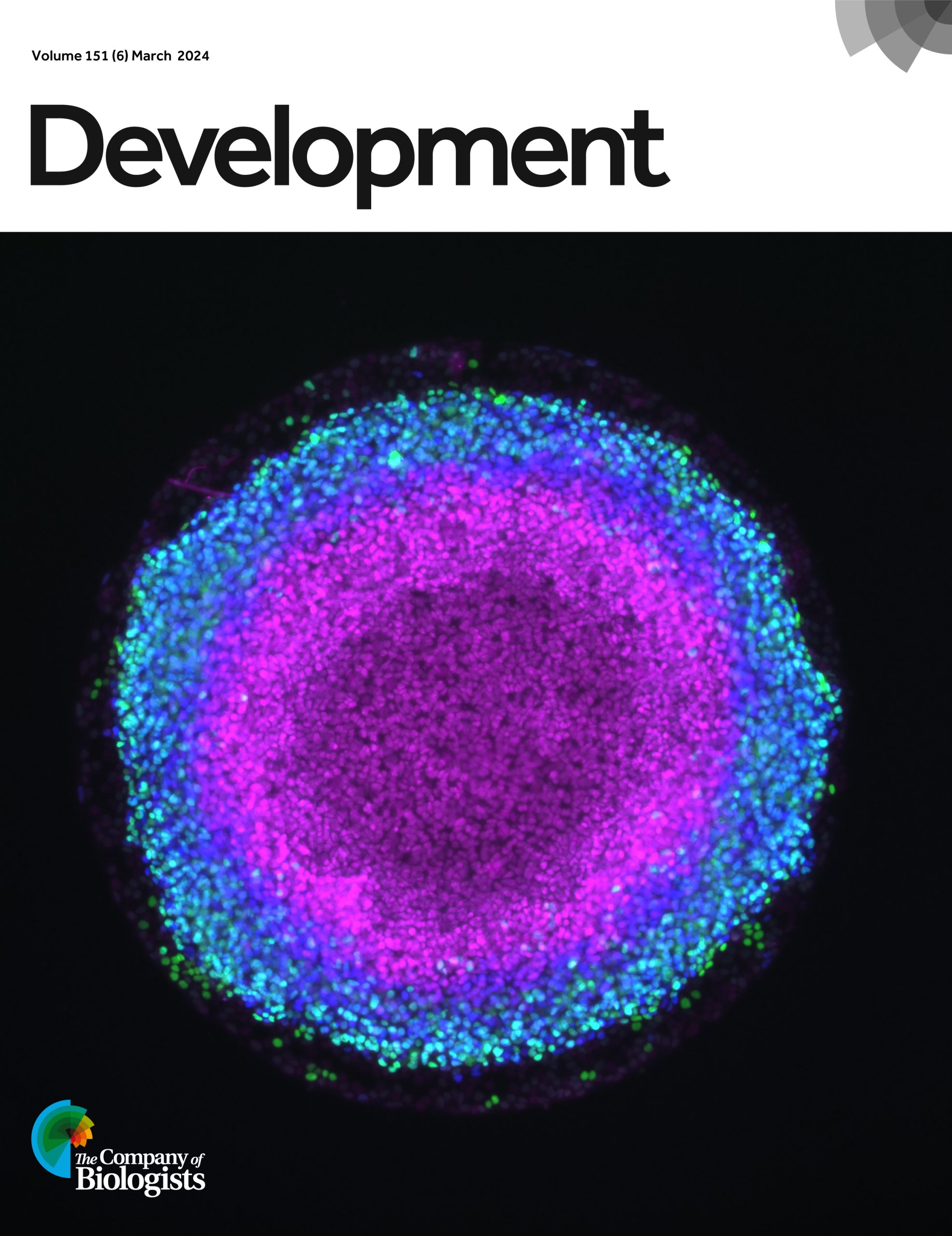
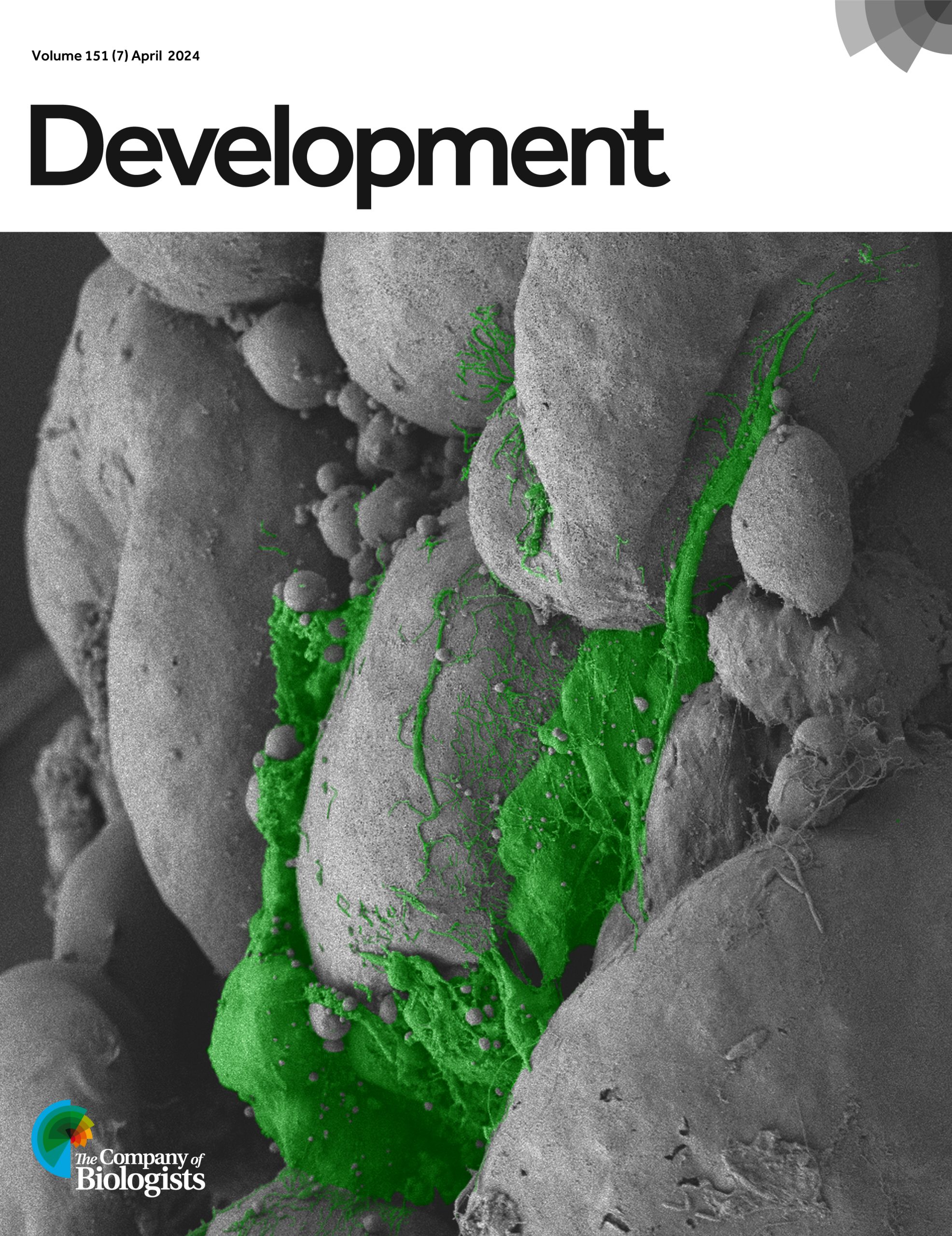
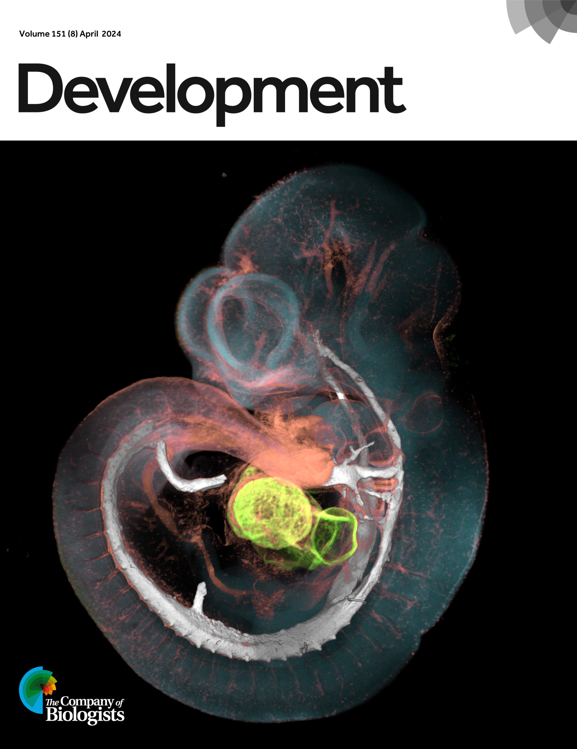
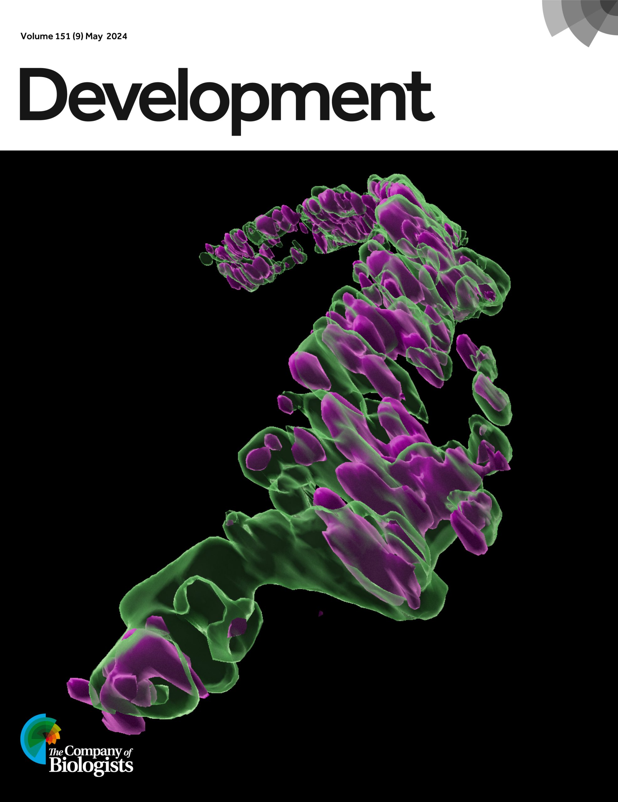
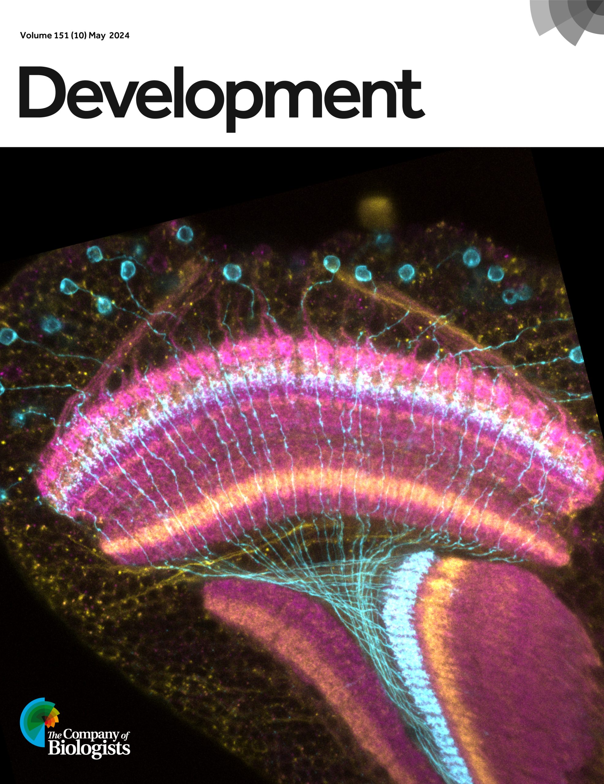
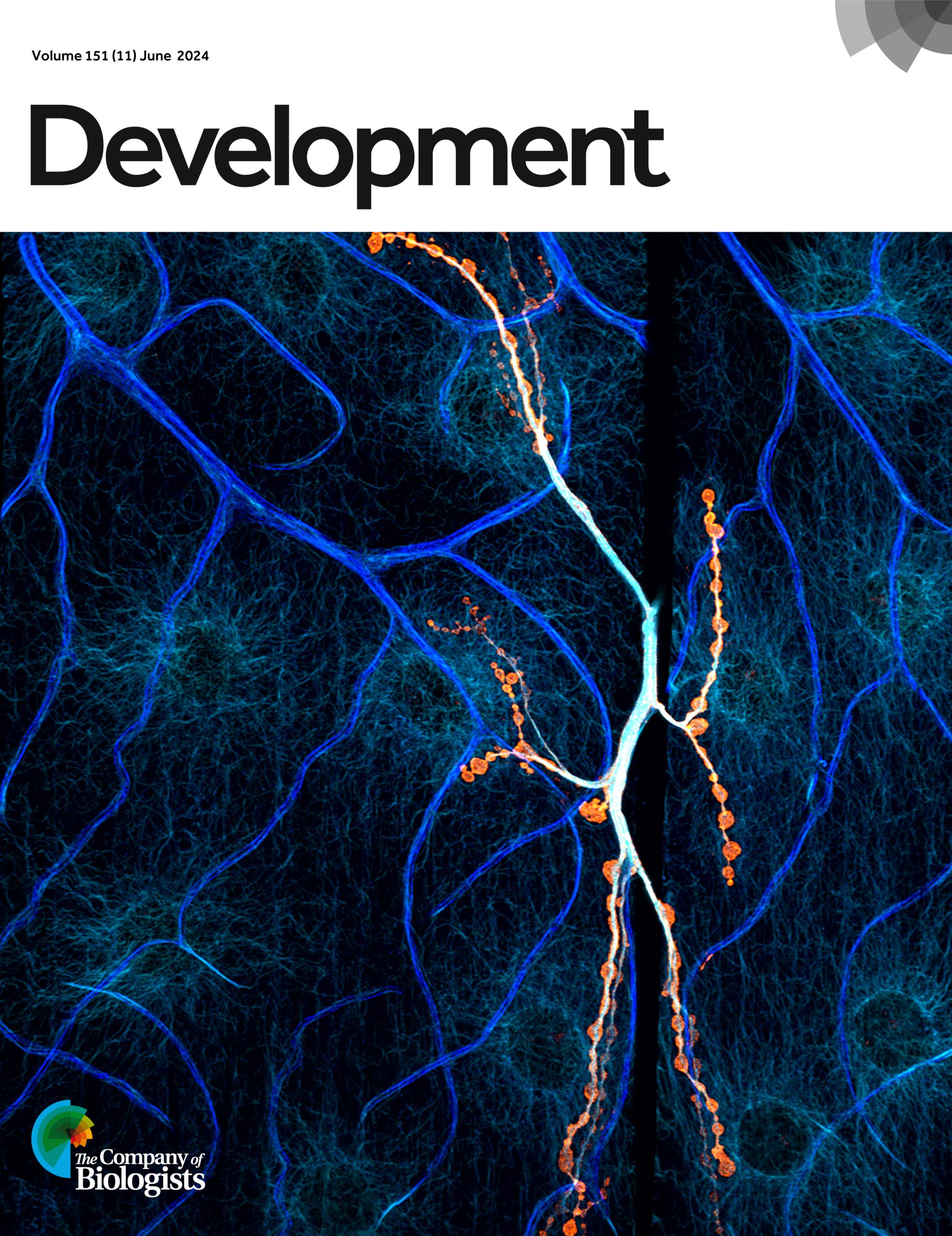
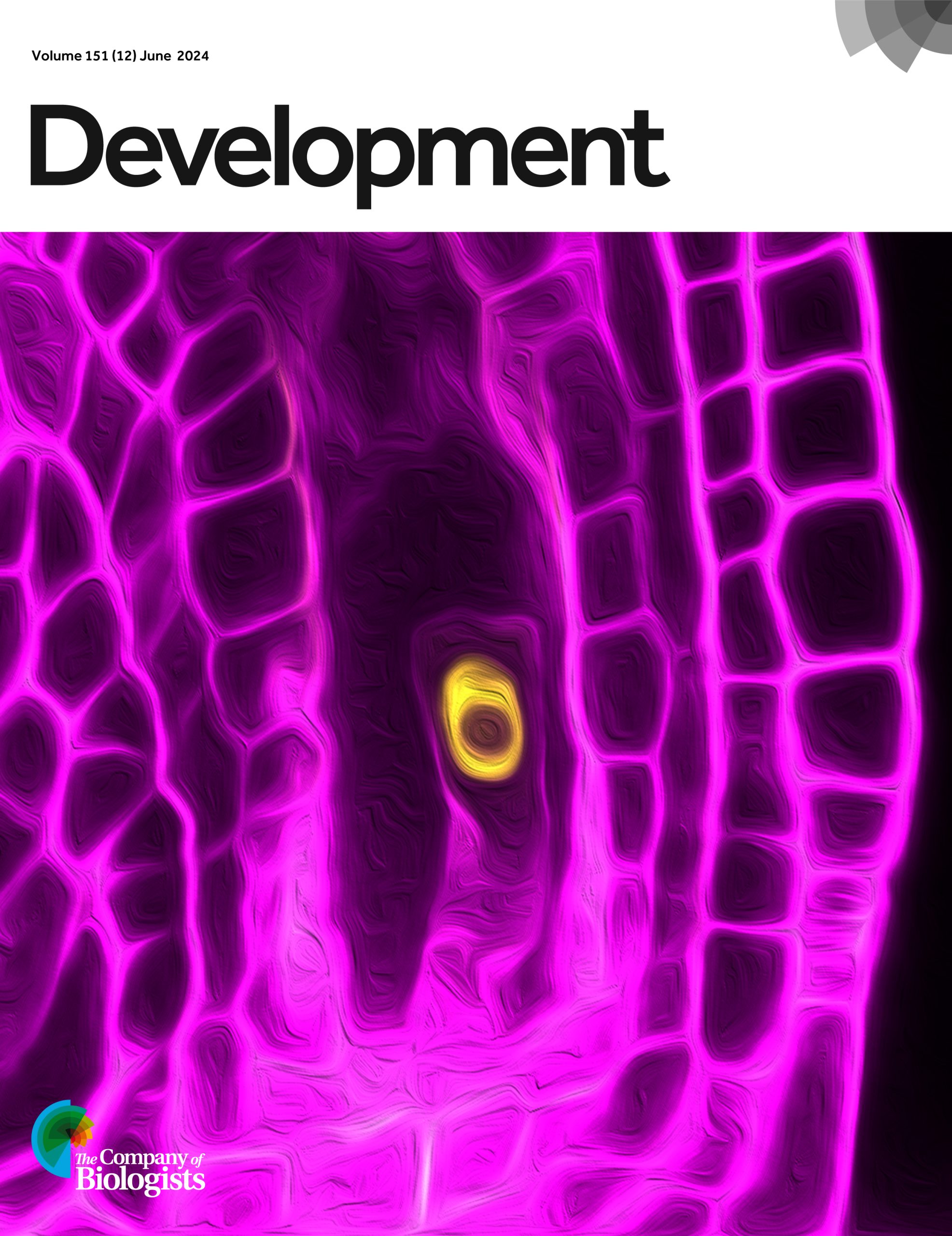
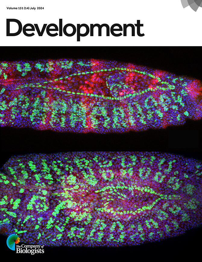
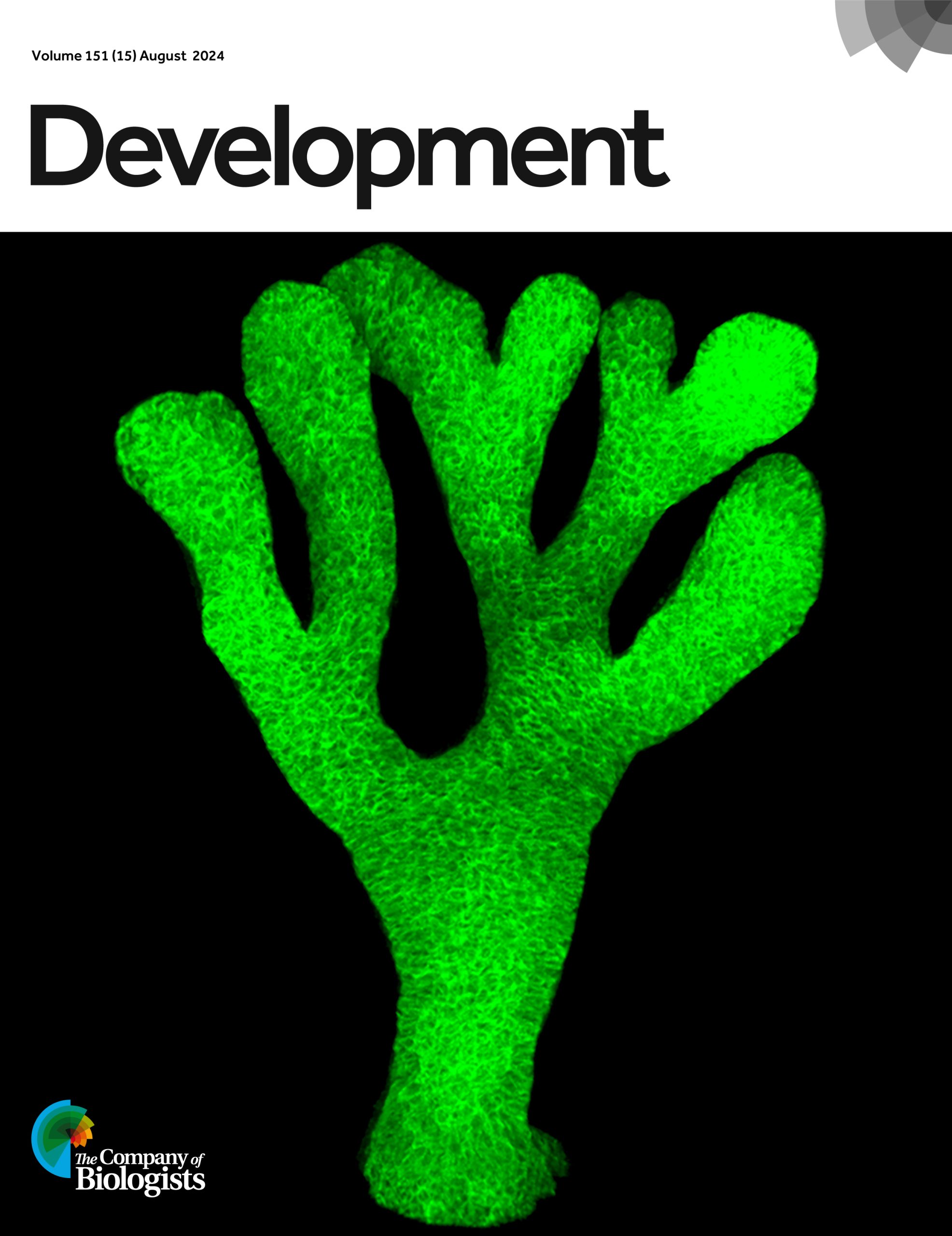
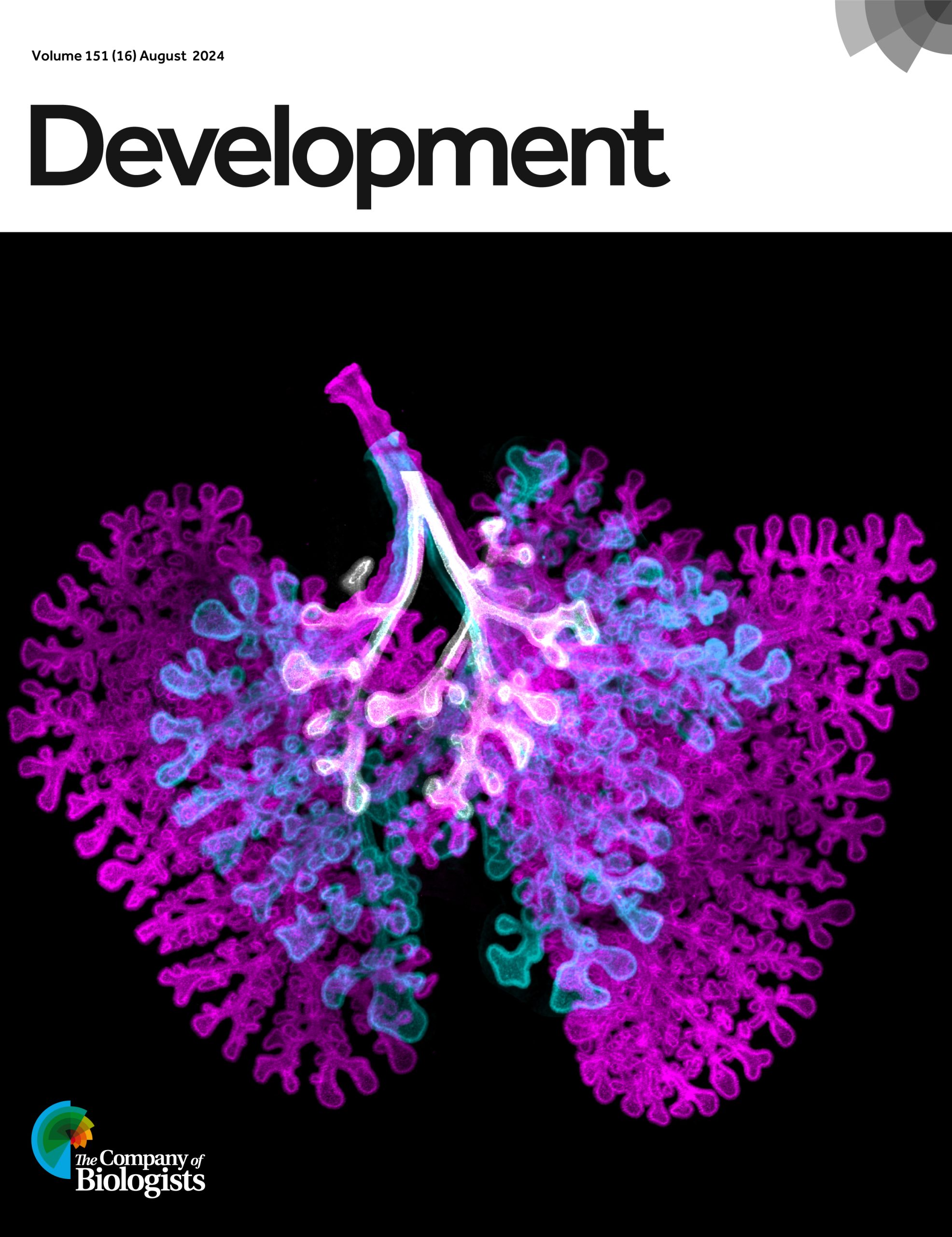
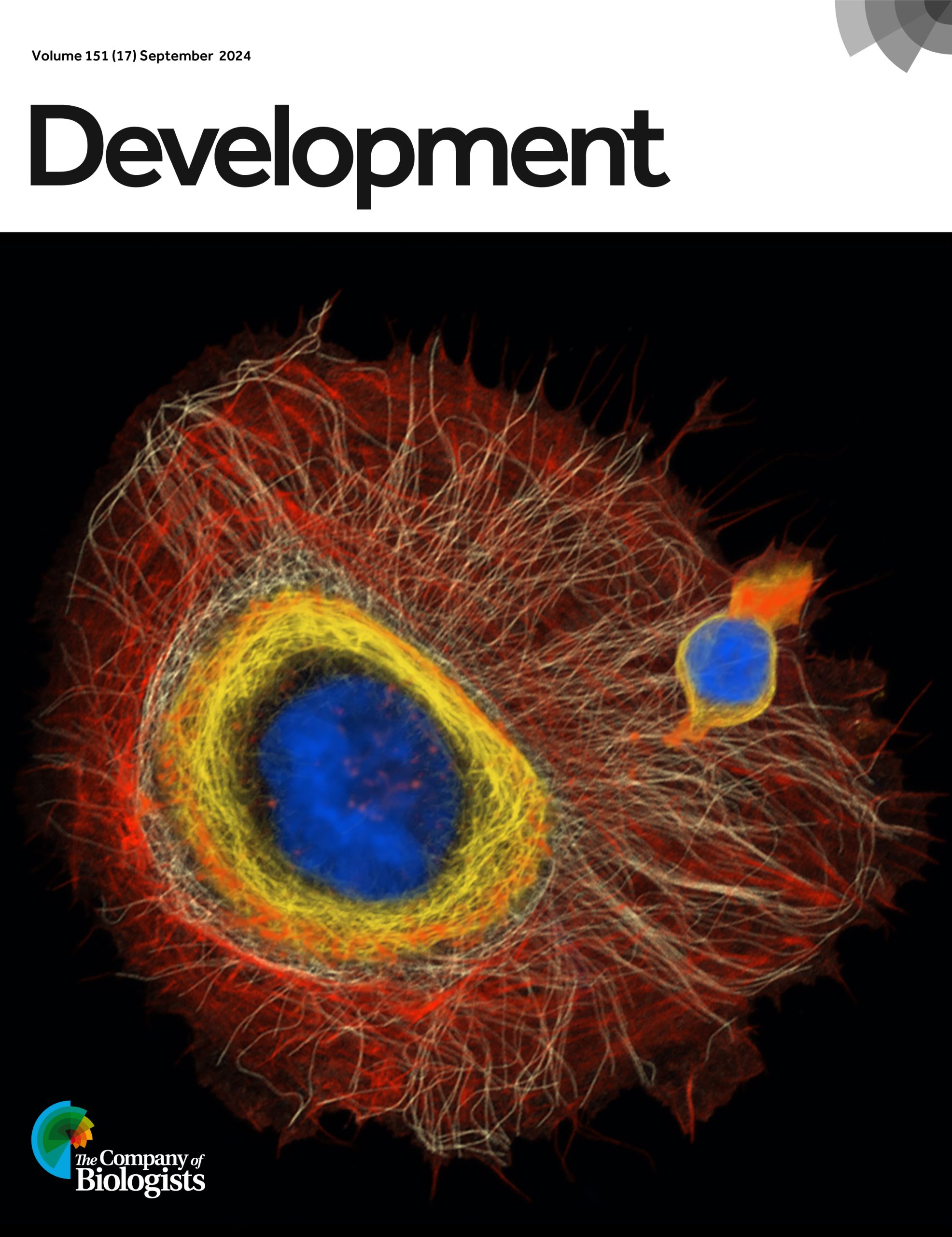
Over the past 12 months, Development has featured 24 cover images. Now, it’s your chance to pick your favourite!
To find out more about each cover image, you can visit Development’s 2024 issue archive. Thank you to everyone who’s contributed to this collection of wonderful images.
*The poll is now closed. Thank you to everyone who voted!*
Issue 1
Issue 2
Issue 3
Issue 4
Issue 5
Issue 6
Issue 7
Issue 8
Issue 9
Issue 10
Issue 11
Issue 12
Issue 13
Issue 14
Issue 15
Issue 16
Issue 17
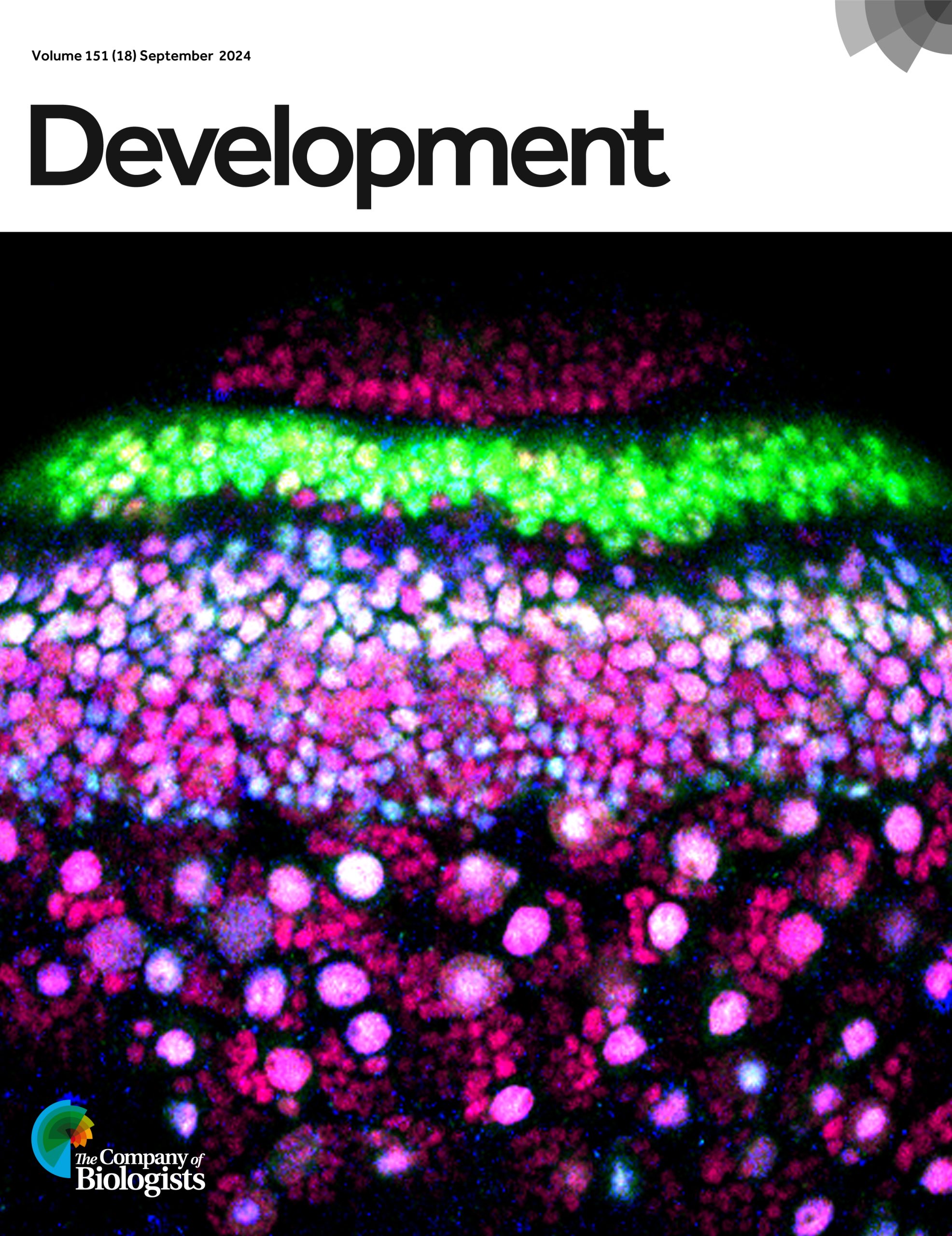
Issue 18
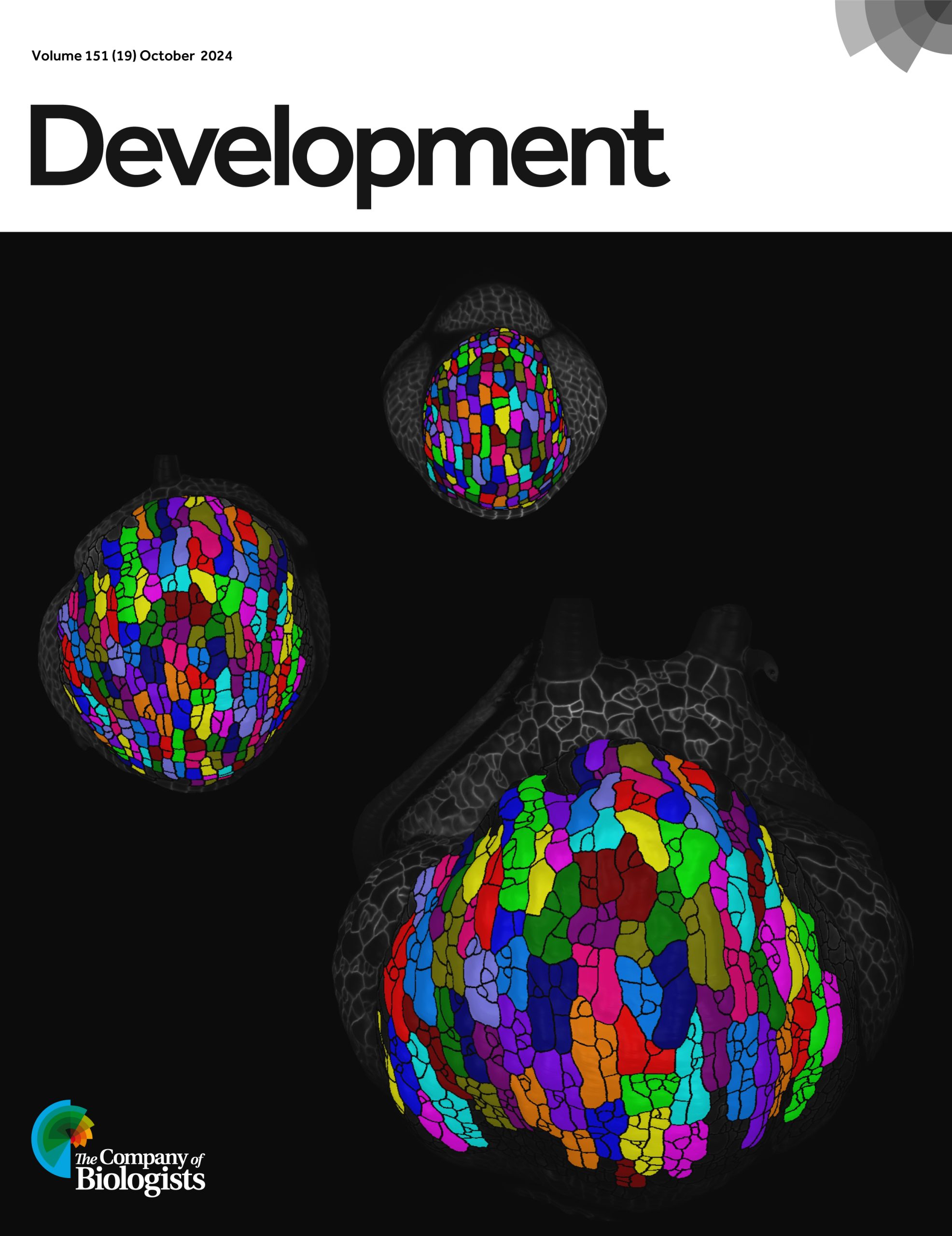
Issue 19
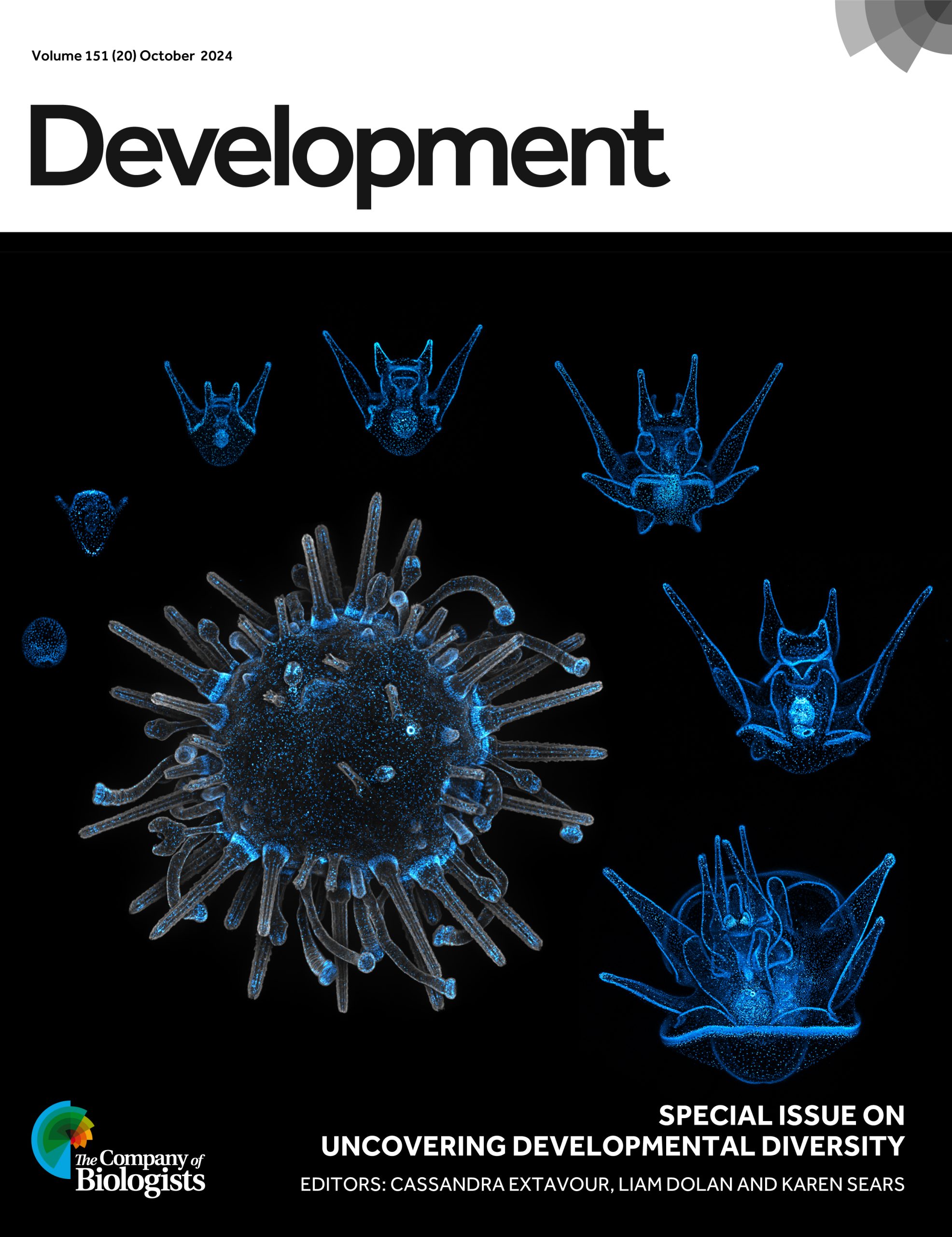
Issue 20
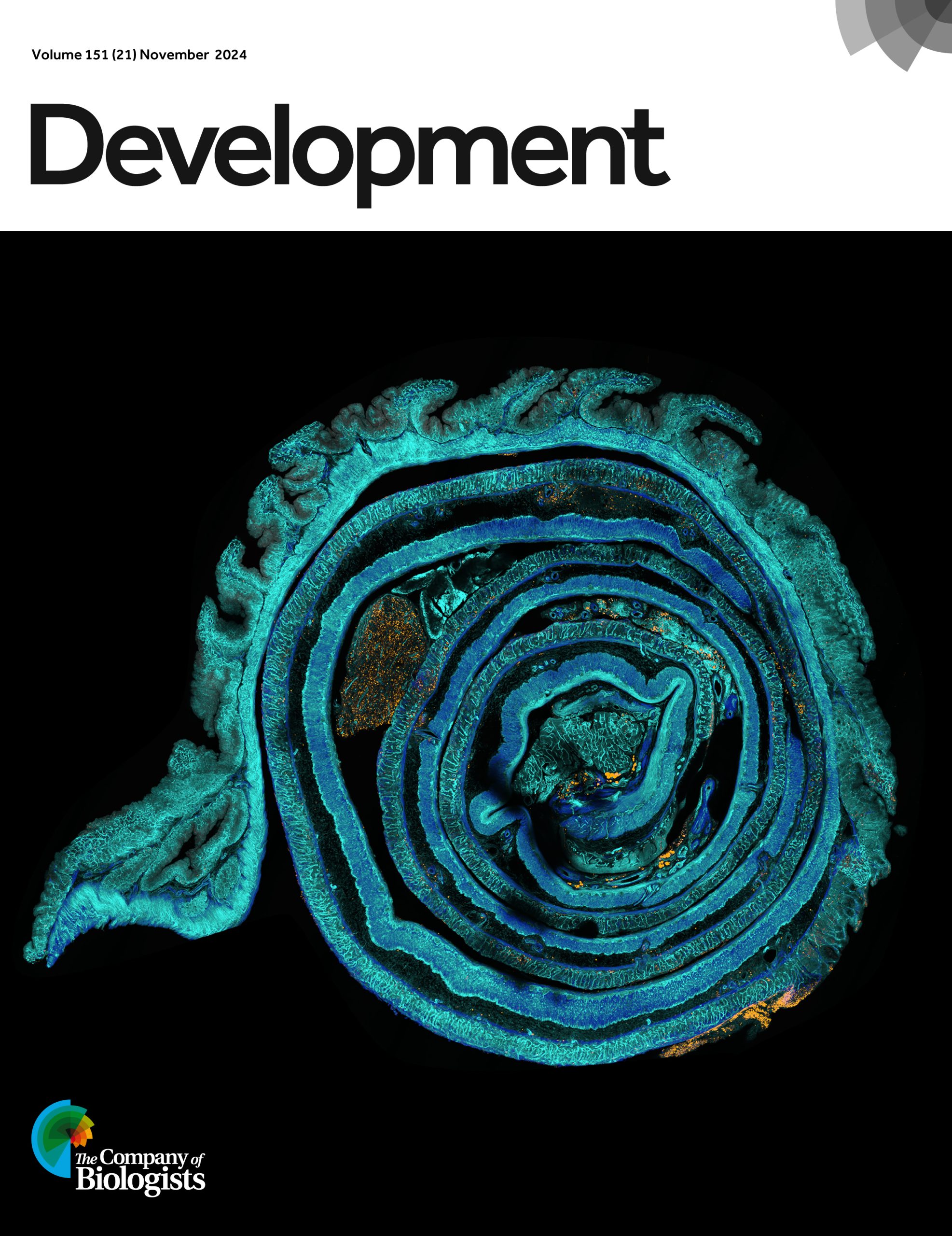
Issue 21
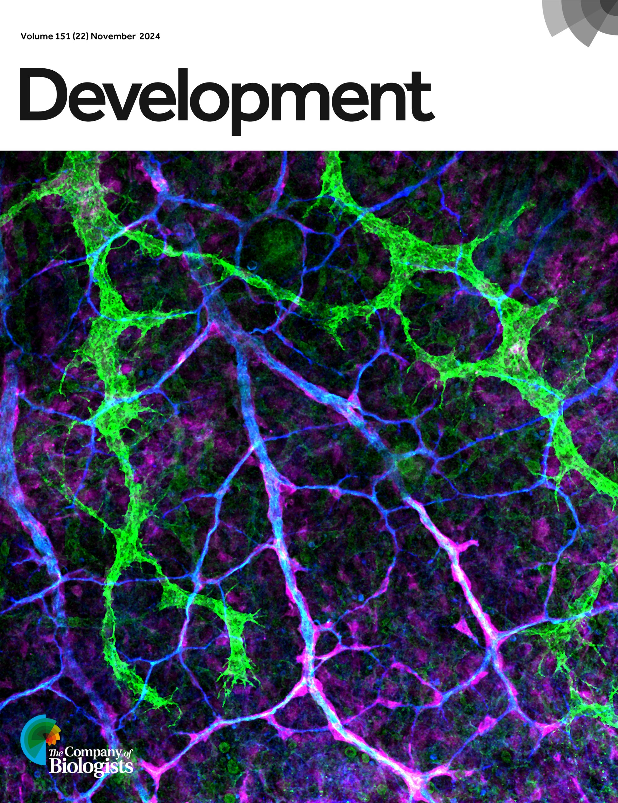
Issue 22
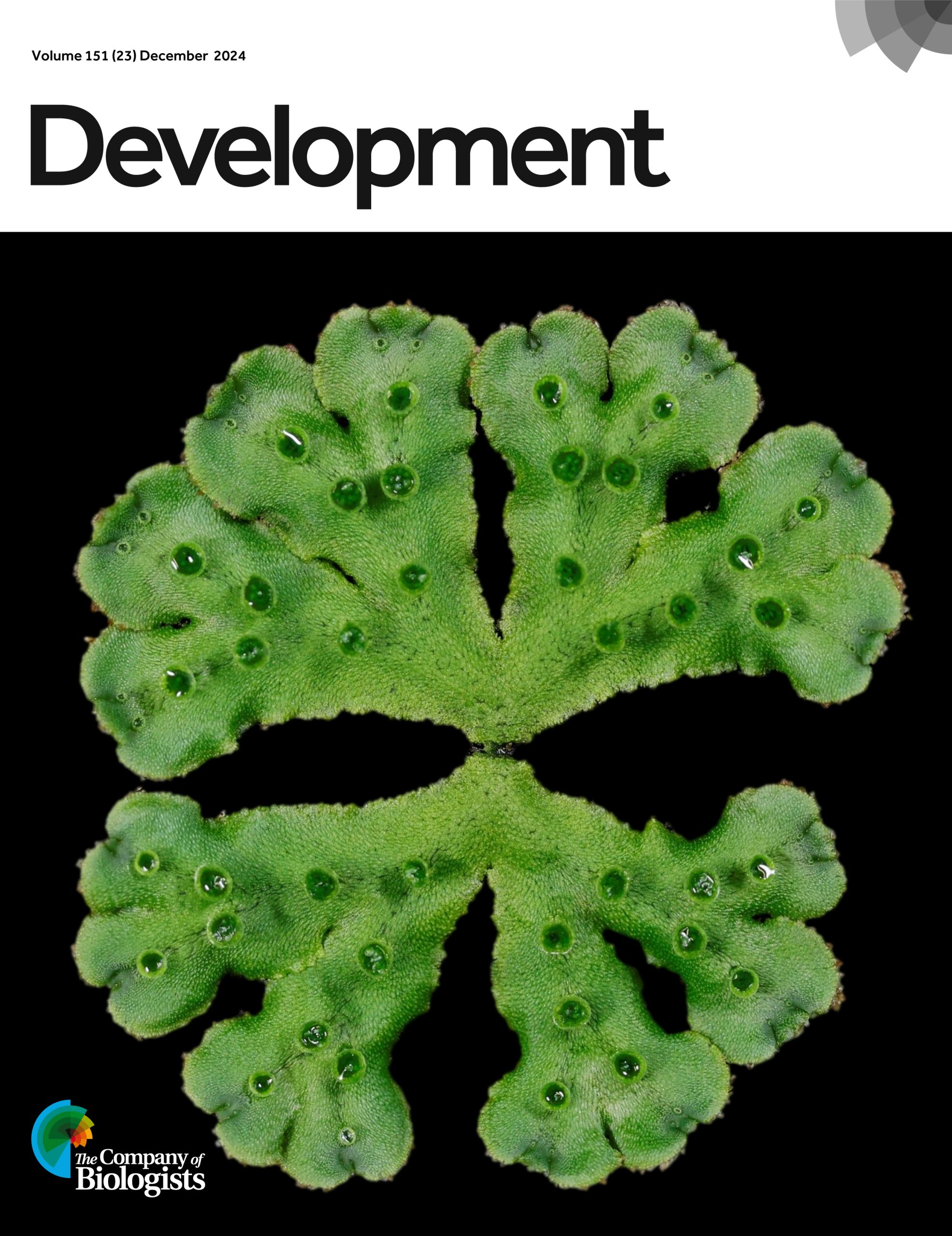
Issue 23
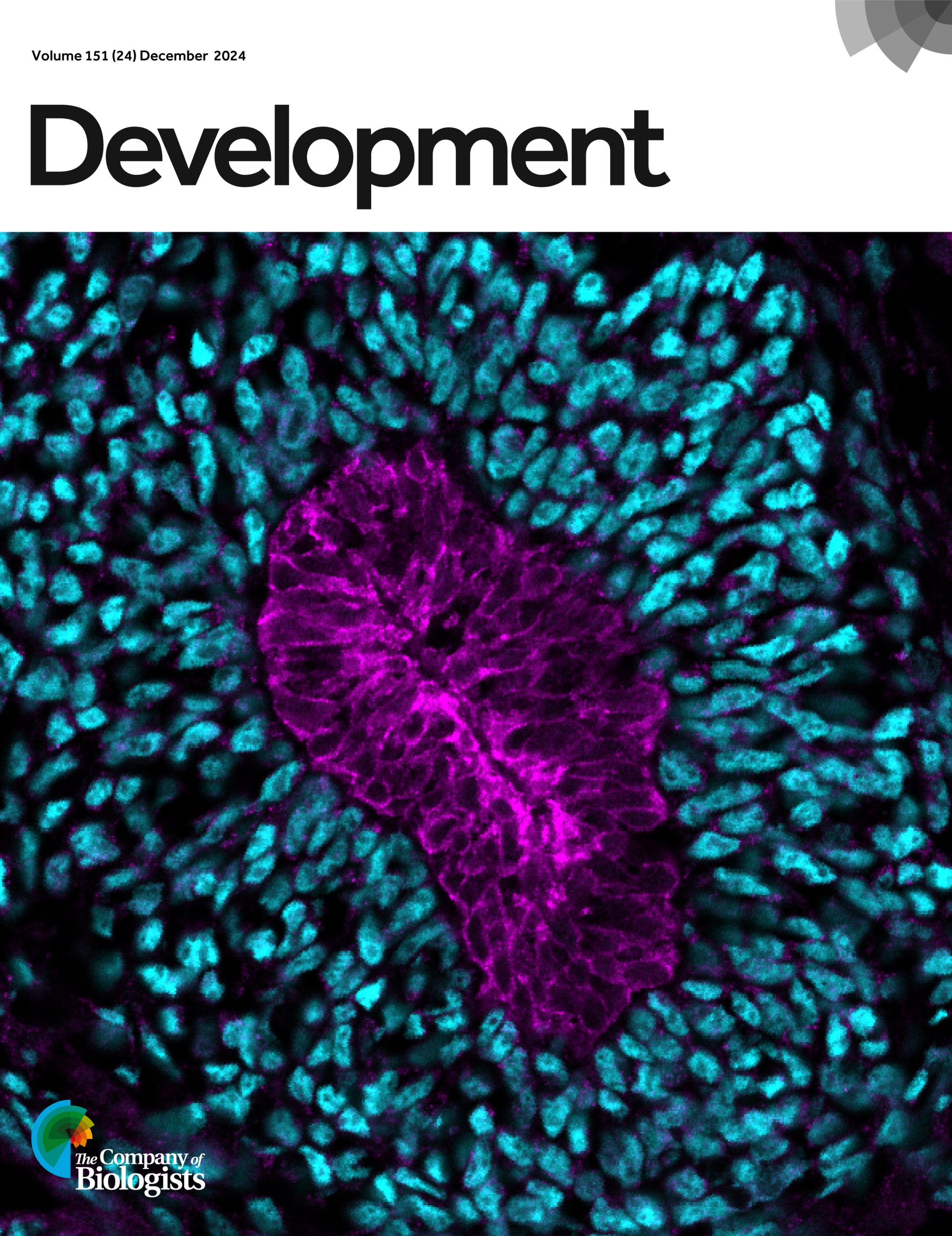
Issue 24
Visit Development’s 2024 issue archive to find out more about each cover image.
Vote for the 2024 Development cover image of the year
*The poll is now closed. Thank you to everyone who voted!*
The final webinar of 2024 featured two early-career researchers working on gene regulation and will be chaired by Development’s Senior Editor, Alex Eve.
PRESS RELEASE: Millions of people around the world are affected by retinal degenerative diseases. In most cases, loss of vision is caused by damage to the macula, a region in the centre of the retina. The macula is rich in cone photoreceptors – cells important for perceiving colour and seeing finer details. Currently, there are no approved treatments to replace the damaged macula, despite its huge impact on the quality of life. Now, a team of researchers from the University of Montreal, led by Professor Gilbert Bernier, found that blind minipigs receiving retinal transplants made from stem cells showed signs of restored vision. They published their study in the journal Development on 5 December 2024.
In this study, Professor Bernier’s team developed a method to coax stem cells into forming sheets of cells that recapitulate the structure of the human retina. The type of stem cells they used are called human induced pluripotent stem cells – immature cells ‘reprogrammed’ from an adult (mature) cell that can differentiate into any type of cells in the body. Using the stem cells, the researchers made ‘retinal sheets’ that are enriched in immature versions of the cone photoreceptor cells, which could become mature cone cells when cultured in the lab.
After successfully creating the retinal sheets in a dish, the researchers tackled the next challenge: transplanting these sheets into minipigs with damaged macula. Professor Bernier explains, “To get as close as possible to human clinical application, we have chosen minipigs because the size of their eyes is near that of humans and the animals are about the same weight as humans. Hence, all surgeries in our study could be performed by a retinal surgeon.”
Upon transplantation, the researchers found that the retinal grafts were able to integrate into the minipig’s damaged retinal tissue. Encouragingly, the minipigs showed signs of restored vision: new neural connections were formed between the grafted photoreceptor cells and the minipigs’ neural cells, and the scientists could detect neural activity of the photoreceptors at the grafted area when the minipigs were placed in a well-lit room.
Given the pressing need to develop therapeutic interventions against vision loss, researchers around the world are testing different ways to repair damaged macula. “Some approaches use dissociated photoreceptor cells; others create micro-dissected retinal organoids, which are lab-grown ‘mini-organs’ in a dish,” says Professor Bernier. “In contrast, our method allows the spontaneous formation of a flat retinal tissue that is already polarised and organised, as in the human embryonic retina.” He adds that their method can generate large yields of retinal tissue for transplantation.
A limitation in this method lies in the difficulty of controlling the placement and orientation of the grafts during surgery. The macula is only 4mm in diameter – about the length of a grain of rice. “To properly orient, place and stabilise the graft in the retina remains a big surgical challenge,” says Professor Bernier. His team are now working to improve the transplantation success rate. They are validating an experimental retinal surgery device to ensure proper orientation and implantation of the graft at the correct retinal disease site. Although many challenges remain, this study demonstrates the potential of retinal sheet transplantation for treating retinal degenerative diseases.
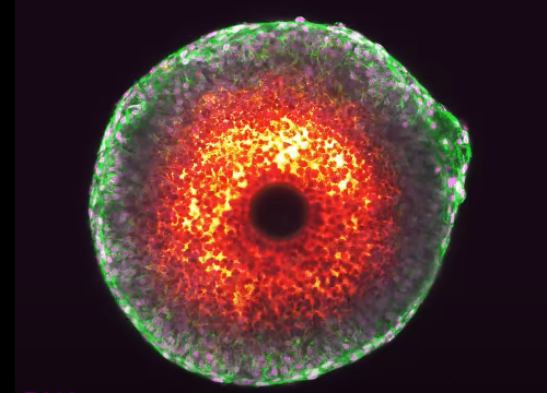
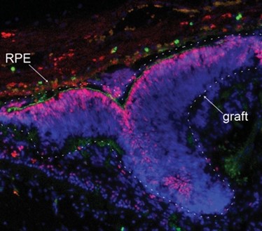
Integration of a human retinal sheet (graft-dashed line) transplanted into a degenerated minipig retina, showing expression of the photoreceptor-specific markers CRX (in red) and PNA (in green) within the graft. Note the graft polarization and close association with the pig’s eye retinal pigment epithelium (RPE). Image Credit: Dr. Andrea Barabino
Enthusiastic about science communication and looking for a chance to broaden your writing experience alongside your research activities? The Node, the community site for developmental and stem cell biologists, is looking to appoint three correspondents who will play a key role in developing and writing content over the coming year.
In 2024, we have been working with Alex Neaverson, who’s used her artistic talents to create illustrations for the Node. You can check out Alex’s illustrations about Rita-Levi Montalcini’s extraordinary life, and the post about the work of Millie Race, winner of the Young Embryologist Network Sammy Lee Award. Thank you Alex for making the Node more colourful!
As a correspondent, you will be expected to contribute around six posts over the course of the year – this could involve creating your own blog series around a theme of your choice, reporting on the latest exciting developments in developmental and stem cell biology, interviewing inspiring scientists, or writing about conferences and other events. We are also open to any other ideas you might have as we would like to shape a programme that both appeals to your interests and benefits the research community.
You will also gain insight into the publishing industry through meetings with the Community Managers and receive regular feedback on your writing. We will help raise your profile as a researcher and science communicator and are also happy to support you by contributing towards conference attendance costs relating to the role, providing reference letters, or in other ways.
Please note, we are also recruiting correspondents for FocalPlane, so when applying you will have the option of choosing to apply for the Node, FocalPlane or both.
We encourage applications from all individuals regardless of sexual orientation, gender identity or expression, religion, ethnicity, age, neurodiversity or disability status. We also welcome applicants from a range of geographic locations.
Please get in touch with us if you have any questions about the programme at thenode@biologists.com
Hopefully some of you will have seen the recent editorial in Development on our approach to peer review. If you haven’t read it yet, please do take a look. In it, James Briscoe (the journal’s Editor-in-Chief) and I discuss some of the initiatives that the journal has taken to try and support authors through the peer review process – including, most recently, encouraging authors to include a ‘Limitations’ section in the discussion of their article, giving you an opportunity to lay out explicitly the scope and extent of your study and, where appropriate, to respond to referee concerns by acknowledging them rather than addressing them experimentally.
Off the back of this editorial, James has also written a blog post that I’d really encourage you to read. Entitled ‘In Praise of Peer Review‘, James sets out why he believes that peer review (in some form) is an invaluable and irreplaceable part of scholarly communication. Alongside the debate that’s been going on around eLife’s exclusion from Web of Science (and subsequent decision to send a partial feed of articles for indexing, the piece has generated some discussion on social media both around whether peer review actually works to guard against publication of fraudulent, sloppy or otherwise dubious papers, and around the degree to which it actually helps to improve papers. I think James has done a great job of setting out the ‘why’ of peer review, but here I thought I’d give my view on the ‘what’: what should a peer review report comprise?
But before I start, let’s remember that – in the majority of cases at least – peer reviewers are both 1) highly knowledgeable in the field of the paper they’ve agreed to review and 2) well-meaning. Yes we all know of cases where papers have been sent to referees that weren’t sufficiently expert or who set out to block publication for political or petty reasons. But these are in the minority – most reviewers are competent to do the job they’ve been asked to, and they want to do it well. And they do it for little or no reward, because they believe that it’s an important part of their responsibility as a member of the academic community. If or how they should be rewarded is a whole other topic that I won’t get into now, but I am incredibly grateful for their dedication.
So, what do I want a referee to do?
Firstly, I want a referee to be respectful. Remember that there are people behind the data and – before hitting the ‘submit’ button on their report – pause to consider the potential impact of your words on the authors, particularly the students and postdocs who’ve actually done the work. At Development, we’re very fortunate that the vast majority of referees do abide by this guidance, but that’s not to say that I’ve not come across the odd report that felt overly combative or dismissive in tone – and that’s not OK.
Secondly, I want the report to be reasonable in terms of the amount of additional work requested. Think about the amount of time (and money!) that might be involved in addressing any particular point and ask how important that point really is to the main story of the paper. Which leads me on to:
Thirdly, I’d ask the referee to focus primarily on addressing the question ‘do the data support the conclusions?’ and not ‘what could the authors do to make the conclusions more interesting?’. While it’s very useful to get expert opinion on how important/relevant/useful/important the paper will be for the community, it’s primarily the editor’s job to decide on whether the paper is – in principle – appropriate for the journal in question.
And finally, I want the referee to be honest about what aspects of the paper they can and can’t (or even did and didn’t) assess. Are you able to judge if the authors have used appropriate statistical analyses? (And if so, did you actually check?!) If the paper contains computational work, do you have the expertise to assess it fully? If the authors deposited data, did you look at it? We fully appreciate that referees can’t always be experts in every area of a paper – particularly an interdisciplinary one – and we try to recruit referees with complementary expertise, but it’s really useful to know what you did and didn’t review.
Most reports I read (and I read a lot!) do largely follow these guidelines, but there is still a definite tendency for a referee report to read a bit like a shopping list of potential experiments and textual revisions. Experienced authors can often read the nuance to decide which points to tackle experimentally, and good editors will (either pro-actively or in response to author queries) help to navigate the revision process. But referees can also do their bit to shepherd papers through the often all-too-painful process of publishing by remembering that there’s both a financial and a temporal limit to how much a group of authors can (and should) do to revise a paper, that a single paper can’t solve a whole research question, and that their opinion isn’t necessarily any more valid than that of the authors (or, for that matter, the other referees).
We could discuss ad nauseam the benefits and problems of pre-publication peer review in its current form (and I frequently do!), and alternative models are beginning to emerge that can act in parallel to, or even replace, our current system. But let’s also think about the little steps that we can take to make the current system less onerous and more constructive – thus easing the path to publication.
The massive presence of disorder and variability challenges the traditional metaphor of the developmental process as a perfectly executed program leading to precise mechanisms at every level [1,2]. Yet, the final outcome —the organism— remains both astonishingly complex and remarkably reproducible. This paradox piqued the interest of Dimitri Fabrèges and Takashi Hiiragi. Back then, around 2017, Takashi was research group leader at the EMBL Heidelberg, and Dimitri a postdoc in his group. They began to explore the idea of disorder and variability from a provoking viewpoint: instead of undermining the precision of the developmental process, randomness and variability might actually act as driving forces that ensure precision and reproducibility.
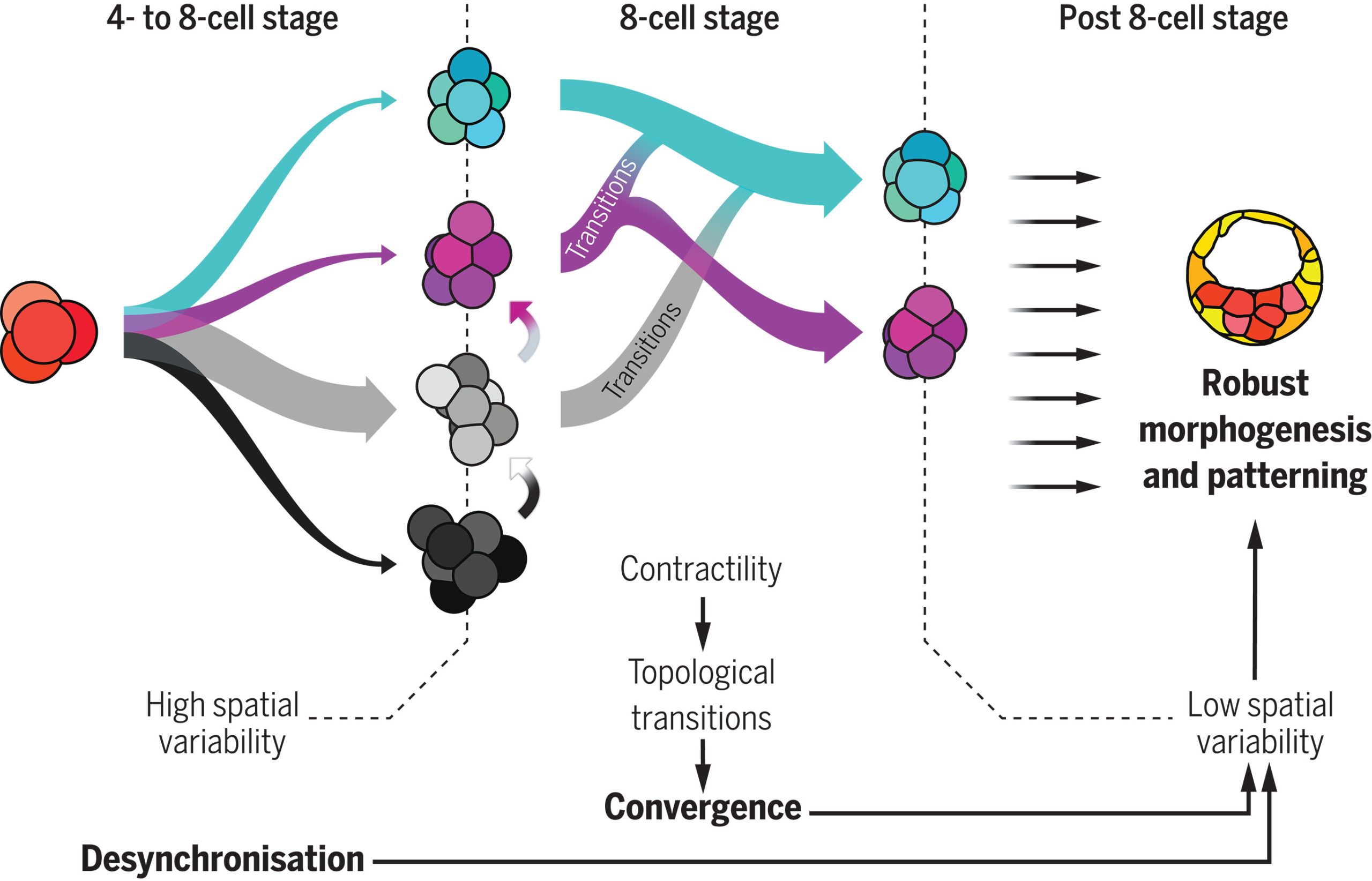
Motivated by this hypothesis, the researchers focused on the early stages of mammalian development; particularly, on the initial cleavage process of mouse, rabbit and monkey embryo, encompassing the first cell divisions post-fertilization up to the 16-cell stage. A first analysis showed that the division times of different cells in the same embryo were progressively being desynchronized. A pivotal moment in this sequence is the 8-cell stage —that is, after 3 division rounds— a moment in which cells already divided in a quite disorganized way. Due to this high variability, researchers found that the beginning of this stage was characterized by a highly heterogeneous set of cell packing configurations. However, such initial variability is smoothly but steadily reduced along the so-called compaction process, leading to a seemingly common, spherical-like structure at the end of the stage. Such a structure guarantees that, in the next round of divisions —i.e., at the 16-cell stage— there will be a suitable proportion of inner and outer cells. Achieving this correct proportion is essential: inner cells will lead to the organism itself, while outer cells form the placenta and extra-embryonic material. This observation raised a challenging question, namely, how can one support the intuitive claim that embryos begin highly heterogeneous but become remarkably similar with a more rigorous foundation.
This was the perfect challenge for Virginie Uhlmann, an expert on biological image processing who, at the time, had just started as research group leader at the EMBL-EBI, in Cambridge. She tackled this question by developing an advanced computational framework able to analyze and track the geometric changes in the embryonic shape in high detail. This approach conceptualized an embryo’s developmental path as a trajectory within a high-dimensional space whose coordinates captured relevant geometrical properties [3]. The key result was that, indeed, the trajectories exhibited significant initial disparity but converged surprisingly by the end of the compaction process in a particular region of the abstract space that characterized the embryo geometry.
Which structure was represented in this region? Why did this particular structure seem to act as the developmental “target”? At the beginning of the winter of 2018, Takashi’s research group organized a retreat in the Catalan coastal town of Sitges, gathering several groups from the ISTA. Among the attendees were Edouard Hannezo, who just opened his research group as PI, and his first postdoc, Bernat Corominas-Murtra —both physicists working on biological problems. The evening was windy and stormy, and a little café was the refuge where they largely discussed with Dimitri and Takashi about the challenge of identifying and explaining the emergent structure. Although no immediate solution came out, Edouard and Bernat concluded that a deeper and simpler structural characterization was necessary —that is, complementing the geometric analysis with a topological one, stripping out all details but the raw structure. Weeks after, they stumbled upon a relatively recent publication showing a key mathematical finding: there are exactly different 13 ways to pack 8 spheres such that none of them exhibit independent movement. This result provided the key to define a classification scheme: either the embryos conform approximately to one of these 13 packing configurations, or they are floppy, meaning that some cells retain independent movement [4]. By establishing a suitable notion of “distance” among sphere packings, the researchers could classify embryos at various developmental time points. Their analysis revealed that, although variability was very high at the onset of the 8-cell stage, as the compaction process progressed, embryos consistently converged towards these similar packing structures along similar developmental pathways. At that point, the target structure was identified: the D2d packing of 8 spheres, in the Schoenflies notation.
Fig 1: 4-cell stage embryos give rise to many shapes at the beginning of the 8-cell stage, during which cell contractility triggers topological transitions. Ultimately, embryos are driven toward the most optimal packing (cyan). In parallel, the cell-autonomous desynchronization progressively increases temporal variability and helps to maintain topological optimality through generations, lowering spatial variability and promoting robustness. Picture taken from (Fabreges et al. 2024).
How does the embryo, without any external help, solve this kind of Rubik’s cube, i.e., transition from an arbitrary cell configuration to a specific optimal only one through successive cell rearrangements? Looking at the empirical data, one observable stood out above the other due to its clear trend: Adhesion was increasing along the compaction. Edouard suggested to challenge the simple hypothesis whether this slight change in the cell adhesion was enough to trigger all the topological rearrangements. The hypothesis has deep consequences. It implies that an increase on the cell adhesion could not only trigger deformations within the cells (i.e., increasing the contact surface, for example), but also qualitative reorganizations of the whole embryonic cell mass in a reproducible way. Computer simulations showed that such a genetically encoded slight increase in cell adhesion, coupled with significant random fluctuations in cell positions —disorder— was paradoxically facilitating the transition from any arbitrary packing of cells to the single optimal configuration. This hypothesis stands out as the simplest and, in the case of the mouse, it enabled even to reproduce in-silico the developmental trajectories of real embryos. In the case of rabbit and monkey, the role of other agents, like the zona pellucida —an external membrane that may exert a compressing force to the cell packing— could not be fully discarded.
At this point, the puzzle of the convergence towards a common, suitable embryo configuration was solved. However, the role of the temporal variability, which was experimentally observed at the starting point of the whole project and inspired it all, remained to be understood. Using several genetic perturbations, the results were surprisingly concluding that initial variability was actually required to achieve precise convergence. In particular, embryos in which cell divisions occurred more synchronously than in the wild-type ones showed a poorer convergence at the end of the compaction process, thereby hampering the further development of the embryo. The provoking hypothesis of Takashi and Dimitri on the role of stochasticity was thus proven to be fully consistent.
The researched path was not easy: Big part of the project was carried out during the COVID-19 pandemic. In turn, during the project, Dimitri and Takashi moved to Utrecht, to the Hubrecht Institute, Virginie to the University of Zurich, and Bernat to the University of Graz. Researchers from several institutions1 provided their bits of knowledge in the multiple challenges that paved the achievement of the results, and, as in living organisms, the sum of different expertises —biology, physics, mathematics and computer science— ended up in something that was much more than the sum of its parts. As in any adventurous interdisciplinary research, moments of joy and concern alternated, sometimes without pause in between… All in all, this research provides a new, constructive interpretation of the striking amount of disorder observed along developmental stages: When coupled to the changes in cell mechanics, the interplay among them can lead to significant and precise reorganization events within embryos, paving the way for a new understanding on how complex geometries and, in general, organization patterns arise in living beings. Disorder, therefore, far from being a problem the system has to deal with, may be one of the leading forces driving the precision of organism development.
Publication:
Dimitri Fabrèges et al. Temporal variability and cell mechanics control robustness in mammalian embryogenesis. Science 386, eadh1145 (2024)
References:
[1] M. Carlson, W. Reeves, M. Veeman, Stochasticity and stereotypy in the Ciona notochord. Dev. Biol.397, 248–256 (2015).
[2] R. Dumollard, N. Minc, G. Salez, S. B. Aicha, F. Bekkouche, C. Hebras, L. Besnardeau, A. McDougall, The invariant cleavage pattern displayed by ascidian embryos depends on spindle positioning along the cell’s longest axis in the apical plane and relies on asynchronous cell divisions.eLife6, 1–23 (2017).
[3] R. Delgado-Gonzalo, N. Chenouard, M. Unser, Spline-based deforming ellipsoids for interactive 3D bioimage segmentation. IEEE Trans. Image Process.22, 3926–3940 (2013).
[4] N. Arkus, V. N. Manoharan, M. P. Brenner, Minimal energy clusters of hard spheres with short range attractions. Phys. Rev. Lett.103, 118303 (2009).
1Other institutions involved:
Institute for the Advanced Study of Human Biology (WPI-ASHBi), Kyoto University, Kyoto, Japan.
Department of Developmental Biology, Graduate School of Medicine, Kyoto University, Kyoto, Japan.
Research Center for Animal Life Science, Shiga University of Medical Science, Shiga, Japan.
INRAE, BREED, Paris-Saclay University, Jouy-en-Josas, France.
École Nationale Vétérinaire d’Alfort, BREED, Maisons-Alfort, France.
PhD position in the Denholm lab at the University of Edinburgh, UK
You will use modern techniques to study the development and/or physiology of one of the most powerful water-conserving systems in nature – the beetle cryptonephridial (or ‘buried kidney’) complex.
Insects can live and thrive in some of the most inhospitable environments on earth, including extremely desiccating conditions such as deserts. Many species possess a powerful water-conserving system called the cryptonephridial (or ‘buried kidney’) complex (CNC), which recovers water from the rectum and recycles it back to the body. This remarkable system even allows water vapour absorption from moist air, providing a novel physiological mechanism for water uptake. It is estimated that >400,000 insect species have a CNC, with CNCs being particularly common in beetles. The broad principles underpinning CNC physiology were laid down half a century ago, and the system has since become a staple textbook example of a countercurrent exchange system. Despite this, next to nothing is known about CNC development, molecular physiology, endocrinological regulation or evolution.
In this project you will use the model beetle species Tribolium and exploit enabling technologies including genomics, single-nuclei RNAseq, informatics, imaging and in-vivo analysis to identify how this system develops and functions. We have catalogued gene expression profiles (using snRNAseq) from the CNC of this species (in both embryo and adult), providing a window into its embryonic development and the molecular players involved in its physiological function at single-cell resolution.
The techniques you will use and be trained in include: (1) Bioinformatics. You will use this to prioritise key genes involved in the development and function of the system. (2) Hybridisation chain reaction fluorescent in situ hybridisation. You will use this to map expression of candidate genes in embryonic, larval and adult CNCs. (3) Gene knock-down: you will use RNAi to knock-down gene activity for each candidate and, use (4) Microscopy (both fluorescence confocal and electron microscopy) and simple physiological assays to establish roles for these genes in CNC development and function.
Results from this project will significantly expand our understanding of one of the most powerful water-conserving systems in nature, one that is fundamental to insect physiology, ecology and evolutionary success.
From the hard, protective scales of reptiles to the soft, insulating fur of mammals, amniotes are equipped with a remarkable range of skin appendages that help them thrive in diverse environments. To fulfil their various functions, such as mechanical protection, thermoregulation, and the provision of camouflage, skin appendages must be precisely arranged (or patterned). For instance, birds display a regular spatial arrangement of feathers which enable flight (1), whereas snakes exhibit a near-perfect hexagonal pattern of scales, which are aligned to their muscles and ribs (2), to aid locomotion.
All of these skin appendages develop from ‘placodes’ (3, 4) comprised of an epidermal thickening, an underlying aggregation of dermal cells, and conserved molecular signalling. It is well-established that the spatial patterning of placodes is self-organised through interactions between activatory and inhibitory morphogens, forming a so-called Turing reaction-diffusion system (5-7). In other words, the resulting self-organised “polka-dot” arrangement of gene expression provides a template defining where skin appendages will develop (1, 3, 4, 8).
Remarkably, the irregular polygon-shaped head scales of crocodiles present a fascinating exception to this paradigm, as they appear to emerge from a mechanical process, i.e., in the total absence of a genetic template (9). However, the exact nature of this process has remained elusive for more than a decade. In our new article published this week in Nature (10), we solve this mystery through a multidisciplinary approach combining experimental manipulations of developing crocodile embryos, cutting-edge light sheet microscopy, and state-of-the-art numerical simulations.
First, we imported a large number of Nile crocodile eggs from South Africa to our laboratory in Geneva, Switzerland. After incubating these embryos until their head scales begin to emerge, we performed precise in-ovo intravenous injections of epidermal growth factor (EGF) protein that, as its name indicates, increases epidermal growth while also promoting keratinisation. As we increased the dose of EGF, we observed the development of an extensive labyrinthine pattern of convoluted folds across the jaws of the crocodile embryo, instead of the normal polygonal scales. These results demonstrate that crocodile head scales develop through compressive folding and that EGF treatment exacerbates this folding. Note that tissue buckling from ‘constrained growth’ is thought to shape other biological forms, including the intricate folds of the human brain (11), villification of the human and chicken gut (12), the surface wrinkling of mucosa (13), and the small polygons on the glabrous skin of the rhinarium (or naked nose) of dogs, cows and ferrets (14). Interestingly, we have recently showed that, for this latter example, the polygonal form of the pattern is imposed by the underlying polygonal pattern of blood vessels that act as ‘mechanical positional information’ (14).
Remarkably, when we allow the crocodiles to hatch four weeks after the EGF treatment has finished, they no longer display a fully labyrinthine pattern, as it partially relaxes into a pattern of smaller polygons. Importantly, this pattern resembles that of another crocodilian species – the spectacled caiman. Therefore, simply by promoting skin growth and differentiation during embryonic development, we can alter the hatchling head scale pattern from that of a Nile crocodile to that of a caiman. Therefore, shifts in the molecular components of an intricate Turing reaction-diffusion system are not required to explain the evolution of head scale patterns in crocodilians. Instead, slight variations in the expression of a very small number of genes affecting skin growth and/or stiffness can clearly explain this macroevolutionary change.
We next used cutting-edge light sheet microscopy to build a realistic 3D computer model of the crocodile embryo’s head before the emergence of scales. This model incorporates the key elements that we suggest determine the mechanical patterning of crocodile head scales. This includes the geometry of the skin (such as the spatial variation of epidermal and dermal thicknesses) and underlying bone, the organisation of collagen fibres within the dermis, and the relative growth rates of the two skin layers. Remarkably, our computer growth simulations accurately reproduce the natural head scale arrangement of Nile crocodiles, which includes elongated units on top of the jaws and smaller polygons on the sides. In addition, increasing both skin growth and stiffness in our simulations replicates the labyrinthine brainy folding seen in our EGF-treated embryos. These results show that the polygonal arrangement of crocodile head scales can indeed emerge from a purely mechanical system of constrained skin growth, in the complete absence of self-organising gene interactions.
Overall, our findings show that the polygonal head scales of crocodiles emerge from compressive folding of the skin. Importantly, natural variations to the skin’s growth and material properties have allowed the evolution of diverse head scale patterns observed across different crocodilian species. Therefore, while gene interactions can drive many examples of embryonic patterning (7), self-organisation in development is not restricted to such chemical interactions. Indeed, self-organised physical processes also contribute to the emergence of intricate biological forms (15-17).
Check out our (hopefully appealing) 3D imaging data and numerical simulations in the following video summarising our results:
English version
———————————————————————
A shorter English version
———————————————————————
The French version
———————————————————————
Versions in other languages (Spanish, German, Japanese and Chinese) will be made available in January 2025.
References
1. W. K. W. Ho et al., Feather arrays are patterned by interacting signalling and cell density waves. PLoS biology17, e3000132 (2019).
2. A. C. Tzika, A. Ullate-Agote, S. Zakany, M. Kummrow, M. C. Milinkovitch, Somitic positional information guides self-organized patterning of snake scales. Sci Adv9, eadf8834 (2023).
3. N. Di-Poï, M. C. Milinkovitch, The anatomical placode in reptile scale morphogenesis indicates shared ancestry among skin appendages in amniotes. Science Advances2, e1600708 (2016).
4. R. L. Cooper et al., An ancient Turing-like patterning mechanism regulates skin denticle development in sharks. Science Advances4, eaau5484 (2018).
5. A. M. Turing, The Chemical Basis of Morphogenesis. Philosophical Transactions of the Royal Society of London. Series B, Biological Sciences237, 37-72 (1952).
6. S. Kondo, T. Miura, Reaction-Diffusion Model as a Framework of Understanding Biological Pattern Formation. Science (New York, N.Y.)329, 1616-1620 (2010).
7. M. C. Milinkovitch, E. Jahanbakhsh, S. Zakany, The Unreasonable Effectiveness of Reaction Diffusion in Vertebrate Skin Color Patterning. Annu Rev Cell Dev Biol39, 145-174 (2023).
8. S. Sick, S. Reinker, J. Timmer, T. Schlake, WNT and DKK determine hair follicle spacing through a reaction-diffusion mechanism. Science314, 1447-1450 (2006).
9. M. C. Milinkovitch et al., Crocodile Head Scales Are Not Developmental Units But Emerge from Physical Cracking. Science339, 78-81 (2013).
10. G. N. Santos-Durán, R. L. Cooper, E. Jahanbakhsh, G. M. Timin, Michel. C., Self-organised Patterning of Crocodile Head Scales by Compressive Folding. Nature, 11 December 2024.
11. T. Tallinen et al., On the growth and form of cortical convolutions. Nature Physics12, 588-593 (2016).
12. A. E. Shyer et al., Villification: How the Gut Gets Its Villi. Science342, 212-218 (2013).
13. B. Li, Y.-P. Cao, X.-Q. Feng, H. Gao, Surface wrinkling of mucosa induced by volumetric growth: Theory, simulation and experiment. Journal of the Mechanics and Physics of Solids59, 758-774 (2011).
14. P. Dagenais et al., Mechanical positional information guides the self-organized development of a polygonal network of creases in the skin of mammalian noses. Curr Biol, (2024).
15. B. Li, Y. P. Cao, X. Q. Feng, H. J. Gao, Mechanics of morphological instabilities and surface wrinkling in soft materials: a review. Soft Matter8, 5728-5745 (2012).
16. A. J. Hughes et al., Engineered Tissue Folding by Mechanical Compaction of the Mesenchyme. Dev Cell44, 165-178 e166 (2018).
17. A. Bailles, E. W. Gehrels, T. Lecuit, Mechanochemical Principles of Spatial and Temporal Patterns in Cells and Tissues. Annu Rev Cell Dev Biol38, 321-347 (2022).
Matthew J. Stower, Richard C. V. Tyser, Shifaan Thowfeequ, Felix Zhou, Marta Portela, Konstantinos Miti, Jacintha Sugnaseelan, Xin Lu, Shankar Srinivas
Xiaofei Li, Rainy Wortelboer, Yi Song, Sahana Balasubramanian, Callie McLain, Alex Hernandez Manriquez, Joseph Suh, Brenton D. Hoffman, Adam V. Kwiatkowski, Glenn L. Radice
Ana R. Hernandez-Rodriguez, Yisha Lan, Fengtong Ji, Susannah B.P. McLaren, Joana M. N. Vidigueira, Ruoheng Li, Yixin Dai, Emily Holmes, Lauren D. Moon, Lakshmi Balasubramaniam, Fengzhu Xiong
Oguzhan F Baltaci, Andrea Usseglio Gaudi, Stefanie Dudczig, Weili Wang, Maria Cristina Rondon-Galeano, Ye-Wheen Lim, James Rae, Anne Lagendijk, Robert G Parton, Alison Farley, Benjamin M Hogan
Sarah Bowden, Magdalena Maria Brislinger-Engelhardt, Mona Hansen, Africa Temporal-Plo, Damian Weber, Sandra Hägele, Fabian Lorenz, Tim Litwin, Clemens Kreutz, Peter Walentek
Laura Lahti, Nikolaos Volakakis, Linda Gillberg, Behzad Yaghmaeian Salmani, Katarína Tiklová, Nigel Kee, Hilda Lundén-Miguel, Michael Piper, Richard Gronostajski, Thomas Perlmann
Hedda B. Somsen, Kristel N. Eigenhuis, Anne L. Kuijpers-Korporaal, Mariana Pelicano de Almeida, Roberto Montoro Ferrer, Kelsey Kats, Mike R. Dekker, Thuur Zuidweg, Zakia Azmani, Mirjam C.G.N. van den Hout, Wilfred F.J. van IJcken, Danny Huylebroeck, Maarten Fornerod, Raymond A. Poot, Debbie L.C. van den Berg
Daniel M. Snell, Wazeer Varsally, Aurélien Courtois, Sergio Menchero, Prabhakaran Munusamy, Richelle Rietdijk, Obah A. Ojarikre, Stephanie Strohbuecker, Haskan Kaya, Mahesh N. Sangrithi, James M.A. Turner
Victoria Osorio-Vasquez, Jan C. Lumibao, Kristina L. Peck, Kathryn Lande, Jonathan Zhu, McKenna Stamp, Shira R. Okhovat, Hyemin Song, Satoshi Ogawa, Jasper Hsu, Yang Dai, Angelica Rock, Chelsea Bottomley, Ethan Thomas, Alexandra Fowler, T’Onj McGriff, Siri Larsen, Muhamad Abdulla, Phil Greer, Jessica Gibson, Michael Downes, Ronald Evans, Jingjing Zou, Andrew M. Lowy, David C. Whitcomb, Rebekah White, Melena Bellin, Herve Tiriac, Dannielle D. Engle
Karen Bellec, Lynsey R Carroll, Kathryn AF Pennel, Yuanliangzi Tian, Yachuan Yu, Alexander R Cameron, Fabiana Herédia, Alisson M Gontijo, Joanne Edwards, Rippei Hayashi, Julia B Cordero
Angela Armento, Inga Sonntag, Ana-Cristina Almansa-Garcia, Merve Sen, Sylvia Bolz, Blanca Arango-Gonzalez, Ellen Kilger, Ruchi Sharma, Kapil Bharti, Rosario Fernandez-Godino, Berta de la Cerda, Simon J Clark, Marius Ueffing
Nedaa Al_Jezani, Asmaa Affan, Catherine Leonard, Nabangshu Das, Luiz Gustavo Almeida, Daniel Young, Anand O Masson, Antoine Dufour, Paul Salo, Pam Railton, James N Powell, Roman J Krawetz
Jose Blanco-Ameijeiras, Mar Garcia-Valero, Yara El Majzoub, Elena Rebollo, Javier Macho-Rendon, Jorge Corbacho, Juan Ramon Martinez-Morales, Elisa Marti
Anupama Rao, Andrew Russell, Jose Segura-Bermudez, Charles Franz, Rejenae Dockery, Anton Blatnik, Jacob Panten, Mateo Zevallos, Carson McNulty, Maciej Pietrzak, Joseph Aaron Goldman
Dorsa Toghani, Sanika Gupte, Sharon Zeng, Elmir Mahammadov, Edie I. Crosse, Negar Seyedhassantehrani, Christian Burns, David Gravano, Stefan Radtke, Hans-Peter Kiem, Sonia Rodriguez, Nadia Carlesso, Amogh Pradeep, Alexis Georgiades, Fabienne Lucas, Nicola K. Wilson, Sarah J. Kinston, Berthold Göttgens, Le Zong, Isabel Beerman, Bongsoo Park, Derek Janssens, Daniel Jones, Ali Toghani, Claus Nerlov, Eric Pietras, Marion Mesnieres, Christa Maes, Atsushi Kumanogoh, Thomas Worzfeld, Jin-Gyu Cheong, Steven Z Josefowicz, Peter Kharchenko, David T. Scadden, Antonio Scialdone, Joel A Spencer, Lev Silberstein
Davide Cinat, Rufina Maturi, Jeremy P. Gunawan, Anne L Jellema-de Bruin, Laura Kracht, Paola Serrano Martinez, Yi Wu, Abel Soto-Gamez, Marc-Jan van Goethem, Inge R. Holtman, Sarah Pringle, Lara Barazzuol, Rob P. Coppes
Soomin Cho, Emilia Servián-Morilla, Victoria Navarro Garrido, Beatriz Rodriguez-Gonzalez, Youxi Yuan, Raquel Cano, Arjun A. Rambhiya, Radbod Darabi, Robert S. Haltiwanger, Carmen Paradas, Hamed Jafar-Nejad
Mariana A. S. Artur, Robert A. Koetsier, Leo A. J. Willems, Lars L. Bakermans, Annabel D. van Driel, Joram A. Dongus, Bas J. W. Dekkers, Alexandre C. S. S. Marques, Asif Ahmed Sami, Harm Nijveen, Leónie Bentsink, Henk Hilhorst, Renake Nogueira Teixeira
Christine T. Nolan, Ian Campbell, Anna Farrell-Sherman, Bryan A. Briones Ortiz, Kerry A. Naish Ph.D, Veronica Di Stilio Ph.D, James E. Kaldy Ph.D, Cinde Donoghue Ph.D, Jennifer L. Ruesink Ph.D, Takato Imaizumi Ph.D
Salek Ahmed Sajib, Michał Bykowski, Pedro Barreto, Caroline Mauve, Etienne Delannoy, Sophie Blanchet, Rim Chamas, Markus Schwarzländer, Łucja Kowalewska, Bertrand Gakière, Livia Merendino
Silvia Basanta, Daniel J. Stadtmauer, Jamie D. Maziarz, Caitlin E. McDonough-Goldstein, Alison G. Cole, Gülay Dagdas, Günter P. Wagner°, Mihaela Pavličev
Sara Nolbrant, Jenelle L. Wallace, Jingwen Ding, Tianjia Zhu, Jess L. Sevetson, Janko Kajtez, Isabella A. Baldacci, Emily K. Corrigan, Kaylynn Hoglin, Reed McMullen, Matthew T. Schmitz, Arnar Breevoort, Dani Swope, Fengxia Wu, Bryan J. Pavlovic, Sofie R. Salama, Agnete Kirkeby, Hao Huang, Nathan K. Schaefer, Alex A. Pollen
Nikolaos Papadopoulos, Siddharth S. Kulkarni, Christian Baranyi, Bastian Fromm, Emily V.W. Setton, Prashant P. Sharma, Andreas Wanninger, Georg Brenneis
Alexandra T. Lion, Sophie M. Bodine, Kelley R. McCutcheon, Mayank Ghogale, Santhan Chandragiri, Deema Abayawardena, Bikram D. Shrestha, Abigail Descoteaux, Kathryn Alvarez, J’nesse A. Balkman, Breelyn Cocke, Athula H. Wikramanayake, Jennifer Schlezinger, Joyce Y. Wong, Vivek N. Prakash, Cynthia A. Bradham
Maddison L. Graffeo, Joseph Nguyen, Farin Yazdan Parast, D. Jo Merriner, Jessica E.M. Dunleavy, Denis Korneev, Hidenobu Okuda, Anne E. O’Connor, Donald F. Conrad, Reza Nosrati, Brendan Houston, Moira K. O’Bryan
Cláudia C. Oliveira, José Córdoba, John R. Pearson, Elizabeth Guruceaga, Ernesto Marín-Sedeño, Melissa García-Caballero, Juan Antonio Guadix, José M. Pérez-Pomares, Adrián Ruiz-Villalba
Majid Tarahomi, Miriam S. Zagers, Simin Zafardoust, A. Mohammadzadeh, Zohre Fathi, Hasti Sareban, Farnaz Fatemi, Soleimani Fakhr, Geert Hamer, Sjoerd Repping, Femke Schrauwen, Jan P. van Straalen, Frédéric M. Vaz, Madelon van Wely, Sebastiaan Mastenbroek
Alek G Erickson, Sergey Isaev, Artem Artemov, Jingyan He, Bettina Semsch, Aliia Murtazina, Jia Sun, Katrin Mangold, Anthi Chalou, Jonas Frisen, Michael Ratz, Emma Andersson, Peter V. Kharchenko, Igor Adameyko
Julia Naas, Meritxell Balmãna, Laurenz Holcik, Maria Novatchkova, Lina Dobnikar, Thomas Krausgruber, Sabrina Ladstätter, Christoph Bock, Arndt von Haeseler, Christopher Esk, Jürgen A. Knoblich
Cecilia Laterza, Elisa Cesare, Hannah T. Stuart, Martina D’Ercole, Alessia Gesualdo, Maria Grazia La Barbera, Sara Brignani, Carmela Ribecco, Roberta Polli, Roberta Frison, Silvia Angiolillo, Andrea Maset, Onelia Gagliano, Davide Cacchiarelli, James Briscoe, Elly M. Tanaka, Alessandra Murgia, Nicola Elvassore
Theo Perochon, Zeljka Krsnik, Marco Massimo, Yana Ruchiy, Alejandro Lastra Romero, Elyas Mohammadi, Xiaofei Li, Katherine R Long, Laura Parkkinen, Klas Blomgren, Thibault Lagache, David A Menassa, David Holcman
Claudia Feng, Elin Madli Peets, Yan Zhou, Luca Crepaldi, Sunay Usluer, Alistair Dunham, Jana M Braunger, Jing Su, Magdalena E Strauss, Daniele Muraro, Kimberly Ai Xian Cheam, Marc Jan Bonder, Edgar Garriga Nogales, Sarah Cooper, Andrew Bassett, Steven Leonard, Yong Gu, Bo Fussing, David Burke, Leopold Parts, Oliver Stegle, Britta Velten
Jelly HM Soffers, Erin Beck, Daniel Sytkowski, Marianne Maughan, Devarakonda Devasi, Yi Zhu, Beth Wilson, Yu-Chieh David Chen, Ted Erclik, James W. Truman, James B. Skeath, Haluk Lacin


 (1 votes)
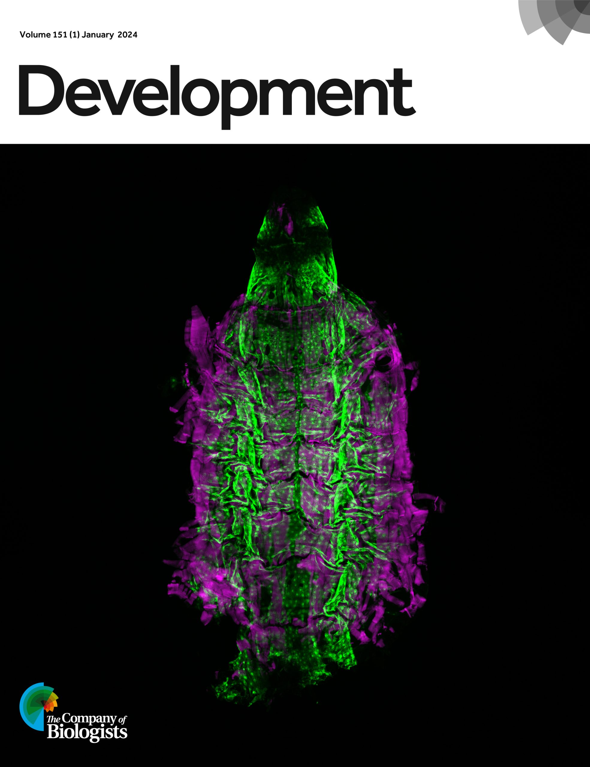

 (No Ratings Yet)