In their recent paper, Maia-Gil and colleagues explored whether and how nuclear properties can influence nuclear positioning in vivo. Their work revealed that in the densely packed retinal zebrafish neuroepithelium, nuclear deformability facilitates apical nuclear migration (Maia-Gil et al. 2024). Here, they share the science and the adventures that led to the development of this project.
What was already known?
The Norden Lab has been focused on understanding the cell and tissue biology behind zebrafish retinal development for 1.5 decades by now. The retina develops from a pseudostratified neuroepithelium, composed of a single, densely packed layer of highly elongated cells. Among other topics, the lab has been investigating a hallmark of such pseudostratified neuroepithelia which is apical nuclear migration before mitosis. This phenomenon is characterised by a fast and directed movement of nuclei toward the apical surface of the tissue. At the start of this project, we already knew:
Why nuclei migrate apically: their apical positioning before cell division ensures tissue integrity (Strzyz et al. 2015).
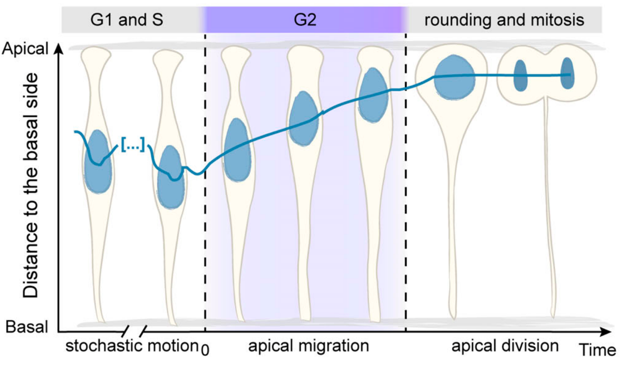
When this migration occurs: during the G2 phase of the cell cycle (Leung et al. 2011).
Schematic representing nuclear apical migration during the G2 phase of the cell cycle (Adapted from Yanakieva et al. 2019).
The big question: how to move through the crowd … and to get to the bar?
One evening, while the lab was out for a social gathering, we found ourselves facing a packed bar, and I was struggling to get my favourite drink – Porto Tónico. Determined to reach the bartender, we started brainstorming different ways to navigate through the crowd quickly and efficiently. In the middle of many creative (and impractical) ideas, someone jokingly suggested: “What if we could deform and squeeze through the crowd – just like nuclei do during apical migration?”
This offhand idea raised a novel question: Does nuclear deformability facilitate apical nuclear migration in the crowded retinal neuroepithelium?
Nuclei (grey) dynamics during zebrafish retina development. (Maia-Gil et al. 2024).
Our approach: stiffening nuclei and tracking their movement during organ development
We already knew that zebrafish retinal nuclei are highly deformable and express low levels of Lamin A/C (Yanakieva et al. 2019), a nuclear envelope protein with expression level that correlates with nuclear stiffness. To test our hypothesis that nuclear deformability helps apical migration, we increased nuclear stiffness by using a previously generated transgenic zebrafish line in which all nuclei overexpress Lamin A (Amini et al. 2022). With crucial support from our collaborators, we used atomic force microscopy (with Elias Barriga and Jaime Espina) and developed a mechanical model that represents confined nuclei as compressible droplets (with Anna Erzberger and Roman Belousov) to confirm that Lamin A overexpression indeed led to stiffer nuclei in the retinal neuroepithelium.
The run to the apical side
With this knowledge and our established tools, we used light-sheet microscopy to image zebrafish retinas and quantify apical nuclear migration in vivo. We aimed to answer several key questions:
Do stiffer, Lamin A overexpressing nuclei reach the apical side? Yes, but in contrast to control nuclei, stiffer nuclei take twice as long to cover equivalent distances. This delay occurs regardless of whether the surrounding nuclei are stiff or normal, suggesting that nuclear deformability facilitates migration in a cell-autonomous manner.
Does the deformability of surrounding nuclei affect apical migration? Yes! Control nuclei surrounded by stiffer nuclei also take longer and show less directed movement. This indicates that the mechanical properties of the environment influence nuclear migration.
Does increased nuclear stiffness impair apical migration in a less crowded epithelium? Here, the effect was less pronounced. Lamin A overexpression in the zebrafish hindbrain, a less crowded neuroepithelium, had only minor effects on apical nuclear migration. This suggests that the impact of nuclear stiffness depends on tissue packing.
Does nuclear stiffness affect other processes requiring cellular deformation? Yes! Control cells took longer to round up before mitosis when in a stiffer environment, showing that nuclear properties can influence mitotic entry in a non-cell-autonomous manner.
Together, these findings demonstrate that nuclear properties influence nuclear positioning and mitotic entry in a tissue-dependent manner during neuroepithelial development.
A personal developmental project…during revisions
My belly was growing, fatigue was setting in, and the time was ticking. The revision email arrived with a challenging to-do list. Priorities had to be redefined and our strategy adjusted. We needed more hands on the bench and had to dig deep into previous data. What could have been an erratic roller coaster turned into a successful and rewarding scientific adventure. I am grateful to have been surrounded by outstanding scientists who also advocate for woman in science. Their encouragement was invaluable – not only for the success of my PhD project but also for the development of the two most beautiful retinas I have ever seen.
References
1. Maia-Gil, M., Gorjão, M., Belousov, R., Espina, J.A., Coelho, J., Gouhier, J., Ramos, A.P., Barriga, E.H., Erzberger, A., and Norden, C. (2024). Nuclear deformability facilitates apical nuclear migration in the developing zebrafish retina. Current Biology 34, 5429-5443.e8. https://doi.org/10.1016/j.cub.2024.10.015.
2. Strzyz, P.J., Lee, H.O., Sidhaye, J., Weber, I.P., Leung, L.C., and Norden, C. (2015). Interkinetic Nuclear Migration Is Centrosome Independent and Ensures Apical Cell Division to Maintain Tissue Integrity. Developmental Cell 32, 203–219. https://doi.org/10.1016/j.devcel.2014.12.001.
3. Leung, L., Klopper, A.V., Grill, S.W., Harris, W.A., and Norden, C. (2011). Apical migration of nuclei during G2 is a prerequisite for all nuclear motion in zebrafish neuroepithelia. Development 138, 5003–5013. https://doi.org/10.1242/dev.071522.
4. Norden, C., Young, S., Link, B.A., and Harris, W.A. (2009). Actomyosin Is the Main Driver of Interkinetic Nuclear Migration in the Retina. Cell 138, 1195–1208. https://doi.org/10.1016/j.cell.2009.06.032.
5. Yanakieva, I., Erzberger, A., Matejčić, M., Modes, C.D., and Norden, C. (2019). Cell and tissue morphology determine actin-dependent nuclear migration mechanisms in neuroepithelia. Journal of Cell Biology 218, 3272–3289. https://doi.org/10.1083/jcb.201901077.
6. Amini, R., Bhatnagar, A., Schlüßler, R., Möllmert, S., Guck, J., and Norden, C. (2022). Amoeboid-like migration ensures correct horizontal cell layer formation in the developing vertebrate retina. eLife 11, e76408. https://doi.org/10.7554/eLife.76408.
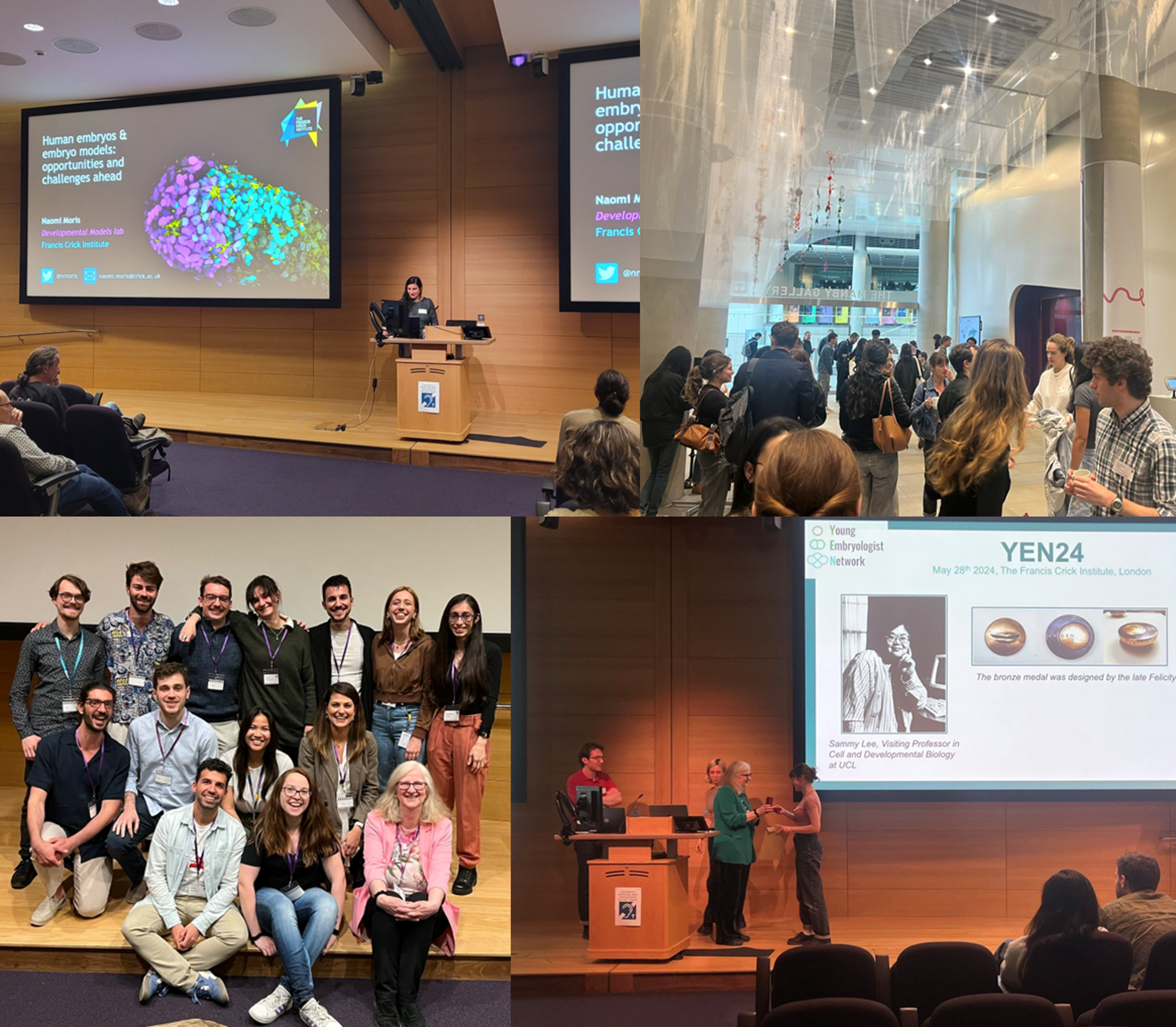
Register now for the Young Embryologist Network meeting – Monday 19 May 2025 at the Francis Crick Institute.
Are you an early career researcher in developmental and stem cell biology? Then come to the 17th Young Embryologist Network Conference! Registration and abstract submission for talks and posters can be accessed here: YEN 2025 Registration form. Abstract deadline is the 13 April 2025.
YEN has a long history of enabling PhD students and early career researchers to share their research and network [1,2], as well as supporting the developmental biology research community [3]. Each year, YEN conferences are organised by students and postdoctoral researchers based in the UK. In recent years, the scope and reach of the YEN conference has grown significantly. We are committed to providing an open and free networking opportunity for developmental and stem cell biologists.
For YEN 2025 we again have a great line-up of speakers, with Nicolas Rivron from the Austrian Institute of Molecular Biotechnology (IMBA, ÖAW) giving the Sammy Lee Memorial Lecture on his work on blastocysts and in vitro embryo models. We will also welcome Peter Rugg-Gunn, Group Leader in Epigenetics of Human Development and Head of Public Engagement at the Babraham Institute (Cambridge UK) and Maud Borensztein, Group Leader at IGMM-Montpellier (France), who is working on X chromosome reactivation during germline development.
We are also pleased to announce this year’s Scientific Perspectives talks, which will focus on ‘Embryology at the frontiers of technology’ with Rob Tetley from Nikon Instruments and Simon Hanassab from Imperial College London, who is working on how AI can improve decision making in IVF.
PhD students and postdocs who would like to give a short talk or present a poster are invited to submit an abstract. We welcome international applications and have secured some funding to support travel grants. Please register using the form at forms.gle/fxtmNHjEziVTFprN6.
As always, we are very grateful to our sponsors including The Company of Biologists, 10X Genomics and Azenta, The Crick for hosting us and to the participants for attending.
Would you like to learn more about how to make your lab greener? We are excited to offer you insight into what sustainable action you can put into practice in the lab.
Jeroen Dobbelaere is our guest author for our newest series of blogs entitled “How to make labs more sustainable”. In this series, Jeroen will introduce you to useful advice and resources that can help you make your lab more environmentally- friendly. It will include aspects such as energy usage, lab equipment selection and procurement.
With Jeroen’s broad experience in both sustainability and academia, his blogs will be a great primer on how to combine these aspects. See more about Jeroen here.
As embryos develop, their cells perform two fundamental tasks: they divide to populate the developing organism, and they specialize into different cell types—skin cells, brain cells, and more—to carry out a variety of essential functions. In our paper, we set out to explore how the process of cell division influences the differentiation of various cell types during early development.
When I joined Allon Klein’s lab, the team had just published single-cell atlases of zebrafish and frog development (Wagner et al. and Briggs et al. 2018), offering a detailed map of cell states over several hours of embryonic development. Allon and I began brainstorming ways to use them to learn new biology beyond cataloging transcriptional states. We had long discussions on project directions and various fundamental developmental processes we could investigate—genome organization, metabolism, or cell division. As I started reading literature, cell division quickly stood out.
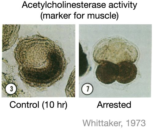
Due to its periodic nature, it has been hypothesized that cell division could act as a clock for developmental events. However, much of the literature does not support the universality of this hypothesis. For instance, studies in ascidian embryos showed that when cell division was blocked at the eight-cell stage, some cells still expressed muscle markers at the correct developmental time, suggesting that cell division was not required for commitment to the muscle lineage (Whittaker et al. 1973, figure below). While several similar studies had tested the impact of blocking division on a few marker genes, a systematic investigation into the role of cell division in forming all major cell types during early development was missing. So, we decided to do just that—in zebrafish embryos.
Zebrafish provided an ideal system for this question because their cells divide and differentiate rapidly in the first day of development (Figure below). Going into my first experiments, I expected to find at least some cell types whose differentiation would depend on active cell cycling. Alternatively, I imagined encountering intermediate “mixed” cell states which I could then follow up on for the rest of my PhD. But, to our surprise, all major cell types differentiated just fine without cell division.
For a while, I was stuck on how to move forward in the project. I spent nearly a year analyzing gene expression changes between control embryos and embryos arrested in the cell cycle. Two key patterns emerged:
Blood cells differentiated more slowly in arrested embryos, indicating a cell type-specific delay in differentiation.
Arrested embryos exhibited a characteristic transcriptional program related to cell cycle arrest that was global and independent of cell type.
While differentiation seemed largely unaffected, cell division also controls the proportions of cells across tissues and organs. We asked how blocking division influenced this. One possible outcome was that cell types that normally divide more frequently would be disproportionately reduced in arrested embryos. Alternatively, the embryos might activate a “compensation” mechanism to maintain normal cell proportions.
To answer this, we needed to estimate how many times each cell type has divided under normal conditions—a challenging problem. Emerging lineage tracing tools will likely solve this soon, but we took a computational approach, inferring cell division numbers from single-cell transcriptome data and lineage trees. As expected, cell types that typically divide the most were the most affected by division arrest. However, quantitatively, the effect was less severe than anticipated, suggesting some level of compensation.
Thus, while cell division is not necessary for differentiation, division influences the timing and proportions of cell types.
References
Whittaker, J. R. “Segregation during ascidian embryogenesis of egg cytoplasmic information for tissue-specific enzyme development.” Proceedings of The National Academy of Sciences 70.7 (1973): 2096-2100.
Wagner, Daniel E., et al. “Single-cell mapping of gene expression landscapes and lineage in the zebrafish embryo.” Science 360.6392 (2018): 981-987.
Briggs, James A., et al. “The dynamics of gene expression in vertebrate embryogenesis at single-cell resolution.” Science 360.6392 (2018): eaar5780
Welcome to Development’s January newsletter. We’ll start by wishing all our readers a happy and productive 2025, which – as highlighted below – marks The Company of Biologists’ 100th birthday.
Celebrating 100 years of The Company of Biologists
Development’s publisher, The Company of Biologists, was founded in 1925 and this year marks our 100th anniversary. You can find out more about the history and ethos of this unique organisation in our January Editorial. We’ll be celebrating throughout the year with content in the journal, our community sites and on social media – check out the #100biologists hashtag on Bluesky and X to find out about some of the extraordinary scientists who’ve been associated with the Company over our long history. We’d also love to hear your stories – how has the Company supported you in your career? Please send us your ‘message in a bottle’ to let us know.
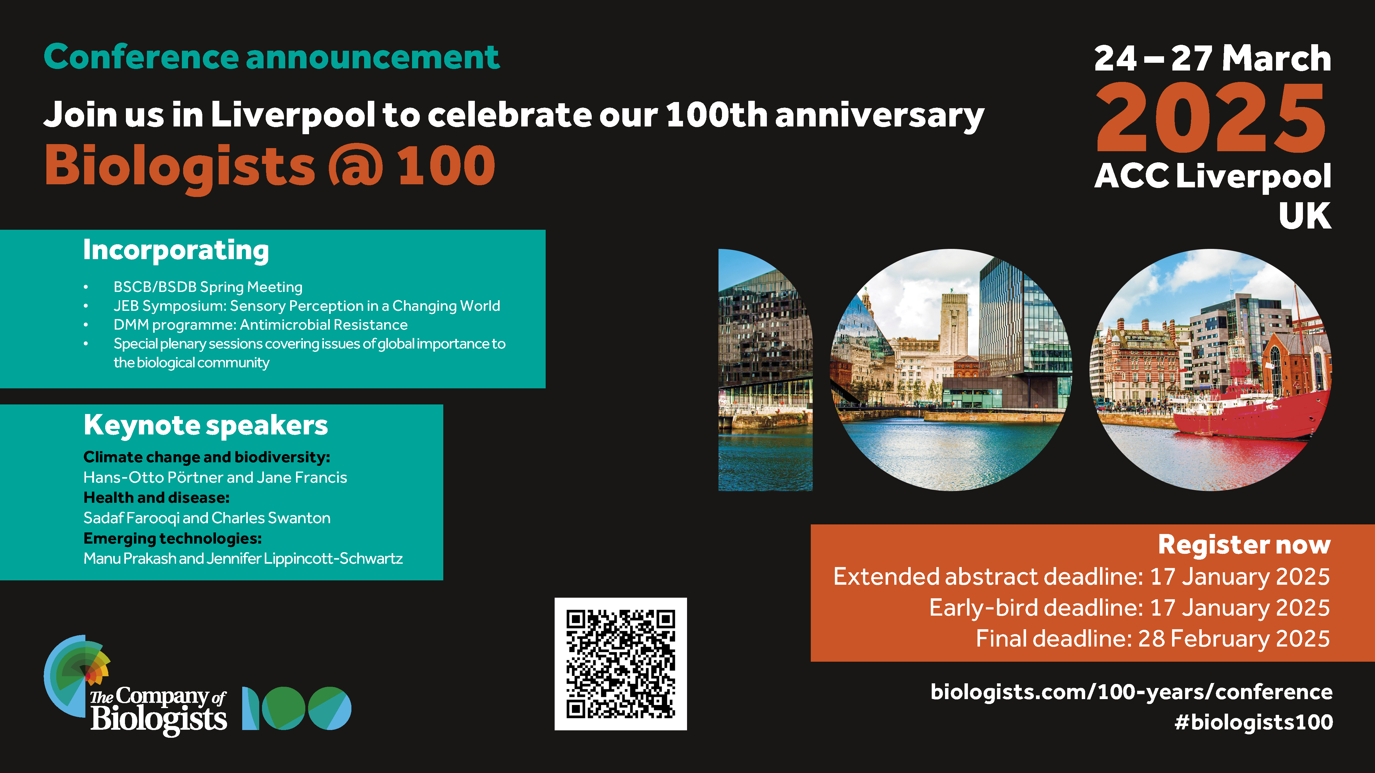
The centrepiece of our celebrations is the Biologists @ 100 conference, being held in Liverpool 24-27 March 2025. We’d love to see you there. The registration deadline is 28 February 2025.
Constructive Critics: Development’s approach to peer review
We all know that the peer review process isn’t perfect and here at Development we’re always trying to find ways to ease the path to publication without compromising our high standards. This Editorial summarises some of the things we’ve done in recent years, including our latest recommendation that authors should include a ‘Limitations’ section in their article – providing the opportunity for frank discussion of potential caveats of the work.
Lifelong Development: the Maintenance, Regeneration and Plasticity of Tissues
We are delighted to announce a call for papers for our 2025 special issue. Guest-edited by Meritxell Huch and Mansi Srivastava, working alongside our team of Academic Editors, this issue will focus on developmental processes beyond the embryo. Full details of the scope of the issue can be found on our website and you’re welcome to send us a presubmission enquiry if you’re unsure whether the scope of your work fits within this issue.
Pathway to Independence programme: call for applications
Are you a postdoc planning to go on the job market this year? Could you benefit from some mentorship, training and networking opportunities? If so, Development’s Pathway to Independence programme could be for you. Now in its third year, this competitive scheme aims to support postdocs as they seek their first independent position: we welcome applications from across the globe and look forward to growing our network of PI fellows.
The Company of Biologists’ Grants and Workshops: upcoming deadlines
Autonomous anteroposterior polarization in aggregates of mouse embryonic stem cells illustrates how alternative initial cell states between the embryo and the aggregates may converge onto similar fates.
The mitochondrial citrate carrier, SLC25A1, regulates trophoblast differentiation and placental development to safeguard embryonic heart formation.
Sign up to Development’s email alerts (such as table of contents alerts) and the journal’s newsletter, to keep up to date on news, including special issues, calls for papers, content highlights/updates, journal meetings and more.
The latest issue of Development (vol 152 issue 2) features a Perspective article by Duygu Özpolat, Swathi Arur (one of Development’s Academic Editors), and Mansi Srivastava (currently serving as a Guest Editor for the journal). The piece represents their views and is not intended as a formal position statement of the journal, but much of what they write resonates strongly with my own opinions and with discussions I’ve had with the editors of the journal over the years.
Back when I was growing up as a developmental biologist, the tables of contents of journals like Development were full of papers with some variation on the title “Gene/protein X controls process Y in organ/organism Z”. Indeed, my first ever paper (proudly published in Development) essentially conforms to this formula. And getting those papers published in ‘top’ journals at that time (and since!) generally meant understanding that ‘control’ at a molecular level – what other genes or proteins does X interact with or regulate? This approach has been hugely important for our field, and we’ve made enormous progress in understanding the logic of developmental processes through delving into molecular mechanisms. But it’s not the only approach or level of understanding at which we can gain profound insights. As someone who’s always been interested in cellular and tissue-level behaviours, I’ve often felt this focus to be too narrow. Indeed, one of my mantras since joining the journal has been that “mechanistic understanding doesn’t necessarily mean molecular mechanistic understanding”. (The other, incidentally, is that “development doesn’t stop at birth” – which is why I’m personally delighted by our current special issue topic!).
It’s notable that, in the latest iteration of the journal’s Aims and scope, we actually removed the words ‘mechanism’ and ‘mechanistic’ from our description of the kinds of papers we seek to publish. That’s not to say we’re not interested in mechanistic work – of course we are! – it’s that we recognise that ‘mechanism’ is all-too-often conceived as being at the molecular/genetic level. Particularly with advances in 4D imaging and in measuring and manipulating forces, we can now gain significant insights into how developmental processes are orchestrated by studying cellular behaviours and without really worrying about what molecules are involved and we’d like those papers to find their natural home in Development. We also need to acknowledge the importance of foundational descriptive work, without which those interested in understanding ‘mechanism’ couldn’t even start their research.
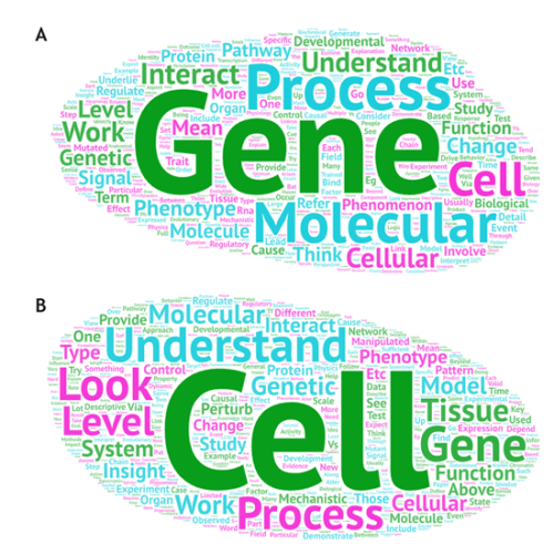
Survey answers to the questions A “In your area of work, what is the most common interpretation of the term ‘mechanism’ as applied to research questions?” and B “When you assess work in your field, what do you look for in terms of a mechanistic understanding of development?”. Taken from Özpolat et al., 2025. The reversal in size of the words ‘Gene’ and ‘Cell’ in the two word clouds exemplifies the mismatch between common perception and personal views.
So, if you’re a referee reviewing a paper for Development (or, frankly, anywhere else!), I’d urge you to avoid asking yourself “does this paper provide new (molecular) mechanistic insights?” and instead to ask “how valuable are these findings for the field?”. I don’t think I can put it better than Duygu, Swathi and Mansi do in their article: “question reductionism, be open-minded about the approach used and the level of mechanism under study, and consider each study relative to what is already known in that system, assessing the potential of the work to advance knowledge”.
The Institut de Biologie du Développement de Marseille (IBDM) is inviting applications for group leader positions. We are seeking innovative researchers who aim to address fundamental questions in biology, including the development, function, and dynamics of complex biological systems.
Our Mission
Research at the IBDM synergistically integrates developmental biology with molecular, cell, and computational biology, as well as evolution, biophysics, neurobiology, physiology, and physiopathology. Affiliated with CNRS and Aix-Marseille University (AMU), the IBDM uniquely fosters interdisciplinarity through strong connections with physicists, computational scientists, and mathematicians (via the CENTURI program). The institute also contributes to major federative programs at AMU, tackling key challenges in Neuroscience, Cancer and Immunology, Rare Diseases, and Imaging.
Our collaborative and international scientific culture, English as the working language, and exceptional location on a campus in the heart of the Calanques National Park make the IBDM a unique place to conduct world-class research.
What We Offer
A generous start-up package.
Access to state-of-the-art core facilities, including advanced light and electron microscopy and top-tier animal facilities (mouse, Drosophila, Xenopus).
A commitment to mentoring, with support to secure a tenured position (CNRS or AMU) and extramural funding (ATIP/Avenir, ERC, FRM, etc.).
A strong emphasis on equality, diversity, and inclusivity in our working environment.
Application Process
Interested candidates should submit a single PDF file containing:
A cover letter outlining their motivation to join the IBDM.
A CV, including the date of PhD defense.
A summary of main research achievements (maximum 2 pages).
A detailed research project (maximum 5 pages).
Contact details for three references.
Applications and queries should be sent to the search committee at ibdm-call@univ-amu.fr.
Application Deadline: March 30, 2025
Selected candidates will be invited for in-person interviews scheduled for June 2025.
Join Us!
Be part of a dynamic research community, advancing knowledge at the frontiers of biology in one of the most inspiring environments in the world. Apply today and help shape the future of science at the IBDM!
In their paper recently published in Evolution & Development, Vanessa Spieß, Rannyele P. Ribeiro and colleagues explore the regenerative abilities of the marine segmented worm Syllis malaquini. Their research reveals that a small piece of this tiny worm can regenerate its entire body forming a whole new individual. Now, co-first and co-corresponding author Rannyele P. Ribeiro offers insights behind this fascinating discovery.
How did the project get started?
During my PhD, supervised by Dr. M. Teresa Aguado, professor at University of Gottingen, I discovered a new species of segmented worm, Syllis malaquini, living in an aquarium. This worm has a segmented body, which means that between the head and tail there is a trunk formed by repeated body units called segments. The trunk of the worm also has a regionalized digestive tube, with foregut and gut regions. My thesis characterized morphological and cellular dynamics of S. malaquini regeneration, showing that the worm could restore the missing body part after amputation of half of its body1. That means splitting a worm into two pieces generates two new worms that are clones of one another. This breakthrough revealed a critical research challenge: identifying the smallest body fragment that retains the potential for whole-body regeneration. To answer this question, Master’s student Vanessa Spieß conducted many experiments isolating body fragments with different segment numbers, and from different gut regions along the antero-posterior axis of the worm. At that time, I transitioned to do my postdoctoral research with Dr. Duygu Özpolat, assistant professor at Washington University in St. Louis. However, I continued to collaborate with Vanessa Spieß and Dr. Aguado to analyze and interpret the acquired data, culminating in the recently published paper in Evolution & Development2.
Why did you choose Syllis malaquini as your research organism?
Syllis malaquini was discovered serendipitously while performing experiments to investigate regeneration in segmented worms collected from an aquarium located at the University of Leipzig, Germany3. While working with what was thought to be Typosyllis antoni, I observed an unexpected ability to regenerate the anterior body, of which T. antoni is incapable4,5. This observation led to a careful examination of the morphology and DNA sequence of my experimental worms, revealing a new species that we named S. malaquini. This species amazed me in many ways. During an experiment, I successfully cultured multiple fragments from a single individual in a Petri dish, with each fragment regenerating into a complete clone, multiplying the worm culture. This astonishing ability suggests a form of near immortality by continuous regeneration and self-cloning. I think that their regenerative mechanisms probably rely on powerful mechanisms that maintain cellular health and proliferative capacity. Therefore, this worm is an excellent model for studying not only whole-body regeneration but also fundamental mechanisms of cellular integrity maintenance.
Can you summarize the key findings of the paper in one paragraph?
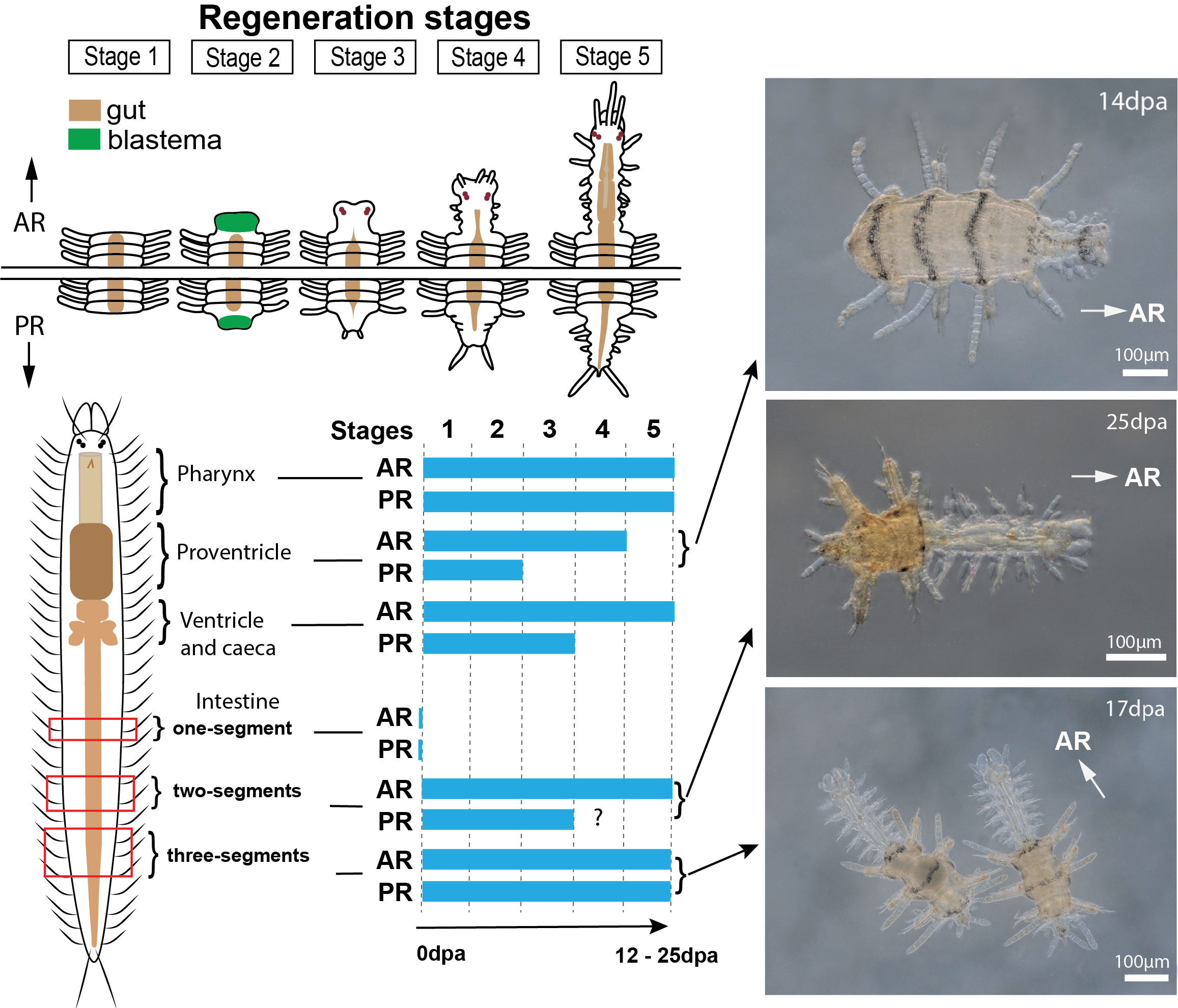
Our research revealed the minimum body size for whole-body regeneration in Syllis malaquini, assessing regenerative success and failure. We not only confirmed the worm’s remarkable regenerative capacity but also demonstrated its ability to achieve whole-body regeneration from extremely small fragments of the trunk, specifically from the intestinal region. We discovered that, while a piece of trunk with just two segments (that is around 300 μm long) can initiate head and tail regeneration, it cannot restore the whole-body. However, a fragment with three segments can successfully regenerate an entire new individual (Fig. 1). The research also uncovered that regenerative capacity varies depending on the gut region, with fragments of the foregut being less regenerative than of the ones from the intestinal region. This discovery opens new avenues for understanding the influence of the gut regions in successful regeneration in segmented organisms.
Were there any other unexpected results and challenges, especially associated with working with a non-model research organism?
Working with Syllis malaquini presented unique challenges, particularly in controlling sexual reproduction, which hindered our ability to perform transgenesis and genome editing. While we successfully maintain asexual reproduction in aquariums, the lack of control over egg and embryo production limits genetic tractability in this species, as generating transgenic segmented worms typically relies on egg injection techniques. To overcome these obstacles, I’m currently developing gene knockdown systems using RNAi in segmented worms. This approach offers a promising avenue for investigating the molecular basis of S. malaquini’s regenerative ability, despite limitations in traditional genetic editing techniques. By adapting and refining these methods, we aim to unlock new insights into the mechanisms underlying whole-body regeneration in this fascinating species.
What’s next for this story?
A direction to be followed next in S. malaquini research is identifying the crucial factors that enable successful regeneration in amputated fragments with three segments but are absent in fragments with only two segments. We will keep this question in mind exploring anterior-posterior molecular patterning and specific cell-cell signaling as candidate components playing a vital role in successful regeneration. My current postdoctoral research on Platynereis dumerilii explores the systemic effects of regeneration on developmental transitions, gametogenesis, and lifespan, potentially revealing parallels with S. malaquini. Leveraging comparative research across multiple species, we aim to unravel unique regeneration mechanisms and adaptations in segmented worms.
Figure 1. Regenerative capabilities of Syllis malaquini. A body fragment containing two segments can regenerate both head and tail structures but does not exhibit further growth. Body fragments with three or more segments can regenerate into complete individuals. This demonstrates the remarkable regenerative plasticity of S. malaquini, with segment number being a critical factor in determining regenerative outcomes. Figure obtained from Spieß et al. 20242.
Last year I became the mother of fraternal twins. I love being a parent, but in a profession rife with gender inequalities, I am also aware of how my transition to motherhood will affect my career. What I didn’t realise, however, was how being a scientist would help me to be a good mother to my two children.
We often hear about the difficulties facing women (and mothers) in academia. At my main university in Denmark, men still hold more than 75% of professorships, 85% of top management positions and receive 76% of the total funds paid out from Denmark’s Basic Research Fund1. To exist in this environment, women often need to do better and work harder than their male colleagues. So how does one fit in starting a family?
Even in the most balanced relationships, women pay a higher price for their children. During pregnancy, most women experience some level of illness, sleep deprivation and pregnancy-related complications. They take time off for scans, classes, medical appointments and prescribed rest. Safety concerns for the developing foetus can interrupt or stop laboratory and field work. After birth, many women experience the “hidden” medium-term and long-term complications of pregnancy and birth, including depression (11-17%), urinary incontinence (8-31%), anal incontinence (19%) and lower back pain (32%)2. When women do return to work, they may need to schedule time for pumping into their workday and often come to work exhausted from nighttime feeding. Attending and presenting at conferences as an expectant or new mother is often not possible3. Moreover, women typically carry a larger mental load than their spouse and can be unfairly criticised for this with the label “mommy brain”4. Sadly, many department chairs—aware of the unequal load of parenthood—do not appropriately recognise career interruptions during hiring and tenure evaluations, and seem genuinely flabbergasted when there are then fewer women candidates available for recruitment at the more senior levels5.
Becoming a mother undoubtedly impacts our careers as scientists. But women also receive messaging that being a scientist influences their ability to be a good mother. We can be made to believe that we have left it too late6, returned to work too soon and that the time demands of the job are simply incompatible with the traditional view of motherhood. What I have come to realise over the last few months, however, is that being a scientist has laid the groundwork for me to be a fantastic mother to my two children. By sharing my experiences, I hope I can help to write a new narrative and highlight to women scientists the unique strengths and abilities that will prepare them for this stage of their journey.
—
As a cell biologist, I am accustomed to repetitive tasks. Recurrent cycles of feeding, changing and putting my babies to sleep during the first few months of their lives therefore felt somewhat familiar and manageable, rather than overly burdensome and monotonous. Having spent most of my adult life formulating and testing hypotheses in the lab, my mind easily came up with new theories for why things may be going wrong or why the babies may be being fussy, and I have been able to experiment with possible solutions. Importantly, I know how to persevere with a hypothesis and not give up on a good idea too soon, a problem that many desperate and time-poor new parents can fall victim to. I am accustomed to working extremely long days, late nights and weekends. My friends are used to long stretches of time without seeing me and me turning up late to social events. Being a scientist has even helped me to develop a taste for cold coffee, which only makes me smile when I order a flat white and both babies instinctively begin crying.
I am particularly good at multi-tasking and managing my time. My many years at the bench have made me incredibly proficient at opening bottles with one hand, sterilising and labelling things, and protecting myself from spills. I can forecast, plan and mitigate risk better than most parents. I am almost always prepared and when I’m not, I learn quickly and pay attention to the serendipitous wins. I understand how drugs work, when vaccines are due and when to be worried about a fever. Contrary to the conventional view of scientists as cold, unemotional beings, most of us are extremely creative and playful, a trait that has obvious benefits when raising young children. Those of us who engage in teaching are accustomed to teach Socratically, which I believe will be helpful as my children begin to ask questions about the world around them.
—
Gender inequalities in academia are a huge problem—for all women—and this requires urgent attention from our university leaders5. But to those women scientists apprehensive about the kind of mothers they may be, my message is simple: your efforts in the laboratory are likely to help you in ways you may not have yet imagined.
Acknowledgements
I am supported by grants from the National Health and Medical Research Council of Australia (NHMRC, #2003832), the Novo Nordisk Foundation (#NNF20OC009705) and the International Brain Research Organization (PG24-9230796649).
References
1. Mænd og kvinder på de danske universiteter – Danmarks talentbarometer 2019. (2020).
2. Vogel, J. P. et al. Neglected medium-term and long-term consequences of labour and childbirth: a systematic analysis of the burden, recommended practices, and a way forward. The Lancet Global Health12, e317–e330 (2024).
3. Chalmers, S. B. et al. Towards inclusive and sustainable scientific meetings. Nat Cell Biol25, 1557–1560 (2023).
4. Callaghan, B. L., McCormack, C., Kim, P. & Pawluski, J. L. Understanding the maternal brain in the context of the mental load of motherhood. Nat. Mental Health2, 764–772 (2024).
5. Davis, F. M., Elias, S. & Ananthanarayanan, V. Scientists with intersecting privilege must work towards institutional inclusion. Nat Cell Biol25, 789–792 (2023).
6. Nowogrodzki, J. PhD parents: the pros and cons of having a child during your doctorate. Nature637, 749–751 (2025).
Stem cell models as laboratories to study self-organization
My road from physics to developmental biology began in a journal club during my PhD in Adam Cohen’s lab at Harvard. We were discussing the first reports from Madeline Lancaster, Jürgen Knoblich, and their co-authors(Lancaster et al., 2013) describing the self-organization of cerebral organoids from stem cells. I was instantly fascinated by the images they reported: while no one would mistake these in vitro structures for a real brain, they still showed remarkably complex patterns of gene expression and tissue morphologies. In the physics community, there is longstanding interest in self-organizing systems: patterns that do not follow an external blueprint, but rather emerge from feedbacks in interactions between a system’s components. These stem cell models demonstrated the power of biological self-organization, and offered simplified ‘physics laboratories’ for decoding how multicellular programs arise from basic interactions between cells.
After completing my PhD, I moved to the Lewis-Sigler Institute (LSI) at Princeton as an independent fellow to study stem cells and organoids. The scientific community at the LSI is deeply interdisciplinary and has a long tradition of excellence in both developmental biology and biological physics. Much of this success came from leveraging the fruit fly embryo (D. melanogaster) as a quantitative system for studying fundamental principles of pattern formation. Fly embryos are highly amenable to quantitative measurement and genetic perturbation, and they can be produced at scale to achieve statistical power. I hoped that stem cell models could offer similar advantages, while opening new questions in developmental biophysics.
Choosing a problem: symmetry breaking in the gastruloid
I arrived at Princeton in January 2021 – a challenging time to start a postdoc anywhere, but my transition was made easier by the collegial and collaborative nature of the Princeton community. I was broadly interested in stem cell self-organization, and I began by searching for a self-organizing program to ‘decode’. I found a collaborator and mentor in Jared Toettcher. Jared is a bioengineer and molecular biologist whose group (together with Stas Shvartsman’s group, also at Princeton) had done pioneering work applying ideas from signal processing to the role of the Erk signaling pathway in fly development. Recently, he and his student Evan Underhill had started studying a stem cell model called the gastruloid(van den Brink et al., 2014).
The gastruloid recapitulates aspects of gastrulation: specifically, the formation and morphology elongation of an anterior-posterior axis. Evan and Jared were studying how FGF signals produced in the posterior end of the gastruloid activated downstream Erk and Akt signals to control patterning and elongation. I became interested in an earlier aspect of gastruloid formation: how does the gastruloid break symmetry to establish a posterior pole in the first place?
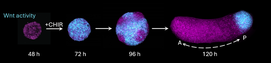
Evolution of patterns of Wnt signaling activity during gastruloid development. Wnt goes from being active throughout the spheroid (following CHIR stimulation), to breaking into ‘patchy’ domains, to eventually polarizing at the posterior end of the gastruloid.
The mystery stems from the lack of explicit spatial cues which the gastruloid receives. In vivo, gastrulation is patterned by spatial cues provided by extra-embryonic tissues, including Wnt signaling molecules provided to the posterior epiblast. Gastruloid formation is triggered by activating Wnt signaling activity everywhere with the small molecule CHIRON-99201 (‘CHIR’). Gastruloids eventually form a polarized domain of Wnt activity in the posterior, just as the embryo does – but how does this local domain form when all cells receive the same stimulus?
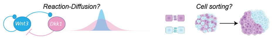
Theories of self-organization and pattern formation offer candidate explanations. One possibility is that gastruloid polarization could be an example of Alan Turing’s reaction-diffusion theory of pattern formation(Turing, 1952): that is, Wnt-dependent feedback in the production of diffusing activators and inhibitors of Wnt signaling can amplify small differences and eventually spontaneously restrict activity to one local domain. Another possibility is that cells spatially rearrange to sort themselves into different domains as they change signaling levels. Discerning between these candidate models is extremely challenging: both could explain the patterns of Wnt signaling activity we observe during symmetry breaking and morphogenesis.
Theoretical models of symmetry breaking. In a reaction-diffusion mechanism, feedbacks in long-range chemical signals amplify initial asymmetries to generate polarization. In a cell sorting mechanism, local interactions guide rearrangements into polarized domains.
Recording signals to decode self-organization
Ideally, we could trace the history of signals that cells see and ask: when can we predict cell fates based on early signaling states? By measuring this kind of ‘fate information’, we could identify when the gastruloid breaks symmetry, and test predictions of mechanistic models. But this is also a very hard problem: cells communicate through many different signaling pathways, and optically tracking cells through divisions and migrations over several days of gastruloid morphogenesis would be extremely challenging.
To trace signaling histories of cells, we turned to synthetic biology. When I was in graduate school, I had followed work from Alex Schier’s group (then at Harvard MCB) developing new technologies for recording cells’ lineage within the genome(Farrell et al., 2018; McKenna et al., 2016). I realized that if this ‘molecular recording’ strategy were adapted to record signaling states (rather than lineage), it could offer us a tool to reconstruct signaling histories without solving the live-imaging cell tracking problem. (As an aside: many other groups have shared this same insight; signal recording is now a robust area of biotechnology development!)
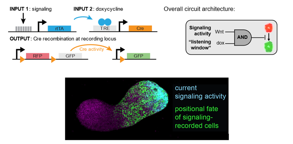
Fortunately, Michelle Chan had just joined the LSI and Molecular Biology Department at Princeton. Michelle is an expert in lineage tracing and had done pioneering work building molecular lineage recorders in the mouse embryo(Chan et al., 2019). With her advice and guidance, we were able to weigh tradeoffs between different design strategies. After prototyping a few flavors, we settled on a recombinase-based strategy. Recombinases (the most famous of which is an enzyme called Cre) can make site-specific modifications at target sites: for example, excising a red fluorescent protein from the genome to permit expression of a green one. We adapted this classical lineage tracing strategy by embedding Cre within a genetic circuit that only expresses in the presence of two inputs: (1) signaling activity in a pathway of interest, and (2) a small molecule (doxycycline, or ‘dox’) used to gate a ‘listening window’ in which the circuit is queried. This logic allows us to relate the output of the Cre recording activity in a target signaling pathway within a known time window.
Design of a signal-recording genetic circuit. Top-left: genetic components of signal recording circuit. Top right: equivalent circuit abstraction. Bottom: example of comparison of previously recorded (green) and current (cyan) Wnt activity in a gastruloid.
In many ways, this approach is quite old-school compared to other emerging technologies for signal recording. As designed, each recombinase only can record one bit of information in a single signaling pathway. But while our design sacrifices in information bandwidth, it gains exceptional sensitivity and fidelity: we could reliably resolve signaling states with high confidence within developmentally relevant temporal windows (3-6 hours). While this approach may not scale to the unbiased screening of all signaling pathways, in many developmental contexts we have a strong prior to focus on a few signals of outsized importance. We viewed this tradeoff of channel bandwidth for sensitivity and fidelity as favorable for our application.
As a practical matter, the main challenge in building these designs into cells was tuning the sensitivities of relevant components. There is a strong ‘Goldilocks principle’ at play: if signal recording is too sensitive, we may get ‘leaky’ background activity even in the absence of signaling activity or dox. But if it is not sensitive enough, then we may not be able to resolve signaling activity effectively. Ideally, the circuit sensitivities should be balanced to be just right.
One way to alter component sensitivities is via the genomic context around the integration site of our synthetic genes. We developed a cell engineering pipeline to screen libraries of randomly integrated parts and then select-out the candidates which landed in the Goldilocks zone. We ultimately identified high-performing cell lines which record each of three canonical signaling pathways that orchestrate gastrulation: Wnt, Nodal, and BMP. The latter two were crucially enabled by work from Ken Zaret’s lab, which validated a panel of pathway-specific sentinel promoters(Serup et al., 2012). This work is a wonderful resource for the community, and we are grateful that they shared constructs of for Nodal- and BMP-responsive elements (AR8 and IBRE4).
Putting theories of self-organization to the test
With our signal-recording cell lines in hand, we set out to systematically map when early Wnt, Nodal, and BMP signaling states predict future cell fates. I won’t belabor all of our results here – please read our paper for the complete story! But I will describe my favorite experiment. One surprising observation was that when we recorded Wnt activity during a ‘patchy’ state – that is, when signaling activity was locally correlated into domains, but not yet globally polarized – we could already predict future cell fates along the final A-P axis. It seemed that cell rearrangements (and *not* reaction-diffusion feedbacks) were sorting the patchy domains into a single pole. But we still weren’t completely certain of our interpretation – more complex reaction-diffusion models could in principle still be consistent with our data.
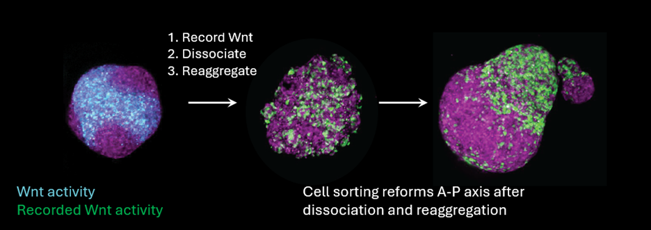
We put our model to the test by testing its predictions more explicitly. Under a strong cell-sorting model, we should be able to record Wnt states with our signal recorder, and then scramble cell positions by dissociating (with Trypsin) and then reaggregating to form new spheroids. We did the experiment, and the result was clear as day – the red and green cells managed to find each other and phase-separate into different domains! We also incidentally observed that reaggregating sometimes made smaller ‘satellite’ gastruloids that sorted into a scaled-down pattern (which we then confirmed by deliberately making smaller reaggregate gastruloids with a flow cytometer). Scaling follows nicely from a cell sorting model: as long as you have the right balance of cell types, sorting gives you scaling for free.
Example of signaling recording – dissociation – reaggregation experiment. (Left) Wnt activity at 96 h (cyan) is recorded and labeled (green). (Middle) Following dissociation and reaggregation, positions of Wnt-recorded cells are randomized. (Right) Wnt-recorded cells sort to form a regenerated posterior domain.
Our synthetic biology approach provided us with an integrated description of how the gastruloid self-organizes and allowed us to interrogate fundamental theories of pattern formation. Many interesting questions remain: for example, what are the physics of gastruloid cell sorting? Are there even earlier physical or biochemical cues which contribute to symmetry breaking? I am also excited about opportunities to use synthetic biology to control signaling states in the gastruloid, and to use this synthetic biology toolbox to decode self-organization in other organoid and embryoid models. In January 2025, I moved to Yale to start a new research group to chase these questions. Please reach out if you are interested in joining the adventure!
References
Chan, M. M., Smith, Z. D., Grosswendt, S., Kretzmer, H., Norman, T. M., Adamson, B., Jost, M., Quinn, J. J., Yang, D., Jones, M. G., et al. (2019). Molecular recording of mammalian embryogenesis. Nature 570, 77–82.
Farrell, J. A., Wang, Y., Riesenfeld, S. J., Shekhar, K., Regev, A. and Schier, A. F. (2018). Single-cell reconstruction of developmental trajectories during zebrafish embryogenesis. Science 360, eaar3131.
Lancaster, M. A., Renner, M., Martin, C.-A., Wenzel, D., Bicknell, L. S., Hurles, M. E., Homfray, T., Penninger, J. M., Jackson, A. P. and Knoblich, J. A. (2013). Cerebral organoids model human brain development and microcephaly. Nature 501, 373–379.
McKenna, A., Findlay, G. M., Gagnon, J. A., Horwitz, M. S., Schier, A. F. and Shendure, J. (2016). Whole-organism lineage tracing by combinatorial and cumulative genome editing. Science 353, aaf7907.
Serup, P., Gustavsen, C., Klein, T., Potter, L. A., Lin, R., Mullapudi, N., Wandzioch, E., Hines, A., Davis, A., Bruun, C., et al. (2012). Partial promoter substitutions generating transcriptional sentinels of diverse signaling pathways in embryonic stem cells and mice. Disease Models & Mechanisms 5, 956–966.
Turing, A. M. (1952). The Chemical Basis of Morphogenesis. Philosophical Transactions of the Royal Society of London. Series B, Biological Sciences 237, 37–72.
van den Brink, S. C., Baillie-Johnson, P., Balayo, T., Hadjantonakis, A.-K., Nowotschin, S., Turner, D. A. and Martinez Arias, A. (2014). Symmetry breaking, germ layer specification and axial organisation in aggregates of mouse embryonic stem cells. Development 141, 4231–4242.

 (6 votes)
 (No Ratings Yet)