This month on the Node- November 2013
Posted by the Node, on 1 December 2013
This month saw many interesting posts on the Node, in addition to several job and PhD studentship adverts in our jobs page. Here are some of the highlights!
Node series
Our two series continued at full steam this month:
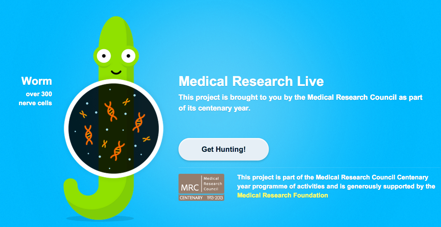
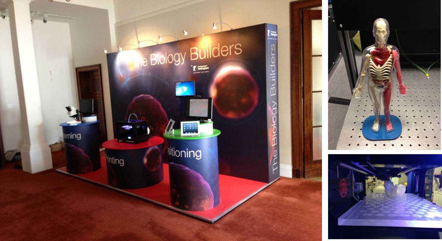
– In our outreach series, the Cosy Science team presented their ongoing project of bringing science to the relaxed environment of the pub, while Worm Watch Lab is a citizen science project in which the public helps scientists study egg laying in C.elegans. The Biology Builders participated in our series by sharing their experience of organising a stand in a science festival, as well as suggesting an easy outreach activity involving ping-pong balls!
– In our ‘A day in the life’ series Stephen Freeman described a day in the life of a chick lab, while James Lloyd wrote about the mysteries of working with moss.
Research
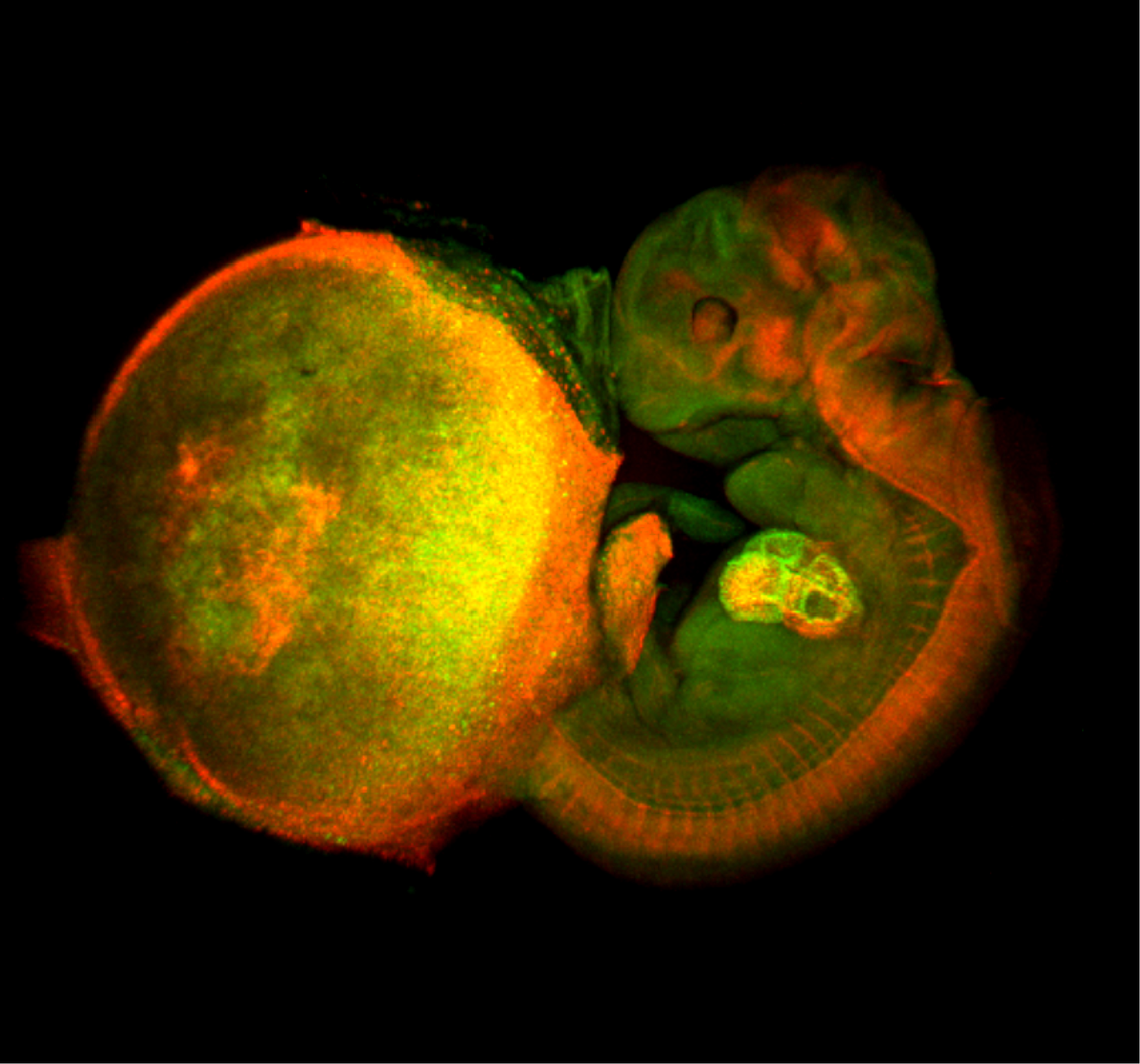 – Atsushi Miyawaki and colleagues wrote about their recent paper in Development using Fucci technology to comparatively characterise endoreduplication and endomitosis.
– The Chicago Journal club is back and their first post of this academic year was a Cell Stem Cell paper on the importance of cell sorting in spatial patterning.
– and Christele considered a recent paper assessing the role of dickkopf-1 (dkk1) in neural progenitors.
Meeting reports
– Megan attended ComBio, the largest annual life sciences conference in Australasia, and wrote about her highlights.
– Lauren and Ioanna reported from the UPMC/Curie Developmental Biology course 2013.
– the Node attended the first joint meeting of the French Society for Developmental Biology and for Genetics.
Also on the Node
 – We interviewed cardiovascular developmental biologist and Development editor Benoit Bruneau.
– Olivier introduced Manteia, a database that allows the comparison of embryological, expression, molecular and etiological data from human, mouse, chicken and zebrafish simultaneously
– and the deadline for the next round of Company of Biologists Travelling Fellowships, to help cover the costs of visiting another lab, is fast approaching
Happy Reading!
 (1 votes)
 (No Ratings Yet)