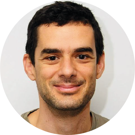
In this SciArt profile, we meet Philipp Dexheimer, who combines his research background and love for art to effectively communicate complex ideas in science to a broad audience. As an artist at heart and a scientist in mind, Philipp’s creations are inspired by the concepts and molecular aesthetics of nature. He works with diverse techniques and media, from Molecular Graffiti to scientific illustrations and videography.
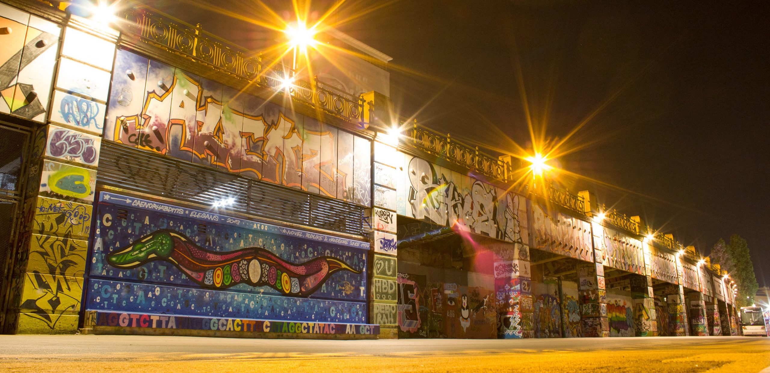
C. elegans Graffiti in Vienna
Can you tell us about your background and what you work on now?
As a scientist, I have always been fascinated by the beauty and complexity of life. During my PhD at the IMP Vienna, I focused on RNA Biology, studying the functions of microRNAs in early animal development using C. elegans as a model system. For my Postdoc, I stayed with the nematodes but shifted my focus to research protein aggregation in the context of myopathies. Our results put protein misfolding on the map as a critical contributor to certain myopathies and suggest that caloric restriction is a promising treatment strategy in this context. My postdoc paper is about to be submitted, and I very recently left the lab to go all-in with Science Art, so it’s the beginning of an exciting new professional chapter for me.
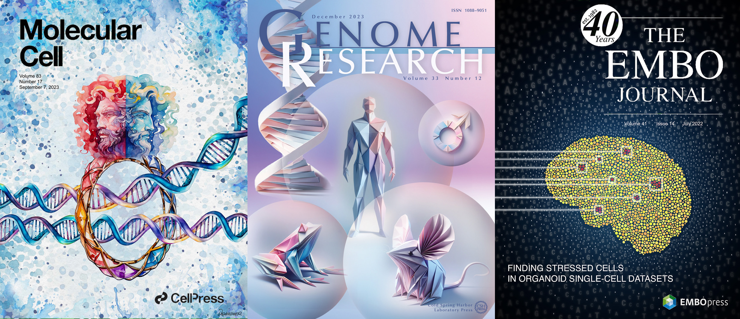
Journal Covers for Molecular Cell, Genome Research and Embo Journal
Were you always going to be a scientist?
I always had a love for biology – Life is, after all, the most fascinating phenomenon in the universe – and the decision to study Molecular Biology after school came very intuitively without having to think about it much. Once I set foot in the lab for the first time as an undergrad, I figured that this is what I want to be doing. Next to the intellectual joy of being a detective investigating the intricacies of nature, the scientific community is just great company; one gets to meet so many inspiring people from all over the world. That’s what made me really feel at home in the institutes of this world, and I would not want to miss a single day of my time as an active researcher.
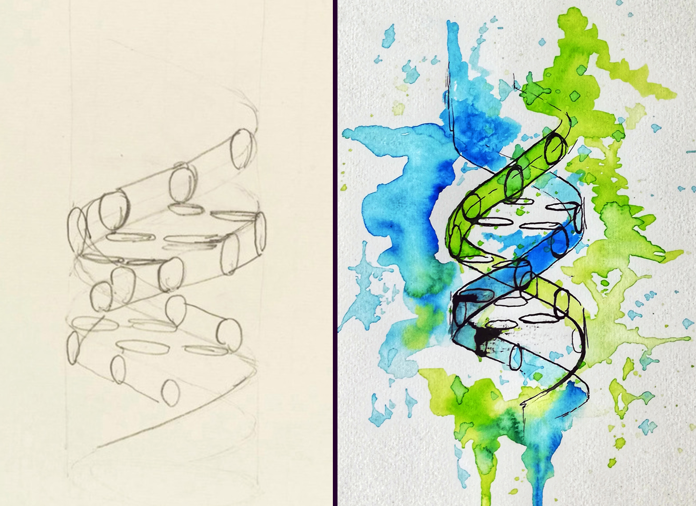
Re-imagining Francis Cricks’ DNA sketch using Ink and watercolor
And what about art – have you always enjoyed it?
In school, I always liked to doodle around when class wasn’t particularly interesting, but things didn’t start moving until I ended up picking up a spray can at the age of 15. I spent a lot of time at the skatepark and on basketball courts which, together with a general interest in Hip Hop culture, led me to start my artistic development by painting Graffiti. Since then, urban art continues to be a passion of mine. Once I got into the lab, I started to draw my inspiration more and more from biology, and by now the main topic of my creations revolves around nature.
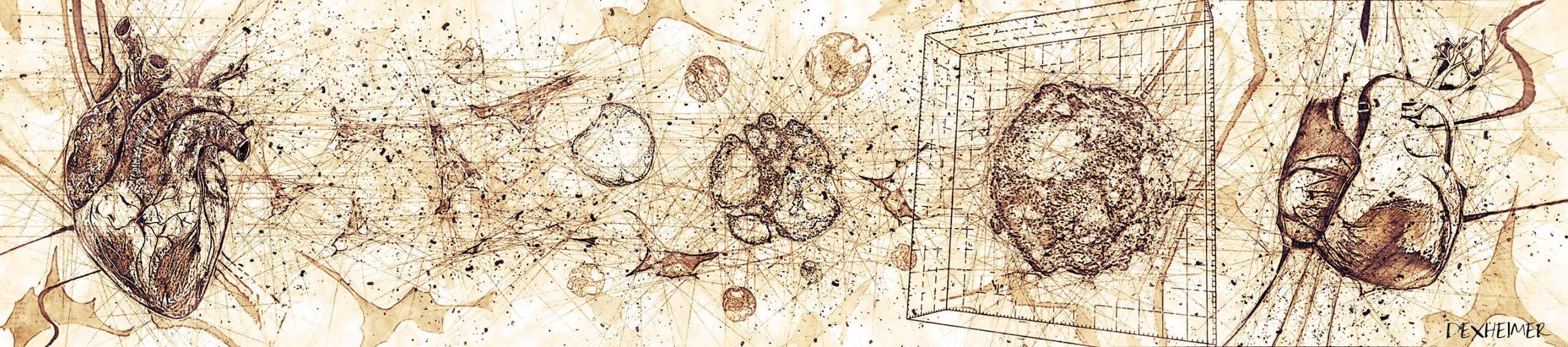
Heart organoids drawn in Leonardo Da Vinci’s style
What or who are your most important artistic influences?
My style has definitely been influenced a lot by urban art. In addition, I love comics and animated cartoons; they continue to be a source of inspiration for my work. More recently, I started to get into watercolor and ink drawings, which I love because the medium is very organic – just like biology itself. David Goodsell is one of my science art heroes; his paintings of the molecular world in scale are to me the most outstanding drawings of the molecular world.
Ubiquitin Graffiti in Vienna
How do you make your art?
When I don’t hold a spray can in my hand, I love working with Photoshop to create digital artwork for journal covers, scientific posters, or biotech homepages – finding a way to illustrate complex concepts in a way that resonates with the human mind & soul never fails to excite me. More recently, I also started to experiment with video creation and editing. Short videos are a great medium to make science accessible to a large audience, and I am convinced that they will become a more important part of the science communication toolbox in the future.
In addition, I think that AI tools are in the process of revolutionizing the way we design illustrations and other art forms – my digital workflows have become increasingly complex and by now involve hopping back and forth between many different programs. I would never take the direct output of any AI tool and use it as a final artwork, though. I want to have a personal touch in there, otherwise it just doesn’t feel like my own creation. AI-generated designs are mostly somewhat generic and lack the certain edge that makes art so interesting after all. My belief is that the interplay of AI prototyping and “classic” manual editing is the future of digital artwork.
Infographic depicting biodegradable spectral conversion films
Does your art influence your science at all, or are they separate worlds?
Both science and art draw a lot from the ability to extract the abstract essence of reality that manifests itself in concrete forms in the world surrounding it. While there is no direct influence of my artistic endeavors on the science I do in the lab, I think that training the cognitive ability of abstraction comes in handy when contemplating biology. In the end, great science aims not at describing a particular phenomenon in a certain organism which happens to be the subject of our studies, but rather at finding the bigger concepts behind the workings of Life.
What are you thinking of working on next?
There are many projects in the pipeline at the moment; my personal favorite goal for the upcoming years is to travel the world and paint large-scale murals on the walls of research institutes and public buildings. I think Molecular Graffiti represents a unique way of porting scientific concepts into public space and has a lot of potential to spark curiosity about the life sciences among a large audience, rendering biology accessible also to non-experts. So if you, dear Reader, happen to have an idea for a wall that could use a beautiful science mural, feel free to reach out!
What better place to hold a conference than a castle? Well, the 25th Conference of the German Society for Developmental Biology (GfE) was held in the historic castle of Osnabrück (see picture below) together with the Dutch Society for Developmental Biology (DSDB) and provided an excellent location to celebrate research. The four-day event brought together researchers from Germany, the Netherlands and around the world to share the latest findings and foster collaboration in the field of developmental biology, including areas such as stem cell biology, pattern formation, regeneration and disease.
The conference was held in the castle of Osnabrück.
Opened by the current president of the GfE, Prof. Kerstin Bartscherer, the meeting started with a keynote talk from Melina Schuh about the beginning of life ‘New insights into meiosis in mammalian oocytes’. After an unscheduled change to the program, due to various strikes and associated delays, and the pre-scheduled talk by Erez Raz on ‘The role of the Dead end protein in controlling the spatial organization and function of RNA molecules within zebrafish germ-cell granules’ the first two sessions ‘Stem cells and fate decisions’ gave broad overview about findings in stem cell dynamics in the plant Arabidopsis and in early mouse embryos, the specification during cardiac development and natural variation in cardiac regenerative capacity, nervous system development in Nematostella, and the skull development in the mouse. The sessions on stem cells were concluded by Jochen Wittbrodt, who gave a talk entitled ‘Towards the genetics of individuality’ using a population genetics approach to show genetic basis of quantitative cardiac phenotypes of two Japanese rice fish models. One of them has elevated heart rates associated with ventricular hypoplasia and impaired cardiac function, which may be related to loss-of-function mutations in candidate genes. For me, this was a highlight of the meeting, as it showed me once again that good research takes time.
The second day began with a special keynote lecture. 2024 is a very special year for developmental biologists: We celebrate the 100th anniversary of the Spemann-Mangold experiment. Christof Niehrs paid tribute to the famous Spemann-Mangold organization experiment and emphasized the importance of this discovery for developmental biology. Accordingly, the following sessions also dealt with ‘Emergence and maintenance of patterns’. Here, we heard how plants use MAP kinase signaling for early cell polarization and the generation of cell wall patterning in the plant vasculature, the self-organization of mucociliary epithelia in Xenopus and, in four insect talks, about feedback loops in the segmentation clock of the red flour beetle, as well as branching during neuronal dendrite differentiation, a role of the ECM receptor Dystroglycan is important for the blood-testis barrier formation and my own talk about basement membrane remodeling in organ formation, all using Drosophila.
In the afternoon we continued with the session ‘When location matters’ where we looked at the role of a RhoGEF in neural crest migration, mechanisms regulating cardiomyocyte invasion of collagenous tissue during zebrafish heart regeneration, apical constriction and cell polarity in cranial neural tube closure, and a proteomics approach to identify cell polarity regulators in plants. Later, the session ‘Genetic and epigenetic control of development’ illustrated how expression patterns are precisely regulated in time, new insights into the role of Pitx2 in cardiac pacemaker development and arrhythmogenesis, a novel function of the hox gene Antennapedia during muscle development, a neuronal subtype specification of spinal projection and motoneurons by a common temporal sequence, and the epigenetic regulation of seed development and plant speciation.
The third day began with a keynote by Susana Coelho on the origin, evolution, and regulation of sexual development through an “Algal views on evo-devo of sex determination”. The following section, “Quantifying and Modeling Development,” ranges from quantifications at the subcellular level, such as the specification of founder cells in lateral root formation, Semaphorin/Plexin Signaling in Collective Cell Migration or the orientation of microtubules in dendritic pruning, to single cell-resolution with studies on lineage-specific genetic modules during cranial development, muscle stem cell heterogeneity and alterations in the thymic niche to the development of reproductive tracts and even the regulation of whole body size. The section ‘Evolutionary adaptions’ took us to a journey across the genome size and gene family expansion in the genus Hydra and the β-catenin-driven endo-mesoderm specification as a Bilateria-specific novelty, but also how leaves adapt during evolution their phenotypes and the tolerance of mouse embryos on ectopic retroviral activity.
Of course, there is one thing that should not be missing at a conference. Day two and three were supplemented with extensive poster sessions in which scientific projects were hotly debated.
In a special session, three distinct awards were granted. Michael Brand holds the laudatory speech for Christiane Nüsslein-Volhard, who was honoured by the GfE with the Klaus Sander Lifetime Achievement Award. In an exploration of color and pattern in the animal kingdom, she elaborated on “Animal Beauty: Function and Evolution of Biological Aesthetics. She addressed the origin and relevance of what humans find beautiful in the animal kingdom. She introduced that color patterns in the animal kingdom have important functions in communication, e.g. in mate choice, but can also develop rapidly and with high variability, which is of great importance in terms of evolution, natural and sexual selection. We know relatively well how invertebrates develop their color patterns, but much is still unknown about vertebrates. Fish are interesting models for studying the development and evolution of color patterns in animals, because they have beautiful patterns made up of a mosaic of differently colored cells in the skin. Fittingly, since this work came from the Nüsslein-Volhard lab, the second award, the PhD award of the GfE goes to Marco Podobnik. In his talk “On the Genetic Basis of Pigment Pattern Diversification in Danio Fish” addressed the question which genes contribute to patterning differences between species. The third award, the GfE Hilde-Mangold Prize 2024, an award for young scientists, went to Daniel Wehner for his work on neuroregeneration after spinal cord injury. He showed how Small leucine-rich proteoglycans inhibit CNS regeneration by altering the structural and mechanical properties of the tissue in the lesion environment. In the evening, the networking event took place at a local nightclub, providing a top destination to make new connections. The poster award winners were also announced here.
The final day was opened by the keynote talk of Anna Akhmanova. She explained how microtubule dynamics control cell polarity and migration. The meeting was completed by two sessions addressing ‘Regeneration and disease models’ with talk that use organoids to model human heart or liver development, study cardiac injuries or regeneration in marine annelids, fish heart and the fish fin. Finally, Hugo Snippert talked about genetic heterogeneity in tumors and how he is studying this in patient-derived colon cancer organoids on the single-cell level.
The joint GfE/DSDB 2024 meeting was an absolute highlight for me. It always reminds me that the development of organisms is what interests me most in biology, and I strongly believe that developmental biology is the foundation for understanding human disease. I was lucky enough to attend the GfE meeting as a student and to have given a talk now as Postdoc is still incredible to me. I’m already looking forward to the 2026 GfE meeting in Potsdam.
Eloise Dries, Yannick Meyers, Daniel Liesner, Floriele Gonzaga, Jakob Becker, Eliane E Zakka, Tom Beeckman, Susana M Coelho, Olivier De Clerck, Kenny A Bogaert
Maria Oorloff, Adam Hruby, Maxim Averbukh, Athena Alcala, Naibedya Dutta, Toni Castro Torres, Darius Moaddeli, Matthew Vega, Juri Kim, Andrew Bong, Aeowynn J. Coakley, Daniel Hicks, Jing Wang, Tiffany Wang, Sally Hoang, Kevin M. Tharp, Gilberto Garcia, Ryo Higuchi-Sanabria
Devon E. Mason, Paula Camacho, Megan E. Goeckel, Brendan R. Tobin, Sebastián L. Vega, Pei-Hsun Wu, Dymonn Johnson, Su-Jin Heo, Denis Wirtz, Jason A. Burdick, Levi Wood, Brian Y. Chow, Amber N. Stratman, Joel D. Boerckel
Samantha M. Barnada, Aida Giner de Gracia, Cruz Morenilla-Palao, María Teresa López-Cascales, Chiara Scopa, Francis J. Waltrich Jr., Harald M.M. Mikkers, Maria Elena Cicardi, Jonathan Karlin, Davide Trotti, Kevin A. Peterson, Samantha A. Brugmann, Gijs W. E. Santen, Steven B. McMahon, Eloísa Herrera, Marco Trizzino
Dmitry A. Kretov, Leighton Folkes, Alexandra Mora-Martin, Noreen Syedah, Isha A. Walawalkar, Kim Vanyustel, Simon Moxon, George J. Murphy, Daniel Cifuentes
Isabel Zhang, Giulia LM Boezio, Jake Cornwall-Scoones, Thomas Frith, Ming Jiang, Michael Howell, Robin Lovell-Badge, Andreas Sagner, James Briscoe, M Joaquina Delás
Rossella Debernardis, Katarzyna Palińska-Żarska, Sylwia Judycka, Abhipsa Panda, Sylwia Jarmołowicz, Jan P. Jastrzębski, Tainá Rocha de Almeida, Maciej Błażejewski, Piotr Hliwa, Sławomir Krejszeff, Daniel Żarski
Keren Cheng, Yasunari Seita, Eoin C. Whelan, Ryo Yokomizo, Young Sun Hwang, Antonia Rotolo, Ian D. Krantz, Maninder Kaur, Jill P. Ginsberg, Priti Lal, Xunda Luo, Phillip M. Pierorazio, Rebecca L. Linn, Sandra Ryeom, Kotaro Sasaki
Stefano Comazzetto, Daniel L. Cassidy, Andrew W. DeVilbiss, Elise C. Jeffery, Bethany R. Ottesen, Amanda R. Reyes, Sarah Muh, Thomas P. Mathews, Brandon Chen, Zhiyu Zhao, Sean J. Morrison
Christopher Zdyrski, Vojtech Gabriel, Oscar Ospina, Hannah Wickham, Dipak K. Sahoo, Kimberly Dao, Leeann S. Aguilar Meza, Abigail Ralston, Leila Bedos, William Bastian, Sydney Honold, Pablo Piñeyro, Eugene F. Douglass, Jonathan P. Mochel, Karin Allenspach
Olivia Sniezek Carney, Kodi William Harris, Yvonne Wohlfarter, Kyuna Lee, Grant Butschek, Arianna Anzmann, Steven M Claypool, Anne Hamacher-Brady, Markus Andreas Keller, Hilary J Vernon
V. Pragathi Masamsetti, Nazmus Salehin, Hani Jieun Kim, Nicole Santucci, Megan Weatherstone, Hilary Knowles, Jane Sun, Riley McMahon, Josh B Studdert, Nader Aryamanesh, Ran Wang, Naihe Jing, Pengyi Yang, Pierre Osteil, Patrick P.L Tam
Xin-Min Li, Hannah Jenke, Sören Strauss, Yi Wang, Neha Bhatia, Daniel Kierzkowski, Rena Lymbouridou, Peter Huijser, Richard S. Smith, Adam Runions, Miltos Tsiantis
Ranjita Thapa, Karl H. Kunze, Julie Hansen, Christopher Pierce, Virginia Moore, Ian Ray, Liam Wickes-Do, Nicolas Morales, Felipe Sabadin, Nicholas Santantonio, Michael A Gore, Kelly Robbins
Florian Laurent, Simon Maria Bartsch, Anuj Shukla, Felix Edgardo Rico Resendiz, Daniel Couto, Christelle Fuchs, Joel Nicolet, Sylvain Loubery, Henning J Jessen, Dorothea Fiedler, Michael Hothorn
Bastienne Zaremba, Amir Fallahshahroudi, Céline Schneider, Julia Schmidt, Ioannis Sarropoulos, Evgeny Leushkin, Bianka Berki, Enya Van Poucke, Per Jensen, Rodrigo Senovilla-Ganzo, Francisca Hervas-Sotomayor, Nils Trost, Francesco Lamanna, Mari Sepp, Fernando García-Moreno, Henrik Kaessmann
Shatha Salameh, Devon Guerrelli, Jacob A. Miller, Manan Desai, Nicolae Moise, Can Yerebakan, Alisa Bruce, Pranava Sinha, Yves d’Udekem, Seth H. Weinberg, Nikki Gillum Posnack
Semih Bayraktar, James Cranley, Kazumasa Kanemaru, Vincent R Knight-Schrijver, Maria Colzani, Hongorzul Davaapil, Jonathan Chuo Min Lee, Krzysztof Polanski, Laura Richardson, Claudia Semprich, Rakeshlal Kapuge, Monika Dabrowska, Ilaria Mulas, Shani Perera, Mina Patel, Yen Ho, Xiaoling He, Richard Tyser, Laure Gambardella, Sarah Teichmann, Sanjay Sinha
James Cranley, Kazumasa Kanemaru, Semih Bayraktar, Vincent Knight-Schrijver, Jan Patrick Pett, Krzysztof Polanski, Monika Dabrowska, Ilaria Mulas, Laura Richardson, Claudia Semprich, Rakeshlal Kapuge, Shani Perera, Xiaoling He, Siew Yen Ho, Nadav Yayon, Liz Tuck, Kenny Roberts, Jack Palmer, Hongorzul Davaapil, Laure Gambardella, Minal Patel, Richard Tyser, Sanjay Sinha, Sarah Teichmann
This is part of the ‘Lab meeting’ series featuring developmental and stem cell biology labs around the world.
Where is the lab?
The Welshhans lab is located at the University of South Carolina, which is in Columbia, South Carolina, USA.
Research summary
The Welshhans Lab works on neural development. In particular, we are interested in the process by which neural connectivity is formed. This process is mediated by a highly dynamic sensory and motor structure located at the ends of developing axons, called the growth cone. Much of our work focuses on local translation, which is the process by which a subset of mRNAs is localized to and locally translated within growth cones to regulate axon guidance. We study how this molecular mechanism and others regulate typical development. Furthermore, we study how the dysregulation of various molecular mechanisms, including local translation and adhesion, contribute to the phenotypes of Down syndrome. We use mouse models and human-induced pluripotent stem cell-derived neurons and brain organoids to study these processes.
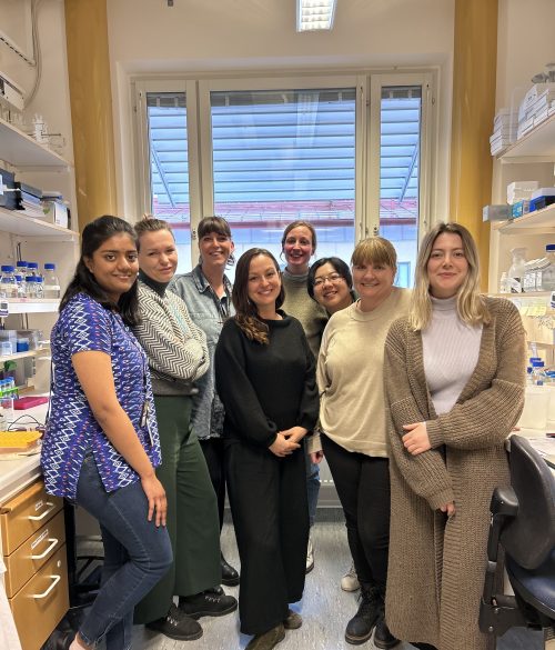
Can you give us a lab roll call?
Katelyn, PhD Candidate: My project examines the local translation of b-actin and how it regulates axon guidance through adhesion-based mechanisms during nervous system development.
Nikita Kirkise, PhD candidate: I am a 4th year PhD student in Kristy Welshhans’ lab. My project investigates the role of extracellular matrix proteins, specifically laminins, in regulating the local translation of mRNAs in growth cones of developing mouse cortical neurons.
Jordan Headen, PhD candidate: I am a second-year PhD student investigating the role of adhesion and the local translation of candidate mRNAs during the development of the nervous system.
We also have three undergraduates in the lab who are studying how adhesion and local translation are altered in Down syndrome. For their research, they are using human fibroblasts as a model.
Favourite technique, and why?
Kristy: My favorite technique is anything involving microscopy and living cells. I find it fascinating to watch living cells under high magnification. It doesn’t matter whether we are using brightfield, fluorescent translation reporters, or some other fluorescent tagging method, I can stare at these movies for hours and always find something interesting!
Apart from your own research, what are you most excited about in developmental and stem cell biology?
Kristy: I am most excited about some recent advances that are improving the quality of life for individuals with neurodevelopmental disorders (including non-pharmacological, pharmacological, gene therapy, and stem cell-based treatments). Many disorders still have no treatment, but recent advances in some of these areas are opening new doors that I hope will continue to gain momentum.
How do you approach managing your group and all the different tasks required in your job?
Kristy: I am a pretty organized person and have a never-ending (but prioritized) to-do list, so that helps me stay on top of things. Every Friday, I time block my calendar with all the activities I need to accomplish in the week ahead. In addition, I meet one-on-one with everyone in the lab every week. Overall, the most important thing to me is the success of my lab members, which means something different for each individual. So, I prioritize that and then fit in everything else around it!
What is the best thing about where you work?
Kristy: In our Department, there is a very supportive group of four faculty who all work on axon biology. This makes it an optimal environment not only for me but for my lab as well. I am also part of a larger group at the University, which is the Carolina Autism and Neurodevelopment (CAN) Research Center. This is a multidisciplinary group composed of faculty and their labs that study neurodevelopment and related disorders. We are composed of people from very diverse disciplines, including Biology, Psychology, Public Health, Computer Science, etc., which has allowed me to think and collaborate with others on my research in novel ways.
Katelyn: The supportive and collaborative environment of both the lab itself, and throughout the University.
Nikita: The best thing about my work is the lab itself. We have such supportive and fun lab members (including our super supportive mentor) that it makes the PhD journey a little less daunting.
Jordan: I like the collaborative and supportive nature of the Welshhans Lab and the department as a whole.
What’s there to do outside of the lab?
Kristy: I love that Columbia is only two hours from the beach and two hours from the mountains. I love to go mountain biking, hiking, and generally spend time outdoors with my family all year round.
Katelyn: Columbia has many parks and lakes to enjoy and is not far from beautiful beaches and the Blue Ridge Mountains. Columbia also has great breweries to explore while enjoying the nice weather.
Nikita: Columbia has many parks and trails, most of which are along the riverside, where you can catch the most beautiful sunsets. We also have a lot of restaurants to try around the University.
Jordan: The Riverwalk is nice for walking and biking throughout the whole year and tubing during the summer. Folly Beach is also enjoyable around the summer months for being in the water and trying the different restaurants near the beach.
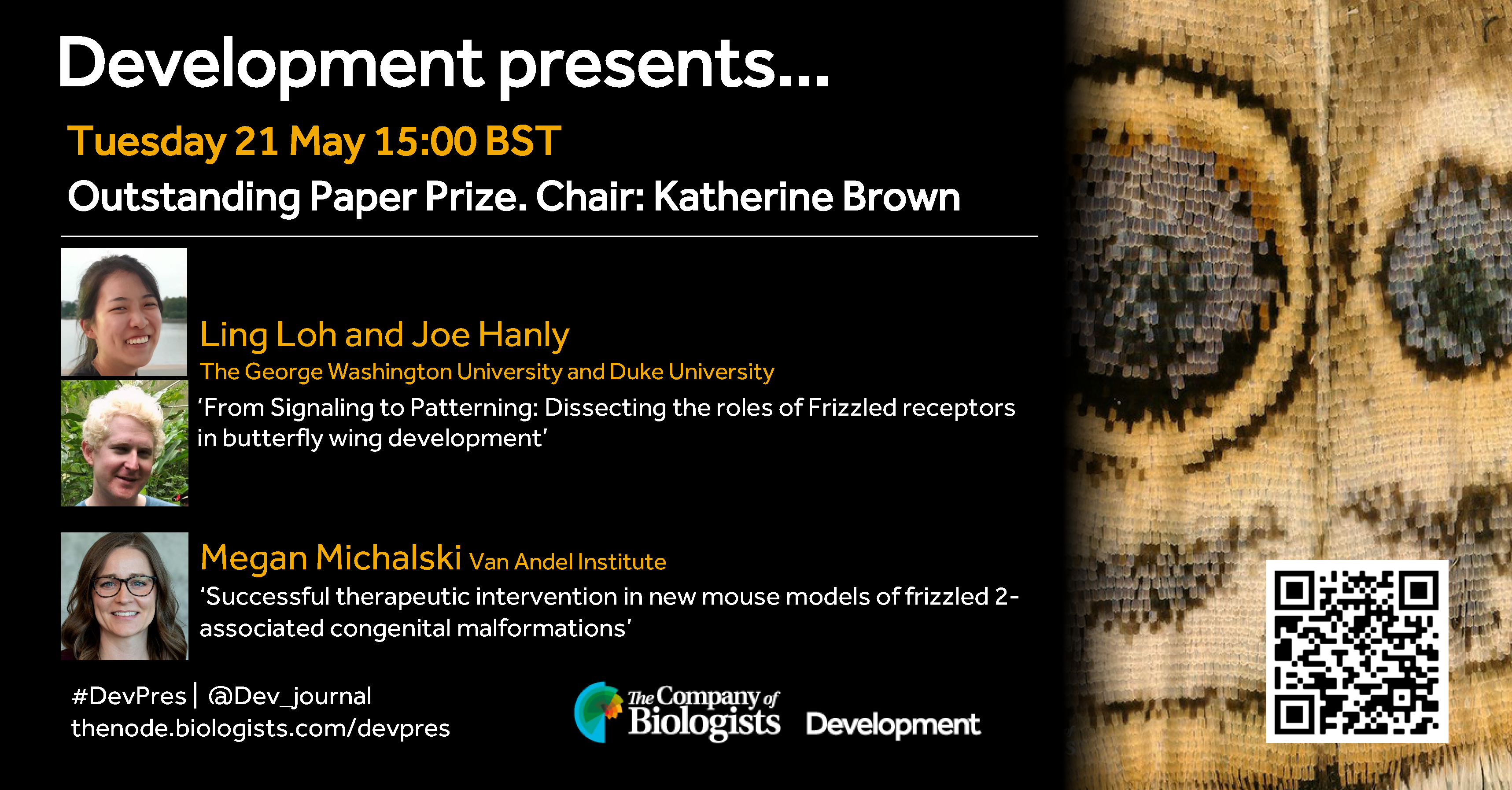
In May, we celebrate the winners of Development’s 2023 Outstanding Paper Prize, hearing from the authors of two papers describing the roles of the Frizzled receptor. Chaired by Development’s Executive Editor, Katherine Brown.
Tuesday 21 May – 15:00 BST
Ling Loh (The George Washington University) Joe Hanly (The George Washington University and Duke University) ‘From Signaling to Patterning: Dissecting the roles of Frizzled receptors in butterfly wing development’
Megan Michalski (Van Andel Institute) ‘Successful therapeutic intervention in new mouse models of frizzled 2-associated congenital malformations’
At the speakers’ discretion, the webinar will be recorded for viewing on demand. To see the other webinars scheduled in our series, and to catch up on previous talks, please visit: thenode.biologists.com/devpres
Earlier this year, my co-correspondent for the Node Brent Foster and I published a pot pourri-style interview article asking biology researchers about their work with non-model organisms (NMOs). As they all had many fascinating details and insights to share about their work with their respective NMOs, we decided to publish the full interviews as well. Below is the interview with Dr Michalis Averof, whose lab works on the shrimp Parhyale hawaiensis at the Institut de Génomique Fonctionnelle de Lyon in France.
Could you give me a three sentence introduction to what you do?
In the lab we study almost exclusively regeneration using this small crustacean [Parhyale hawaiensis] as our model. Τhe origin of the lab is evo-devo. Ever since my PhD, I was interested in comparative developmental biology, in what different organisms can tell us about mechanisms of development and how those mechanisms evolve. This is how we started to work with crustaceans, which I have been working with since then. Then gradually regeneration, which was a side project, became our main focus.
Does Parhyale exclusively regenerate its limbs?
Yes, as far as we know it is the only structure that arthropods can regenerate, basically the appendages.
What are the benefits and the specific challenges of using crustaceans as an organism of study?
There are two reasons for starting to work on a new organism, let’s say on regeneration. The first one is the evolutionary interests. Regeneration is a process that is widespread in the animal kingdom. There are also many species that don’t regenerate. And it’s still an open question to what extent this is an ancient capacity that all animals inherited from their ancestors, which was lost here and there, or a capacity that has emerged during evolution multiple times. The evolutionary dynamics of this process are not well understood at all. Studying different groups of animals allows us to make comparisons and to see to what extent animals use similar mechanisms to achieve this. Hopefully, as we accumulate more information from different branches of the tree of life, a picture will emerge. I have to say, so far, no clear picture has emerged. It’s still not clear how regeneration has evolved and what the earliest origins were. But that’s one motivation for starting [on] a new model.
The other motivation is that sometimes moving into a new system gives you new possibilities, new experimental opportunities. Our animal, for example, is a very bad system to pick if you wanted to do a genetic screen like you would do in C. elegans or in flies. But it turns out that it’s a very good system if you want to image what happens during regeneration. You can image the whole process [at] cellular resolution from beginning to end. So individual organisms, because of technical advantages mostly, offer new opportunities. In this case, the reason why we can image regeneration so well is that these animals as adults are transparent. They’re small enough that we can just image through their legs with a confocal microscope. We can make transgenics so we can label the cells; and we can immobilise them, which can be a big challenge [in other organisms]. With embryos, you can put an egg under a microscope, and it will mostly stay there, and you can do live imaging. With a regenerating adult, this is very difficult. You cannot anaesthetise a zebrafish for an entire week without killing it. In our system [though], what allows us to [image the entire process of regeneration], is the fact that arthropods are encased in a chitinous exoskeleton. We can use simple surgical glue to stick those animals on a cover slip. And they will stay there, they can’t go away until they molt. This is a small, technical thing that makes the system suitable for live imaging. And unless you have an animal that is surrounded by cuticle, it’s very difficult to find a way to do that.
It’s small features like that which make different model organisms valuable and provide new opportunities. The other thing to keep in mind is that regeneration is a process that is very poorly represented in the best models that we have. Mice, flies, C. elegans are very poor at regenerating. So in general, we don’t have great genetic models for regeneration. Zebrafish is the only exception to that.
Was that the motivation to move into the more regenerative aspect of studying Parhyale?
The motivation was simple curiosity. We had developed genetic tools, such as transgenics, we could overexpress genes, and we could label cells with GFP. We had developed these tools for other reasons; at the time, we were studying Hox genes and how they contribute to body plan evolution in the arthropods. That was the reason for generating those tools. But once we had them, it became possible to visualise what is happening. So it was a rather opportunistic thing. Often, you see something that is interesting, and you are drawn in that direction.
So getting into studying crustaceans, was there something that convinced you that they’re really special or as you say, was it more of a chance path?
I was convinced they’re special before, when I was studying evolution. Crustaceans are the closest relatives to the insects, but they have very different body plans, a different organisation of segments in their bodies. So it was an attractive group for studying body plan evolution, how segmental specialisations evolve, and whether the Hox genes might be driving those changes. So that was the reason for going into crustaceans, they were very attractive for that reason. Then, as I said, moving to regeneration was rather serendipitous, because of the tools we had already made and the fact they’re transparent.
How was the discovery made about limb regeneration?
It’s been known for a long time that many crustaceans and other arthropods have the capacity to regenerate their legs. There were some classic studies in the 70s that were using cockroaches as models for studying regeneration.
In a similar vein, then, is there at the tip of your tongue a study in Parhyale that you think is really interesting, that you would recommend to someone if you wanted to get them interested in this model organism?
I’m not sure there is a big breakthrough that has been made in Parhyale yet. I think it’s exciting to be able to see the process of regeneration, that is quite unique. Usually you see snapshots, because it takes very long, it takes weeks or months in some organisms. Having the continuous process on time lapse, where you can see how individual cells are actually behaving and dividing is very exciting, even though in terms of understanding the process, it has not really yet revolutionised the way we see things. There is no paper where I would say, this is a big discovery we’ve made in Parhyale which was very unexpected, and it changed our views.
You have to realise it’s a very small community, there are maybe 20 or 30 people working on the animal. So there’s not so much history and so much that has been done on them. But it’s nice that you have different groups of people with different interests coming in. One of the latest papers to come out this year in Current Biology is [by] people who study biological rhythms, and have studied how Parhyale regulate their daily activities in relation to tides. Our animal is an intertidal species, and it seems it has an endogenous clock that runs with the tidal cycle rather than with a day-night cycle. Well, they have both, but somehow the two interact in a complex way.
Is this something that you can disrupt by removing them from their native tidal environment and putting them in a tank in a building?
Yes. When we keep them here in tanks, we don’t really give them an artificial tide. People who study circadian rhythm had noticed that there were two peaks of activity, one in the morning and one in the evening. And the intertidal cycle is a little bit longer than 12 hours. So that might reflect the fact that they have a 12-hour cycle rather than a 24-hour cycle. But of course, in nature, the tidal cycle comes slowly out of sync with the day-night cycle. And that is not observed in the lab.
It’s exciting that people are beginning to study phenomena that were not accessible before in the standard models, like tides and regeneration. There are new aspects of biology that become accessible once you have a new system.
Considering this community is so small, and considering that the genome of Parhyale might not be as familiar, maybe there’s not 15 different genome iterations like there are for the mouse, how does data analysis and data sharing between these labs work? And what are the interactions like?
You would imagine that small communities are very well interconnected. We are connected, but not very tightly. I think it mostly has to do with the fact that we are on different continents and we study different questions. We talk to each other and we share tools and genetic resources. For example the genome sequencing and assembly was a collective effort.
We all use the same population of Parhyale. It’s a population that has been kept in the lab for more than 20 years. There are a few people beginning to isolate new populations from the wild. The funny thing about the population we share is that it was picked up in an aquarium in Chicago, about 20-25 years ago, and we don’t know which part of the world it came from originally.
It was a pest in the Chicago aquarium. And since Parhyale hawaiensis has been described to be a tropical species that lives all around the world, from Hawaii, to Brazil, to India, we don’t actually know where that particular population came from. We all use it, the genome has been sequenced from, and all our transcriptomic work is based on, that population. Those are the kinds of genomic resources that we all share.
For transgenesis and CRISPR, we more or less use the same protocols. We don’t share transgenic lines so often, but that’s mostly because we have different interests, and each of us develops our own lines for the particular questions we’re asking. The other issue is, we haven’t yet figured out an easy way of sending these animals across the world without having problems with customs. That is a bit of a barrier.
On the other side, as there are so few people working on shrimp, I assume that there’s only one person per department per University per country. Is it difficult to engage with people working on other things? How useful do you find it?
It’s not difficult because each of us belongs to different scientific communities. It’s not that you have to work on the same species to engage with people. The regeneration community, for example, is very large. We talk to that community, we talk to the evolutionary biology community. Even though I have worked with crustaceans, I was always close to the Drosophila developmental biology community. The labs where I did my PhD and my postdoc were fly labs. We don’t feel lonely. We are more isolated in terms of technology, in the sense that for every project, we have to develop our own tools, there isn’t this big community behind you generating Gal4 drivers or Cre lines that are shared, like you have in other systems. When you start a project, you have to generate those tools by yourself. And that is a major limitation when working with our kind of peripheral models. The critical mass of the community is important for generating and sharing tools, and we don’t have that.
Would you say it takes a slightly more adventurous researcher to decide to go down that path?
Definitely, it takes a different kind of researcher. To work with these animals, you have to realise that research is going to move much forward more slowly, because you will have to start many things from scratch. The benefit, on the other side, is that in almost anything you study, you’re going to make new discoveries, because no one has studied that before. So you’re entering a virgin field. It takes a lot of effort to discover something, but whatever you discover is new knowledge. You have the opportunity to shape your research field to a larger extent.
Thank you very much. Is there anything else you’d like to add?
Perhaps I should tell you what my motivation behind all this is, besides the specific questions that we’re asking. Biology has focussed on model organisms for very good reasons, because model organisms give us the tools to go deeper and to study mechanisms. But, over the years, we have developed this idea that model organisms will reveal universal mechanisms, and that we can study most of biology through the model systems that we have chosen.
But I am convinced that there is an enormous amount of biology that we are missing, if we rely only on the established models. There are simply biological phenomena which are not represented in this handful of organisms. Regeneration is partly one such example. But there are other topics, like developmental plasticity, where you have different casts of animals that develop depending on the environment, there are no established genetic models for that kind of study. There are organisms that eliminate half of their genome in somatic cells, they break their chromosomes apart and shed half of the genome during development, and they only keep the full complement in the germline. There is a significant number of organisms, spread all around the tree of life, that do that. There is no way to access this kind of phenomenon and to understand its importance in established models. I see these like new continents of biology that are still unexplored. New model organisms will allow us to explore these. Of course, it’s going to be difficult, and it’s going to take time, and it’s going to take development of tools. But that’s for me the major motivation for going into different systems, because I think there’s biology that we haven’t discovered yet.
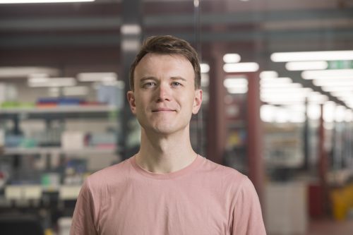
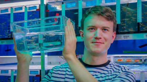
The 2024 German Society for Developmental Biology (GfE) PhD award recipient was Marco Podobnik, who worked on pigment pattern diversification in Danio fish species at the Nüsslein-Volhard laboratory in Tübingen. We caught up with Marco to learn more about his background, his PhD work and his research interests as a postdoc at the Australian Regenerative Medicine Institute.
First of all, congratulations on receiving the 2024 GfE PhD award! What does this award mean to you?
Thank you. I am only half-way joking when the first thing that comes to my mind is, hey, now I can fill out the line that asks for “prizes” in grant applications. Being recognized by the German Society for Developmental Biology is a huge honour. The fact that my work was so collaborative makes it important to draw the attention to my colleagues I worked with over the years. The Max Planck Society creates permissive environments for basic research we conducted, a privilege I am aware of. I wish these opportunities would be more accessible.
Let’s go back to the beginning. When did you first become interested in science?
I believe the way we construct stories of the scientific process is a symptom of the humane desire to tell good stories. This also applies to the story of people’s life history. When I was a kid, I always liked to be out in the forests, trying to identify birds by their sounds. Although I was never really excited about maths, I deliberately chose all science subjects in high school. I had an excellent chemistry teacher, Holger Mummert, who did his PhD in Tübingen. In my final year I joined his class on fossils which I found beautiful. I had to become a biologist, right?
How did you come to do a PhD at the Nüsslein-Volhard laboratory?
I think this destination was the consequence of a random process paired with a developing passion. When I still studied biology at the University of Cologne, I had to find a lab for an internship. So I made a list with shiny labs working on topics I mostly never heard of and brought it to my mentor, Matthias Hammerschmidt. He had done his PhD with Christiane (Janni) Nüsslein-Volhard in Tübingen. In the early 1990s, he participated in the genetic screens in zebrafish [1], a heroic effort approaching the Nobel Prize work in flies in the 1980s [2]. When I showed Matthias the list, he smiled and refused to send me anywhere. Instead, he offered me to take a look into his zebrafish lab. Seeing live zebrafish embryos under a stereomicroscope for the first time was quite a fascinating moment. After that he suggested me to go to the Max Planck Institute for Developmental Biology in Tübingen (now the MPI for Biology).The real deal was the weekly meetings in Janni’s small office, where everybody had to hunch shoulders to fit in. Tiny zebrafish summits regularly joined by Patrick Müller who led a group at the Friedrich Miescher Laboratory next door.
Back in Cologne I obtained my undergraduate degree with Sigrun Korsching, who had been a junior group leader in Tübingen before she established her zebrafish lab in Cologne. At some point Uwe Irion offered me a PhD position in Janni’s lab. I had just come back from two expeditions with other students from the Cologne University on microbial communities in South America and in the North Atlantic Ocean. Biodiversity and development were (and still are) exciting to me, so I chose to work with Uwe on pigment patterning in other Danio species originally coming from Southeast Asia.
Can you summarise your PhD research?
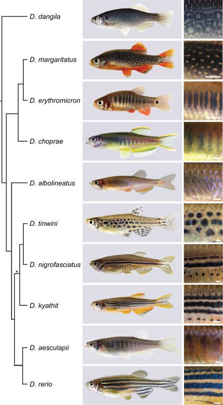
Although Janni’s lab had always been interested in Danio species other than the zebrafish [3, 4], the pioneering work came from David Parichy and colleagues over the last two decades [5, 6]. A milestone was the release of a reliable phylogeny of the genus by Braedan McCluskey and John Postlethwait [7]. I found it fascinating that species most similar in their pigment patterns were not necessarily most closely related, and most closely related species often had most divergent patterns. Zebrafish develop a striking pattern of horizontal blue and golden stripes, while the sister species D. aesculapii conceals itself by forming dark vertical bars that are low in contrast. Given the knowledge about the genetic basis of stripe formation in zebrafish, maybe we could identify the genes that functionally diverged to contribute to patterning differences between species.
Fish species of the Danio genus and their pigment patterns. Phylogeny according to [7]. Scale bars, about 1 mm. Photos by Marco Podobnik and Uwe Irion
We decided to focus on genes that code for proteins mediating direct cell-cell contacts. Uwe had already generated mutants in a handful of these genes and acquired a number of Danio species; a collection I helped to make more comprehensive. We were inspired by the reciprocal hemizygosity test to identify diverged genes and we could meet the requirements to apply it, namely generating null mutants in species and hybrids between species [8]. Such an endeavour in non-model organisms was unthinkable without the CRISPR/Cas9 system, which the lab aims to improve since its adaptation for genetic engineering [9, 10]. Uwe and I started an extensive effort of making hybrids, pairing wild-type and the newly generated mutant species in various combinations. Four years later it all came together: While some genes remained functionally conserved, others diverged across the phylogeny [11].
In the case of the potassium channel gene kcnj13 we identified a repeated and independent functional divergence; this contributed to patterning differences between zebrafish and D. aesculapii [12]. We found that the black melanophores require the kcnj13 function to acquire certain cell shapes within their own population but they also instruct the two other pigment cell types, yellow/orange xanthophores and shiny/blue iridophores, to change their shapes according to their location in the skin. As the D. aesculapii allele is functionally different from the zebrafish one, we propose that divergence in kcnj13 caused changes in the way the pigment cells interact and change their shapes to contribute to the species-specific differences in colour and contrast of the patterns [13].
Your PhD research involves working with a wide range of techniques. Do you have a favourite?
I love genetics for its power to let us understand processes across different levels of biological organisation. During university I learnt about Muller’s classification of alleles [14]. It was beautiful to see how it could be used when we studied pattern development in hybrids between Danio species. The altered patterns in hemizygous hybrids between mutant zebrafish and wild-type D. aesculapii indicated that the kcnj13 allele from D. aesculapii behaves like a hypomorph as it could not compensate the CRISPR/Cas9-induced loss-of-function of the D. rerio allele. The patterns of hemizygous hybrids from the reciprocal cross are similar to the patterns of hybrids between wild-type species. Thus, the wild-type alleles from zebrafish and D. aesculapii cannot be functionally equivalent. Initially we thought that the D. aesculapii allele had lost its function completely but mutant D. aesculapii indicated that the gene function was still required for patterning. I love discovering obvious phenotypes in mutants, as it can be a rare but very profound experience. The suitability of zebrafish for fluorescence imaging in vivo makes it a fantastic model, as you can see cells behaving in real time, sometimes even with subcellular resolution. I can probably make the most meaningful contributions by applying a duet of genetics and in vivo imaging. Often the successful outcome of any experiment relied on the groundwork and expertise of my colleagues. I essentially understand science as teamwork.
Speaking of teamwork, how was your experience collaborating with people across the world for your PhD work?
As you can imagine pigment patterning is a relatively small field, although it hopefully becomes apparent how exciting it is. It’s great that people with expertise in genetics, sequencing methods, protein structure modelling and image analysis collaborate on this topic. I feel there aren’t really boundaries when it comes to common interests and curiosity, it’s just essential to bring passionate people together. It’s important to communicate effectively and to make it fair for the people involved. When it comes to generating ideas, bigger meetings might not always be effective. The most creative ideas emerged during our small lab and one-to-one meetings.
Were there any frustrating times during your PhD? And on the flip side, any particularly memorable moments?
My most exciting moments were the ones when we made discoveries in the lab. Sometimes it took a while to realize them. It requires one to think about observations over and over again. We were all working a lot, basically every day including most weekends, which is an intensity I have decided to reduce. I have fond memories of the garden parties in summer and the yearly Christmas cookie baking at Janni’s house.
You’ve recently moved across the globe to do a postdoc in Melbourne. How was the experience and what motivates your research today?
The Melbourne metropolitan area is a great place for personal life and research, although it can be depressing to think about the fate of the traditional owners of this country. The Australian Regenerative Medicine Institute cultivates broad interests in development, regeneration, evolution and medicine. Our lab headed by Peter Currie explores muscle biology using a spectacular diversity of fish models. It’s stimulating to be part of a bigger team. I find it exciting to help others with their projects, while I am defining my own long-term goals.
Finally, let’s go outside of the lab. What do you like to do in your spare time?
I play the saxophone in various settings for over 20 years now, which is tremendously important to me. I also love hiking. A very memorable trip was the Via Alpina in Switzerland and now very recently a trip to the Victorian Alps in Australia.
Dr. Marco Podobnik Research Fellow Australian Regenerative Medicine Institute, Monash University, Clayton 3800 VIC, Australia
1. Nusslein-Volhard, C. (2012). The zebrafish issue of Development. Development 139, 4099-4103.
2. Wieschaus, E., and Nusslein-Volhard, C. (2016). The Heidelberg Screen for Pattern Mutants of Drosophila: A Personal Account. Annu Rev Cell Dev Biol 32, 1-46.
3. Singh, A.P., and Nusslein-Volhard, C. (2015). Zebrafish stripes as a model for vertebrate colour pattern formation. Curr Biol 25, R81-R92.
4. Irion, U., and Nusslein-Volhard, C. (2019). The identification of genes involved in the evolution of color patterns in fish. Curr Opin Genet Dev 57, 31-38.
5. Parichy, D.M., and Johnson, S.L. (2001). Zebrafish hybrids suggest genetic mechanisms for pigment pattern diversification in Danio. Dev Genes Evol 211, 319-328.
6. Patterson, L.B., and Parichy, D.M. (2019). Zebrafish Pigment Pattern Formation: Insights into the Development and Evolution of Adult Form. Annu Rev Genet 53, 505-530.
7. McCluskey, B.M., and Postlethwait, J.H. (2015). Phylogeny of zebrafish, a “model species,” within Danio, a “model genus”. Mol Biol Evol 32, 635-652.
8. Stern, D.L. (2014). Identification of loci that cause phenotypic variation in diverse species with the reciprocal hemizygosity test. Trends Genet 30, 547-554.
9. Irion, U., Krauss, J., and Nusslein-Volhard, C. (2014). Precise and efficient genome editing in zebrafish using the CRISPR/Cas9 system. Development 141, 4827-4830.
10. Dorner, L., Stratmann, B., Bader, L., Podobnik, M., and Irion, U. (2024). Efficient genome editing using modified Cas9 proteins in zebrafish. Biol Open 13.
11. Podobnik, M. (2023). On the Genetic Basis of Pigment Pattern Diversification in Danio Fish, (Eberhard Karls Universität Tübingen).
12. Podobnik, M., Frohnhofer, H.G., Dooley, C.M., Eskova, A., Nusslein-Volhard, C., and Irion, U. (2020). Evolution of the potassium channel gene Kcnj13 underlies colour pattern diversification in Danio fish. Nat Commun 11, 6230.
13. Podobnik, M., Singh, A.P., Fu, Z., Dooley, C.M., Frohnhofer, H.G., Firlej, M., Stednitz, S.J., Elhabashy, H., Weyand, S., Weir, J.R., et al. (2023). kcnj13 regulates pigment cell shapes in zebrafish and has diverged by cis-regulatory evolution between Danio species. Development 150.
14. Muller, H. (1932). Further studies on the nature and causes of gene mutations. Jones DF, ed. In Proceedings of the 6th International Congress of Genetics. pp. 213-255.
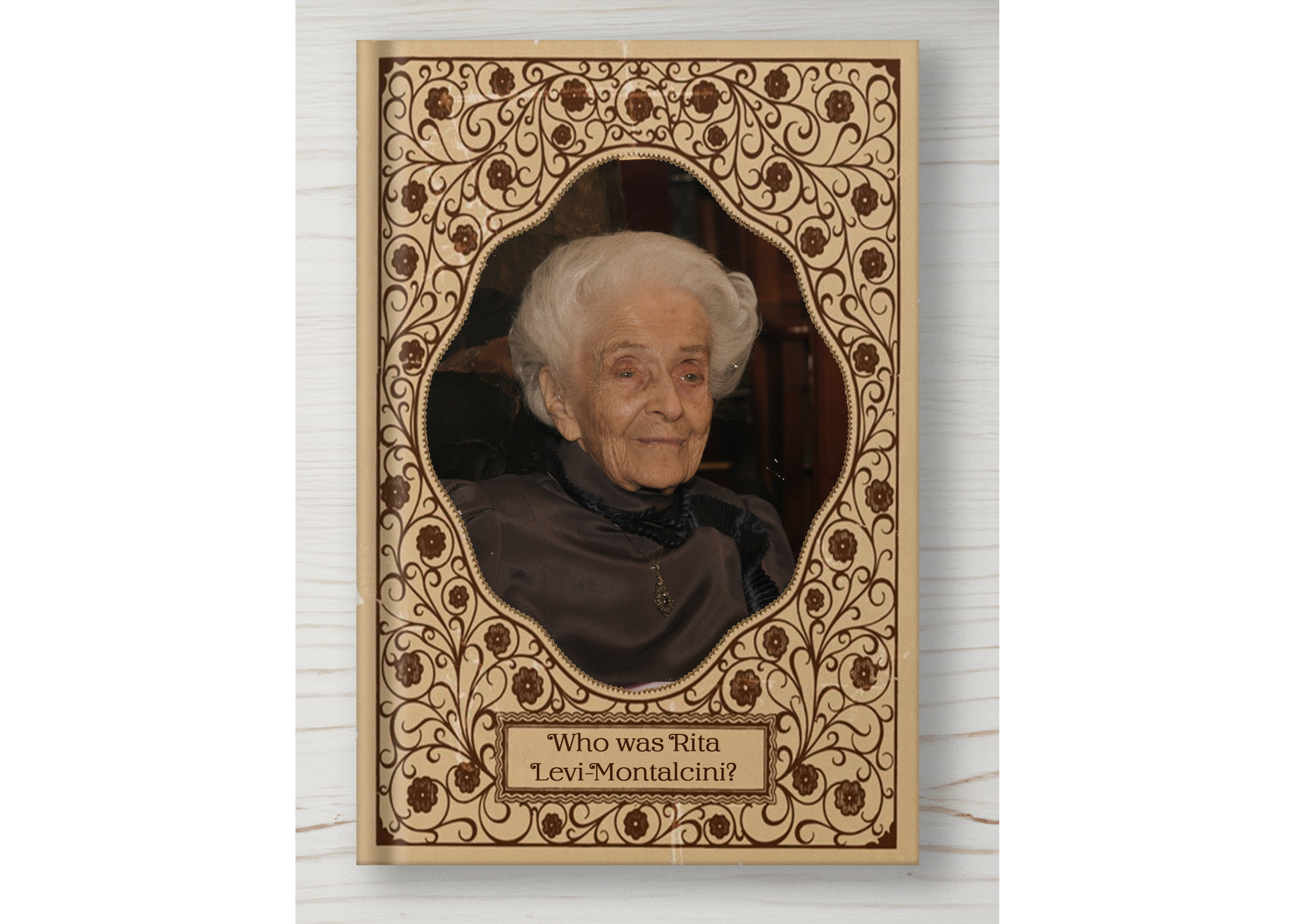
Rita-Levi Montalcini (1909-2012) was an Italian neurobiologist who lived an extraordinary life, and today (22nd April) would have been her 115th birthday. Click on each image to enlarge and read more about her…
Sources: Coutinho, L. and Teive, H.A.G. (2023) ‘Rita Levi-Montalcini: the neurologist who challenged fascism’, Arquivos de Neuro-Psiquiatria, 81(1), pp. 95–98. Available at: https://doi.org/10.1055/s-0043-1761426.
Hamburger, V. and Levi-Montalcini, R. (1949) ‘Proliferation, differentiation and degeneration in the spinal ganglia of the chick embryo under normal and experimental conditions’, Journal of Experimental Zoology, 111(3), pp. 457–501. Available at: https://doi.org/10.1002/jez.1401110308.
Malerba, F. (2022) ‘Why Are We Scientists? Drawing Inspiration From Rita Levi-Montalcini’, Frontiers in Cellular Neuroscience, 15, p. 741984. Available at: https://doi.org/10.3389/fncel.2021.741984.
Here in the Koltowska lab, we are interested in all things lymphatic vessel-related. How lymphatic endothelial cells (LECs) are specified, gain their identity, end up in the right place to form vessels, and how these vessels function.
Lab roll call
Hannah Arnold has been a postdoc in the lab for five years and is interested in lymphatic development, focusing on how LECs migrate and interact to navigate their environment.
Marleen Gloger has been a postdoc in the lab for five years as well and is interested in lymphatic vessel development, specifically LEC cell proliferation, and how these processes are altered in disease conditions such as cancer and metastasis formation.
Di Peng has done a PhD in the group and now continues as a postdoc. She is very fond of observing cellular events during development using different live imaging techniques. Her projects focus on regulation of lymphatic endothelial behaviours.
Faidra Voukelatou has recently started her PhD in the group and is interested in cancer as well as lymphatic vessel research. She enjoys working with zebrafish as an animal model to investigate the dynamics of brain cancer invasion and vasculature.
Renae Skoczylas has been a research engineer in the lab for 6 years and enjoys all things zebrafish and lymphatics. She is particularly happy generating new mutant lines for the lab using CRIPSR technology and being involved in and helping with any other lab members’ projects.
Favourite technique, and why?
KaskaKoltowska: Microscopy! There is something incredibly magical in looking down the microscope and observing life in high magnification. Using microscopy to look at zebrafish heartbeat and blood flowing through the vessels never stops to amaze me!
Apart from your own research, what are you most excited about in developmental and stem cell biology?
KaskaKoltowska: I think how gene expression is regulated and the steps coordinating cell specification is incredibly fascinating. The level of developmental reproducibility in every embryo is just mind-blowing. Biology gets it right almost every time, and if it does not, we can learn something very important.
How do you approach managing your group and all the different tasks required in your job?
Kaska Koltowska: I don’t think I use any specific managing tools. I dedicate time to discussing science with every member of the group regularly. This helps to keep the projects focused. When a project is coming up close to completion I dedicate more time for it. It helps a lot that the team is very efficient and group members can manage themselves very well so my input is minimal. For myself, I often make a weekly prioritisation plan of the most important tasks that need to be done that week and try to stick to it.
What is the best thing about where you work?
We are positioned between two wider communities. That of Vascular Biology, where our lab is located and encompasses ten research groups, and the Uppsala Zebrafish community where our fish are housed alongside five other groups and one service platform.
What’s there to do outside of the lab?
Uppsala is a small but busy student city where you can enjoy restaurants and cafes for a ‘fika’ break. It is located close to nature giving us the opportunity to enjoy the forest for a walk or BBQ in the summer and snow sports in the winter. It also provides an excellent backdrop for walking the boss’ dog. On the other hand, Uppsala is a short train ride to Stockholm so it is easy to enjoy big city life on the weekends and go to museums, theatres or concerts.
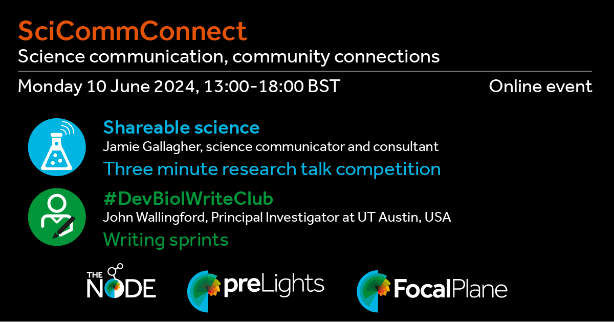
Science communication is an integral part of being a researcher. Want to practice your science writing and presentation skills? Register to attend “SciCommConnect: Science communication, community connections“!
The three community sites supported by The Company of Biologists – the Node, preLights and FocalPlane – are hosting a free, online event on Monday 10 June 2024 from 13:00-18:00 BST, focusing on the different ways in which science can be communicated. We hope this event will present a unique opportunity for you to work on your science writing and presentation skills and connect with your peers across the world in a friendly, informal environment.
Dr Jamie Gallagher is an award-winning science communicator, trainer and consultant. He will share tips and tricks on how to make science talks as interesting, engaging and memorable as possible.
Three minute research talk competition
Similar to the Three minute thesis competition format, this is a chance for you to practice communicating your research in a concise and engaging way.
Present your work for a chance to win a cash prize and to get feedback from Jamie, who is a previous Three Minute Thesis winner. To enter please provide us with short summary of your intended talk (think about how you would advertise your talk in a tweet!)
Applications for the short talk competition are optional and spaces are limited. Deadline for entering into the competition is Sunday 26 May.
Those who do not wish to give a talk will also benefit from listening to people’s talks and Jamie’s feedback. They will also be able to vote for their favourite talk.
#DevBiolWriteClub and themed writing sprints
Prof John B. Wallingford is a Professor at UT Austin. He is passionate about writing and has written on the Node regularly, including the popular #DevBiolWriteClub posts. He will share some excellent writing advice which can directly be applied during the themed writing sprints that will follow.
For the writing sprints, you can pick which group to be in (the Node, preLights or FocalPlane). Each group will work together to brainstorm and draft a piece of writing on a pre-selected topic. Details of the writing briefs for each group will be provided closer to the event.


 (No Ratings Yet)
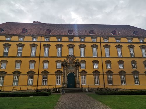
 (3 votes)