In 2023, FASEB will host 22 Science Research Conferences (SRCs). SRCs are multiday, in-person meetings featuring discussion of scientific advances and sharing of cutting-edge research through lectures, posters, informal discussions, and social events. The complete SRC schedule is available on our website. You can sort the schedule by month or topic area. 2023 topics include cell biology, neuroscience, clinical and translational medicine, immunology, genetics and genomics, and many other focus areas. Click on the title of the conference you are interested in to see a description, the location, information about registration fees and deadlines, abstract submission instructions, and other key details. The individual conference websites will be updated with additional information over the next few months. For more information, visit www.faseb.org/meetings-and-events/src-events
Determining the Effects of FOXG1 Mutations on Early Neurodevelopmental Structures Using iPSCs
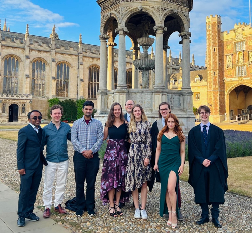
I am an undergraduate Neuroscience student at University College London interested in researching neurodevelopmental and neuropsychiatric disorders. I am grateful for the opportunity that the Gurdon grant gave me to undertake a summer studentship in the lab of Dr Srinjan Basu at the Wellcome-MRC Cambridge Stem Cell Institute under the supervision of Dr Deep Adhya. The lab focuses on imaging organoids to study how chromatin regulators influence stem cell differentiation in the neurodevelopmental conditions autism and epilepsy.
iPSCs and brain organoids as a model for neurodevelopmental conditions
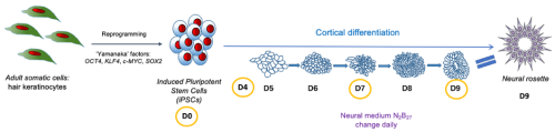
The central nervous system develops from a monolayer of neuroepithelial cells which folds to form the neural tube. Neural stem cells organise themselves within the tube to form neural rosettes, rose-like structures which have been reported both in vivo and in vitro (Hříbková et al. 2018). Recent advances in in vitro neuronal differentiation and organoid technology provide a model system for addressing how normal development of these rosettes is disrupted in conditions such as autism or epilepsy and for dissecting the molecular mechanisms governing these changes. Induced pluripotent stem cells (iPSCs), adult somatic cells reprogrammed back into their pluripotent stem cell stage, can be induced to differentiate into specific cell fates (Fig.1). Using this approach, it has been shown that iPSCs generated from individuals with autism show significant cellular and molecular abnormalities (Adhya et al. 2021). Intriguingly, defects begin much earlier than expected. Autistic iPSCs form abnormal neural rosette structures long before neural stem cells differentiate towards excitatory/inhibitory neurons (Adhya et al. 2021), but how this occurs at the molecular level remains poorly understood.
My project: FOXG1, a transcription factor implicated in epilepsy and autism
The aim of my project was to determine if atypical neural rosette structures form in early cortical organoids from FOXG1-mutant iPSCs. FOXG1 is an essential transcription factor responsible for normal neurodevelopment. Mutations in FOXG1 are significantly associated with syndromic forms of autism and ~90% of FOXG1 syndrome patients show epileptic seizures (Seltzer et al. 2014). Therefore, autism and epilepsy are considered comorbid.
Methods and techniques learned
During my project, I learned how to perform tissue culture (TC): I grew iPSCs and differentiated them into neurons. Working in the sterile TC hood made me a better scientist as I became more conscious about ways to prevent contamination, which is especially important when handling live cell cultures.
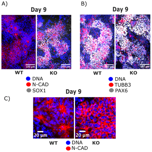
After successfully growing the iPSCs, I fixed them before differentiation and then at several stages during neural rosette formation (Fig. 1). The fixed cells were processed for immunofluorescence (IF) imaging and quantitative polymerase chain reaction (qPCR) to determine morphological changes that take place during cortical differentiation and to see its effect on gene expression, respectively. I isolated RNA using the Mini prep kit, but unfortunately, was unable to continue with the qPCR as some essential components did not arrive in time. Nevertheless, we got interesting results from the imaging alone (Fig. 2). Immunocytochemistry staining was another important lab skill I gained during the studentship. After surveying the current literature, I chose antibodies that reveal morphological features of neural rosettes: SOX1 as an early differentiation marker, PAX6 as a well-established marker of cortical neurons, N-cadherin as a rosette lumen marker, and TUBB3 as a neural cytoskeletal marker. Additionally, DAPI was used as nuclear counterstain.
Fig. 1 shows the outline and timeline of my project. iPSCs were generated by reprogramming adult somatic cells with ‘Yamanaka factors’ into pluripotent stem cells. The iPSCs were subsequently differentiated into neurons by placing them in a neural medium. The cells were fixed and subjected to IF at day 0, 4, 7 and 9 of the cortical differentiation. Neural rosettes form at day 9.
Apart from learning new lab techniques, I was introduced to image analysis softwares such as Fiji and CellProfiler. Furthermore, I was fully immersed into the activities of the research group, including weekly journal club, group meetings and workshops. I had several presentations during the group meetings which helped me improve my ability to discuss scientific concepts and results. All these skills will be useful for the rest of my MSci degree and for a future PhD.
Project outcomes
iPSCs stained with N-cadherin and SOX1 (Fig.2A) or with TUBB3 and PAX6 (Fig.2B) revealed a phenotype for the FOXG1 mutation. Zooming in on the rosette lumen stained with N-cadherin and nuclei stained with DAPI (Fig.2C) showed that neural rosettes do not form at day 9 without FOXG1 while they form in control iPSCs. Interestingly, N-cadherin, PAX6 and TUBB3 stainings reveal that FOXG1-mutant cells at day 9 look like glial progenitor cells, precursors that should form later in the organoid. Altogether, these findings provoke numerous questions about underlying mechanisms of these morphological changes, effect on gene expression and further differentiation into excitatory/inhibitory neurons, that will be researched by the lab in the future. If I had more time in the lab, I would have looked into other gene mutations significant for the comorbidity of autism and epilepsy that may affect the formation of neural rosettes, namely KMT2A and FGFR2. That would help us get a more robust understanding of the shared neurodevelopmental defects of these disorders.
Fig. 2: IF imaging of iPSCs at day 9 of cortical differentiation shows the disruption in neural rosette formation that occurs in FOXG1-mutant cells (KO) while neural rosettes develop normally in control cells (WT).
Summary
Overall, the summer studentship was an amazing experience as I designed my own experiments that I saw through from the beginning to the end. Along the way I gained valuable skills in the lab and research in general. This project introduced me to the exciting field of organoid research that is redefining the way we study neurodevelopmental disorders, and I would like to continue with it in my scientific future. I would strongly recommend applying for the Gurdon grant to students interested in delving deeper into a developmental biology topic and in experiencing what being a scientist entails.
CITED2 is a Conserved Regulator of the Uterine-Placental Interface Marija Kuna, Pramod Dhakal, Khursheed Iqbal, Esteban M. Dominguez, Lindsey N. Kent, Masanaga Muto, Ayelen Moreno-Irusta, Keisuke Kozai, Kaela M. Varberg, Hiroaki Okae, Takahiro Arima, Henry M. Sucov, Michael J. Soares
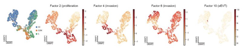
UMAP scatterplots of multiome (snRNA-ATACseq) data of invading trophoblast from Arutyunyan, et al.
Spatially resolved single-cell multiomics map of human trophoblast differentiation in early pregnancy Anna Arutyunyan, Kenny Roberts, Megan A Sheridan, Ilia Kats, Luz Garcia-Alonso, Britta Velten, Regina Hoo, Kevin Troulé Lozano, Louis-Francois Handfield, Luca Marconato, Elizabeth Tuck, Lucy Gardner, Cecilia Icoresi Mazzeo, Iva Kelava, Elena Prigmore, Sarah A Teichmann, Omer Ali Bayraktar, Ashley Moffett, Oliver Stegle, Margherita Y Turco, Roser Vento-Tormo
Cellular Maturation of Oligodendrocytes is Governed by Transient Gene Melting Kevin C. Allan, Tyler E. Miller, Andrew R. Morton, Marissa A. Scavuzzo, Matthew S. Elitt, Benjamin L.L. Clayton, Lucille R. Hu, Jost K. Vrabic, Hannah E. Olsen, Daniel C. Factor, Jonathan E. Henninger, Richard A. Young, Charles Y. Lin, Peter C. Scacheri, Paul J. Tesar doi: https://doi.org/10.1101/2022.11.17.516981
Expression bias in retinoic acid responsive genes defines variations in neural differentiation of human pluripotent stem cells Suel-Kee Kim, Seungmae Seo, Genevieve Stein-O’Brien, Amritha Jaishankar, Kazuya Ogawa, Nicola Micali, Victor Luria, Amir Karger, Yanhong Wang, Thomas M. Hyde, Joel E. Kleinman, Ty Voss, Elana J. Fertig, Joo-Heon Shin, Roland Bürli, Alan J. Cross, Nicholas J. Brandon, Daniel R. Weinberger, Joshua G. Chenoweth, Daniel J. Hoeppner, Nenad Sestan, Carlo Colantuoni, Ronald D. McKay
Adaptation to ex vivo culture drives human haematopoietic stem cell loss of repopulation capacity in a cell cycle independent manner Carys S. Johnson, Kendig Sham, Serena Belluschi, Xiaonan Wang, Winnie Lau, Kerstin B. Kaufmann, Gabriela Krivdova, Emily F. Calderbank, Nicole Mende, Jessica McLeod, Giovanna Mantica, Matthew J. Williams, Charlotte Grey-Wilson, Michael Drakopoulos, Shubhankar Sinha, Evangelia Diamanti, Christina Basford, Anthony R. Green, Nicola K. Wilson, Steven J. Howe, John E. Dick, Bertie Göttgens, Natalie Francis, Elisa Laurenti
Spatiotemporal coordination of stem cell behavior following alveolar injury Maurizio Chioccioli, Sumner Magruder, John E. McDonough, Jessica Nouws, Tao Yang, David Gonzalez, Lucia Borriello, Brian Traub, Xianjun Ye, Caroline E. Hendry, David Entenberg, Smita Krishnaswamy, Naftali Kaminski, Maor Sauler
A human mitofusin 2 mutation causes mitophagic cardiomyopathy Antonietta Franco, Jiajia Li, Daniel P. Kelly, Ray E. Hershberger, Ali J. Marian, Renate M. Lewis, Moshi Song, Xiawei Dang, Alina D. Schmidt, Mary E. Mathyer, Cristina de Guzman Strong, Gerald W. Dorn II
Guillermo Martínez-Ara and colleagues from Miki Ebisuya lab have developed a new optogenetic tool that induces apical constriction in mammalian epithelia. The new tool called OptoShroom3 induces tissue folding in epithelial colonies and provokes changes in curvature and thickness in neural organoids. The authors showed that this optogenetic approach gives spatiotemporal control to manipulate the structure of mammalian tissues.
How did you get started on this project?
This project started before I got to the lab, back when my supervisor, Miki Ebisuya, was based in Japan. Two very talented postdocs spent three years exploring and developing ideas on how to create a tool to control morphogenesis.
I was deeply interested in the creative approaches of synthetic biology and the development of new biological tools, so I did my master’s in systems and synthetic biology. Then, I found about Miki’s lab and thought that their application of synthetic biology to study development was fascinating. Luckily, my interests matched with Miki’s and I was invited to join the project after the lab moved to EMBL Barcelona. For my PhD project, I joined forces with Núria Taberner (one of the talented postdocs) to develop a tool to provoke apical constriction in mammalian systems. I thought this project was super exciting. Using optogenetics and synthetic biology to manipulate tissue structure – I was totally in!
Can you summarise your findings?
Briefly, we developed a new optogenetic tool (OptoShroom3) that allowed spatiotemporal control of apical constriction in mammalian tissues. To demonstrate its potential applications, we showed that OptoShroom3 could be used to alter tissue shape in epithelial cell lines, to induce folding in colonies, and to change the shape of neural organoids. We developed the first tool (to our knowledge) that could be easily applied to manipulate mammalian tissue structure through the control of biological forces.
What are the advantages of using OptoShroom3 compared with the previously existing optogenetic tools used for inducing actomyosin constriction?
Most of the existing tools for the induction of actomyosin constriction relied on plasma membrane recruitment. This means that the factors used to induce contractility (normally related to the Rho pathway), are brought to the cell membrane upon illumination. This, in principle, gives more freedom to induce contractility in any area of the plasma membrane. However, when it comes to inducing tissue shape changes it becomes more challenging. If we want to induce constriction in one area of the cells (say apical surface) we would require very precise illumination of a single plane in the tissue, which requires two photon microscopy (as the lab of Stefano de Renzis had previously done). When the shape of tissues becomes convoluted, like we see in organoids, this becomes even more complicated, if not just impossible. We cannot easily create multiphoton stimulation patterns in 3D shapes, and even if it is achievable, this technique is not easily accessible for most scientists.
The advantage of OptoShroom3 is that it directs constriction specifically to the apical surface. Then, if all the tissue is stimulated, only the apical area of the cells will constrict, inducing selective constriction. This allowed us to induce changes in the shape of organoids with complex structures such as optic vesicle organoids. We stimulated the whole vesicle organoid to provoke localised constriction on the apical side, which led to changes in shape.
Do you have any tips or tricks for researchers using OptoShroom3 in the future?
Whenever someone contacts us with the aim of using OptoShroom3 in their own system, I normally reply something like this:
How well OptoShroom3 works in your system will depend on two things.
1. How epithelial the cell line is, because OptoShroom3 relies on the apical cytoskeletal structures, specifically on the localization of actin along the apical side of cell-cell junctions. When this structure is present, the N-terminal part of Shroom3 will localise there and then OptoShroom3 will be able to induce constriction locally on the apical side. Without epithelialization, there’s no apical constriction.
2. The ratio of expression between both components. Normally, we try to maximise the expression of both the NShroom3 component and the CShroom3 part. However, very high expression of CShroom3 seems to be somewhat toxic, so the optimal levels for the tool to work is high NShroom3-iLID and medium-low SspB-CShroom3 expression.
Finally, I would suggest people to be patient with optogenetic tools. At the beginning, they may not seem easy to use. There are several parameters to be optimised: laser intensity, intervals of stimulation and rest, temporal resolution when imaging… Once you get an idea of how much laser power you need to activate the tool on your microscope, and for how long you need to stimulate to see changes in shape, things will be much easier. I guess this applies to all optogenetic tools. You should give yourself some time to play around with these parameters!
When doing the research, did you have any particular result or eureka moment that has stuck with you?
Yes! I remember clearly the morning when I first saw the deformation of an optic vesicle in the confocal microscope room. The deformation was very clear, and it was finally a direct confirmation that we could use OptoShroom3 to alter tissue shape in organoids, which was one of our main goals! I don’t consider it an ‘eureka’ moment, but rather a happy memory of when an experiment finally works after a lot of effort.
And what about the flipside: any moments of frustration or despair?
There are always ups and downs in academic research. For us, in the first review of the OptoShroom3 publication reviewers were asking for some more experiments and controls. This would normally be ok (we knew it was a possibility), but then one month after getting these requests the confocal microscope I was using for all the experiments suddenly broke. To make it worse, it took three months to get it fixed. During that time, I tested other microscopes around in the building, but because we wanted to compare the new results to the original ones, we needed to make sure that the amount of light used for stimulation was the same. Of course, different microscopes, with different designs, optics and laser sources could not be easily compared. In the end, I spent those three months testing microscopes, planning the new experiments and, perhaps, building patience and resilience. Overall, we had a three-month delay for the project, which back then seemed catastrophic, but later on seems like it was not the end of the world.
Image analysis is a key part of your work, can you tell us about the custom-made pipeline and any tips for researchers looking to use or repurpose the code?
I tried my best to leave clear comments in the code and make it publicly available so that people can extract whatever is useful for them. However, as we mention in the GitHub page, my main aim was to make functional code for the paper, therefore it is possible that some people will struggle to understand it or use it. In that case, my main tip is to contact me! I’m always happy to help and discuss the method with researchers interested in biological image analysis.
Are you testing OptoShroom3 in vivo?
As far as I know, Miki’s lab is not planning on working directly on embryos in the near future. The lab focuses on the use of in vitro systems for synthetic developmental biology. I am also not planning to work in vivo any time soon. However, we do have some collaborators working to create some OptoShroom3 transgenic mice, which is incredibly exciting!
Where will this story take the lab?
As I said, our first aim was to make a tool that we could use to perturb tissue structure using biological forces. Now that is working the logical question is: What do we do with it?
We believe that these types of tools open the door to ask new questions, or at least to ask old questions from a different perspective. We are especially interested in how changes in tissue structure can impact other developmental processes, and we believe that organoids are a great model to ask these questions!
What is next for you after this paper?
While Miki’s lab will continue to explore how OptoShroom3 can be used to study possible feedbacks between shape and other developmental processes, I am getting ready to move to a postdoc position, in which I’m hoping to develop more exciting tools to manipulate or even program tissues!
“As a Christmas treat, we’ve prepared a selection box of some of the best bits from our guests that never made it into the final episodes.”
Dr Sally Le Plage
In the final episode of the Genetics Unzipped podcast, we’re looking back at our favourite genetic stories of the year plus some bonus bits from our interviews that have never been heard before.
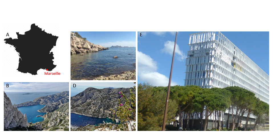
Hi, my name is Marvin Leria and I’m a PhD student, funded by Turing Center for Living Systems (CENTURI), at Aix-Marseille University in Marseille, France. I work in the lab of André Le Bivic under the supervision of Andrea Pasini, and co-supervised by Raphael Clément. Our lab is located in the magnificent Calanques National Park by the Mediterranean Sea, which offers a peaceful environment to do research (Figure 1). The main research topics of our lab revolve around cell polarity, morphogenesis and the evolution of epithelia. Historically, our lab has worked with cell cultures, but we are now developing new models such as marine sponges and more recently placozoans, to highlight conserved features and innovations in epithelial evolution.
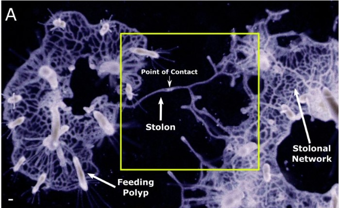
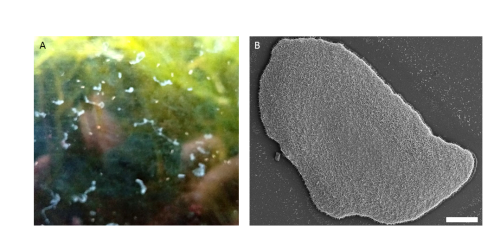
Placozoans are small and flat (around 1-2 mm in diameter, 20-30 µm thick) benthic marine animals (Figure 2.B) that are found around the world, mainly in tropical and subtropical areas such as coral reefs, mangroves, etc. (Schierwater et al., 2021). They are found gliding on rocks and other substrates and they mainly feed upon biofilms containing algae, bacteria and other microorganisms by means of external digestion (Figure 2.A)
Figure 2: The general aspect of placozoans. A: Placozoans, as can be observed on the wall of seawater aquaria. Note the range of diverse morphologies. B: A scanning electron microscopy of Trichoplax sp. H2 (Haplotype 2), scale bar 100µm
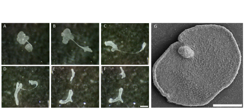
The life cycle of placozoans remains very enigmatic. Reproduction mainly occurs asexually, generally either by binary fission or by budding which produces juvenile animals referred to as swarmers (Figure 3; Thieman and Ruthman, 1991; Eitel et al., 2011; Zuccolotto-Arellano and Cuervo-González, 2020). Little is known about sexual reproduction and embryogenesis even though it is thought to occur in nature (Signorovitch et al., 2005; Eitel et al., 2011).
Figure 3: Placozoan asexual reproduction. A-F: Photos showing reproduction by whole-body fission. The cellular bridge connecting the two halves of a dividing individual become thread-like before breaking. A. The two distal plates are bridged by a cellular thread. B (5min), C (60min) and D (65min): The thread elongates and becomes extremely thinner at its centre. E (70min): The cellular thread broke at its center as the two distinct parts move away from each other. F (70min 30sec) : The remnant broken cellular thread retracts in the two daughter animals. Scale bar 500µm. G: A scanning electron micrograph of a Trichoplax sp. H2 showing a budding structure, scale bar 200µm.
Phylogeny and evolution of Placozoa
The first discovered placozoan species Trichoplax adhaerens was described by the German zoologist Franz Eilhard Schulze in 1883 (Schulze, 1883). Very recently, three other placozoan species have been described, Hoilungia hongkongensis (Eitel et al., 2018), Polyplacotoma mediterranea (Osigus et al., 2019) and Cladtertia collaboinventa (Neumann et al., 2022). Other placozoan haplotypes have been genetically distinguished based on mitochondrial 16S rDNA fragments (Voigt et al., 2004; Signorovitch et al., 2006; Eitel et al., 2013 (review); Osigus et al., 2019; Miyazawa et al., 2021). The genome of Trichoplax adhaerens was sequenced and has been available since 2008 (Srivastava et al., 2008). It is one of the smallest animal genomes. It is composed of ~98 million base pairs and contains about 11,500 protein coding genes. In contrast, Trichoplax mitochondrial genome is one of the largest in the animal kingdom (Dellaporta et al., 2006). The phylogenetic position of placozoans among early-diverging phyla has been enormously controversial and still remains an important topic.
Body plan, cell morphology and physiology
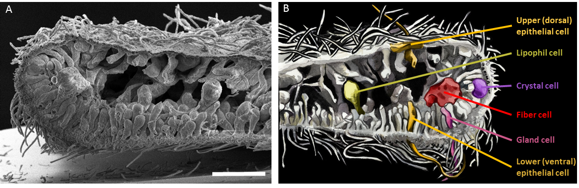
Placozoans have no symmetry axis (only a top-bottom axis), and are devoid of muscles and nervous system. Their simple body plan consists of two monociliated epithelial layers, commonly referred to as the lower (dorsal) epithelium and the upper (ventral) one, with an internal cavity. Six morphologically distinct cell types have been described so far: upper epithelial cells, lower epithelial cells, gravity-sensing crystal cells, digestive enzyme-secreting lipophil cells, mucous-secreting and peptidergic gland cells and internal phagocytic fiber cells (Figure 4; Smith et al., 2014; Mayorova et al., 2019; Mayorova et al., 2021).
Figure 4: The internal anatomy of a Trichoplax sp. H2. A: Scanning electron microscopy cross-section, showing morphologically different cell types in the upper and lower ciliated epithelia and in the internal cavity. Scale bar 10µm. B: An artist view showing the different cell types (drawing courtesy of Pauline Geronimi, Haute École d’Art du Rhin).
Surprisingly, despite the absence of a nervous system, some epithelial gland cells also synthesize neuropeptide-like molecules, which appear to control not only the behaviour of Trichoplax, but also its shape (Varoqueaux et al., 2018). Indeed, placozoans are tiny experts in shapeshifting and are able to adopt an incredible range of diverse shapes (see Figure 2 and Figure 3). How it is possible for placozoans to change their shape while maintaining the integrity of their epithelia is a mystery (see Video 1), and trying to understand this mechanism is the main goal of my thesis work. It is very likely that the shapeshifting ability of placozoans depends on the unusual features of their epithelia, which have no basal lamina and only one type of intercellular junctions similar to adherens junction (Smith and Reese, 2016). This is why most of my work focuses on studying the epithelial organization and will give a comprehensive insight into Trichoplax epithelial evolution and biology.
Video 1: The shape of Trichoplax is changing over time while the animal is moving (the movie is 10-time accelerated).
My day in the lab
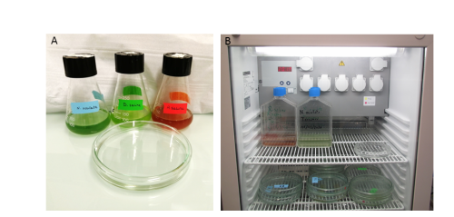
You may have read other contributors to ‘A day in the life of’ describing their adventurous trips to exotic places or wonderful seaside locations to collect their favourite organisms. Things are a bit less fancy for us, since we recovered our Trichoplax from the aquaria of a local tropical fish store in downtown Marseille. To collect animals for our first cultures, we deposited a few glass slides in a sea water tank and leave them there until a biofilm had developed. After several weeks, we took the slides back to the lab and observed some placozoans grazing on them! We now culture our placozoans in the lab in large petri dishes and feed them every week with their favourite red and green microalgae (Figure 5.A). They are kept at 20°C with a dark-light cycle (Figure 5.B). They need to be transferred regularly to new dishes when the old ones get dirty or when their density is getting high. We survey them every day to make sure that the culture living conditions are optimal.
Figure 5: Laboratory culture of Trichoplax. A: Trichoplax are easily maintained in reconstituted seawater in large glass Petri dishes, and fed with a cocktail of different unicellular algae. B: Trichoplax dishes and algal cultures are kept in a 20°C temperature-controlled cabinet under a 12hrs light and 12hrs darkness regime.
For experiments, I transfer the Trichoplax carefully into a drop of sea water on coverslips and let them adhere properly. Once they are ready, I perform immunostaining experiments. Trichoplax are very fragile animals and experiments demand extreme patience and perseverance. After quite some efforts, I have succeeded in setting up fixation protocols that allow me to perform beautiful immunofluorescence staining of the epithelial cells and I can now follow how they change their shapes according to the changes of the whole animal. For this, I mostly use techniques such as confocal microscopy, image analysis and some electron microscopy too.
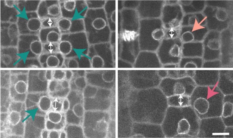
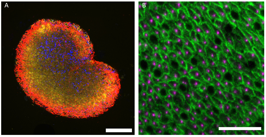
Figure 6: Immunofluorescence studies of the lower ciliated epithelium of Trichoplax sp. H2. A: A reconstituted image from stitched tiles at high-magnification view of the lower epithelium in a whole Trichoplax. The epithelial cell outlines are stained with fluorescent phalloidin (yellow), the nuclei with DAPI (blue) and the cilia with an anti-tubulin antibody (red). Scale bar 50µm. B: Higher magnification view of the lower epithelium. The epithelial cell outlines are stained with an antibody against MAGUKs (Membrane-Associated Guanylate Kinase) (green), the ciliary bases are stained with a basal body marker (magenta). The lipophil cells appear as black dots. Scale bar 10µm.
Challenges and perspectives
Establishing a new model organism is really challenging! Often, there are not many tools available and protocols have to be set up almost from scratch. It took me a while to start getting good results. But it is also very exciting! There are so many things that are yet to be discovered in placozoans which would help us to complete the puzzle of early animal evolution. I am really looking forward to exploring and finding interesting new things!
Eitel M., Guidi L., Hadrys H., Balsamo M., & Schierwater B. (2011). New insights into placozoan sexual reproduction and development. PloS One, 6(5), e19639. https://doi.org/10.1371/journal.pone.0019639
Eitel M, Osigus HJ, DeSalle R, Schierwater B. Global diversity of the Placozoa. PLoS One. 2013;8(4):e57131. doi: 10.1371/journal.pone.0057131. Epub 2013 Apr 2. PMID: 23565136; PMCID: PMC3614897.
Eitel, M., Francis, W. R., Varoqueaux, F., Daraspe, J., Osigus, H. J., Krebs, S., Vargas, S., Blum, H., Williams, G. A., Schierwater, B., & Wörheide, G. (2018). Comparative genomics and the nature of placozoan species. PLoS biology, 16(7), e2005359. https://doi.org/10.1371/journal.pbio.2005359
Dellaporta, S. L., Xu, A., Sagasser, S., Jakob, W., Moreno, M. A., Buss, L. W., & Schierwater, B. (2006). Mitochondrial genome of Trichoplax adhaerens supports placozoa as the basal lower metazoan phylum. Proceedings of the National Academy of Sciences of the United States of America, 103(23), 8751–8756. https://doi.org/10.1073/pnas.0602076103
Mayorova, T. D., Hammar, K., Winters, C. A., Reese, T. S., & Smith, C. L. (2019). The ventral epithelium of Trichoplax adhaerens deploys in distinct patterns cells that secrete digestive enzymes, mucus or diverse neuropeptides. Biology open, 8(8), bio045674. https://doi.org/10.1242/bio.045674
Mayorova, T. D., Hammar, K., Jung, J. H., Aronova, M. A., Zhang, G., Winters, C. A., Reese, T. S., & Smith, C. L. (2021). Placozoan fiber cells: mediators of innate immunity and participants in wound healing. Scientific reports, 11(1), 23343. https://doi.org/10.1038/s41598-021-02735-9
Miyazawa, H., Osigus, H. J., Rolfes, S., Kamm, K., Schierwater, B., & Nakano, H. (2021). Mitochondrial Genome Evolution of Placozoans: Gene Rearrangements and Repeat Expansions. Genome biology and evolution, 13(1), evaa213. https://doi.org/10.1093/gbe/evaa213
Neumann, J.S., Tessler, M., Kamm, K., Osigus, H.J., Eshel, G., Narechania, A., Burns, J., DeSalle, R. & Schierwater, B. (2022). Phylogenomics and the first higher taxonomy of Placozoa, an ancient and enigmatic animal phylum. Frontiers in ecology and evolution. doi: 10.3389/fevo.2022.1016357
Osigus, H. J., Rolfes, S., Herzog, R., Kamm, K., & Schierwater, B. (2019). Polyplacotoma mediterranea is a new ramified placozoan species. Current biology: CB, 29(5), R148–R149. https://doi.org/10.1016/j.cub.2019.01.068
Schierwater, B., Osigus, H. J., Bergmann, T., Blackstone, N. W., Hadrys, H., Hauslage, J., Humbert, P. O., Kamm, K., Kvansakul, M., Wysocki, K., & DeSalle, R. (2021). The enigmatic Placozoa part 2: Exploring evolutionary controversies and promising questions on earth and in space. BioEssays : news and reviews in molecular, cellular and developmental biology, 43(10), e2100083. https://doi.org/10.1002/bies.202100083
Signorovitch, A. Y., Dellaporta, S. L., & Buss, L. W. (2005). Molecular signatures for sex in the Placozoa. Proceedings of the National Academy of Sciences of the United States of America, 102(43), 15518–15522. https://doi.org/10.1073/pnas.0504031102
Signorovitch, A. Y., Dellaporta, S. L., & Buss, L. W. (2006). Caribbean placozoan phylogeography. The Biological bulletin, 211(2), 149–156. https://doi.org/10.2307/4134589
Smith, C. L., Varoqueaux, F., Kittelmann, M., Azzam, R. N., Cooper, B., Winters, C. A., Eitel, M., Fasshauer, D., & Reese, T. S. (2014). Novel cell types, neurosecretory cells, and body plan of the early-diverging metazoan Trichoplax adhaerens. Current biology : CB, 24(14), 1565–1572. https://doi.org/10.1016/j.cub.2014.05.046
Smith, C. L., & Reese, T. S. (2016). Adherens Junctions Modulate Diffusion between Epithelial Cells in Trichoplax adhaerens. The Biological bulletin, 231(3), 216–224. https://doi.org/10.1086/691069
Srivastava, M., Begovic, E., Chapman, J. et al. (2008) The Trichoplax genome and the nature of placozoans. Nature 454, 955–960 (2008). https://doi.org/10.1038/nature07191
Thiemann, M., Ruthmann, A. Alternative modes of asexual reproduction in Trichoplax adhaerens (Placozoa). Zoomorphology110, 165–174 (1991). https://doi.org/10.1007/BF01632872
Varoqueaux, F., Williams, E. A., Grandemange, S., Truscello, L., Kamm, K., Schierwater, B., Jékely, G., & Fasshauer, D. (2018). High Cell Diversity and Complex Peptidergic Signaling Underlie Placozoan Behavior. Current biology : CB, 28(21), 3495–3501.e2. https://doi.org/10.1016/j.cub.2018.08.067
Voigt, O., Collins, A. G., Pearse, V. B., Pearse, J. S., Ender, A., Hadrys, H., & Schierwater, B. (2004). Placozoa — no longer a phylum of one. Current biology: CB, 14(22), R944–R945. https://doi.org/10.1016/j.cub.2004.10.036
Zuccolotto-Arellano, J., & Cuervo-González, R. (2020). Binary fission in Trichoplax is orthogonal to the subsequent division plane. Mechanisms of development, 162, 103608. https://doi.org/10.1016/j.mod.2020.103608
This summer, I was under the supervision of Ashley Libby in James Briscoe’s Lab at the Crick, where they are interested in studying how the spinal cord forms before birth. The lab uses the spinal cord as a model to understand general concepts in embryonic development; research that when better understood can be invaluable in areas such as regenerative medicine. The main aim of my project was to design an in vitro system to study chicken gastrulation.
Embryonic stem cells(ESCs) derived from the blastula of early-stage embryos have the ability to differentiate and give rise to all cell types. Gastruloids are three-dimensional aggregates of ESCs that recapitulate aspects of the organisation of a gastrulating embryo. Gastrulation is one of the most important organising events in the embryo and is responsible for germ layer specification and axis organisation. For this reason, gastruloids are a useful tool for studying cell type emergence in vitro and dissecting unknown aspects of the processes that direct development. However, there is still a lot of research to be carried out to better understand aspects of cell organisation, communication, and signalling for gastruloids to better resemble their target form.
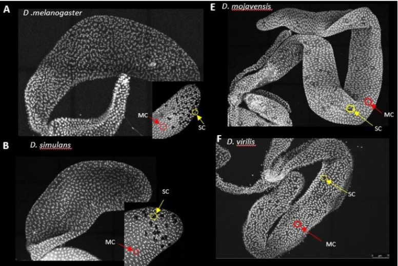
My project began with the exploration of other gastruloid studies in mice, zebrafish, and humans, to develop a method that could be used to replicate this in other animal models. We are able to use such a wide range of models to study gastrulation in humans because of the specific time point in development where embryos of many species share a biological resemblance to each other. The model I tested was the chicken embryo (Figure 1). The first few weeks of my time spent at the Crick involved learning how to dissect the chick embryo and learning how to identify the different ages of each embryo. This process involved practising how to use microscope techniques to harvest embryos.
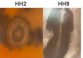
To achieve my end goal of generating a gastruloid, we decided to test Hamburger Hamilton stage 2 (HH2) and Hamburger Hamilton stage 9 (HH9) to provide us with two different stem cell pools. Cells from HH2 embryos have the capacity to differentiate into all cell types. However, the slightly older HH9 embryo has started to differentiate some of its cells but still provides us with the caudal lateral epiblast which we know to be responsible for spinal cord and somite formation driving body axis elongation. My experiments over the summer focused on comparing these two stem cell pools to determine which one is more effective for cell re-organisation following dissociation.
Figure 1. Examples of HH9 and HH2 using dye to view under the microscope.
We designed a protocol to first dissect HH2 embryos or the caudal epiblast from HH9 embryos. Then dissociate the two dissected tissues into a single cell suspension before adding them to a non-adherent 96-well plate.
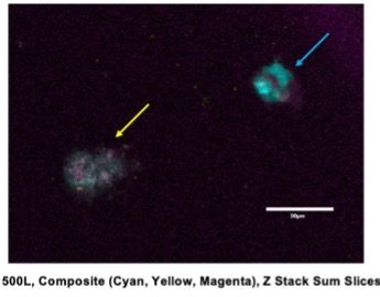
To determine both cell viability and whether organisation occurs within our aggregates, we stained the cells with primary and secondary antibodies for the presence of Brachyury and Sox2. Brachyury is a transcription factor expressed in the developing notochord and primitive streak, whereas Sox2 is a transcription factor required for neural development. We expected that if organization and elongation had occurred, Brachyury and Sox2 would be present in separate domains of the cell aggregate. However, of the various conditions I tested, none were effective in allowing for cell viability for 5 days post-embryo dissociation. This was evident by a lack of fluorescent staining with DAPI that would indicate the presence of DNA, overlapping Sox2 and Brachyury, and difficulty finding real cells that had aggregated and grown after fixing and staining (Figure 2). This raised a lot of questions about what changes needed to be made to make a chicken gastruloid.
Figure 2. Confocal Image of cells stained with primary and secondary antibodies (Yellow Arrow), Cloud of debris (Blue Arrow), Cluster of cells indicated by Cyan DAPI staining, indicating alive cells.
Overall, this summer has been a fantastic learning experience for myself. Being able to work with my supervisor Ashley Libby and learning from her has helped my confidence in the lab and my ability to understand scientific content. Working in the Briscoe Lab has been very enjoyable and has encouraged me to consider a career in research- something I wasn’t as interested in before this placement. For this reason, I would also like to thank the Francis Crick Institute for giving me the chance to carry out this project and also the MRF Rosa Beddington Fund for supporting my project.
As part of the Crick-Calleva program, I had the opportunity to work with Greg Slodkowicz in Margarida Cardoso-Moreira’s lab at the Francis Crick Institute over the past summer, studying the molecular mechanisms underpinning the evolution of the placenta.
The placenta is a temporary organ that facilitates the exchange of nutrients and gases between a mother and a developing fetus. Having emerged around 160 million years ago, the placenta has since diversified across many mammals, and has even arisen independently in other vertebrates, including some snakes and live-bearing fish. Along with its evolutionary diversity, the placenta presents astounding morphological diversity too, showing diverse auxiliary functions in different species. The marked functional diversity of the placenta, along with its recent evolutionary origin, make it a unique model for studying the genetic basis for evolutionary novelty. Over the course of my 9 weeks at the Crick, I used bioinformatic tools to evaluate gene family expansions across mammalian clades. I then triangulated this analysis with in-house expression data from the placenta and decidua, the part of the endometrium that undergoes pregnancy-specific modifications.
To begin, I took annotated genomes from Ensembl, breaking each protein-coding gene into Pfam domains, or domains of function. I arranged these genomes based on sequence order within the gene to create domain architectures that more accurately capture the function of genes. The end goal of this process was to cluster genes into functional families. Expansions in each of these families were detected by comparing gene copy numbers corresponding to these families between mammalian clades using pairwise statistical tests.
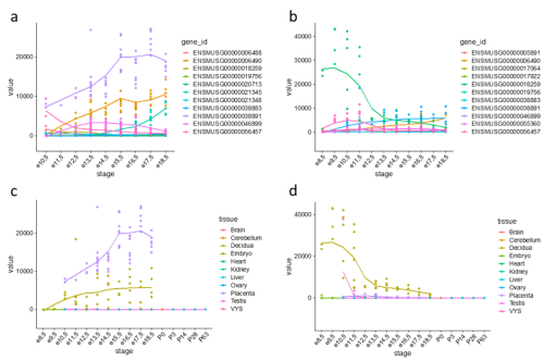
Figure 1: Expression trajectories of PRL (prolactins) in mouse. (a) Trajectories of 11 PRL genes in the placenta. (b) Trajectories of 11 PRL genes in the decidua. (c) Expression trajectory of Prl3b1 across all sampled tissues. (d) Expression trajectory of Prl8a2 across all sampled tissues.
One gene family expansion observed was that of rodent-specific prolactins. Prolactins in humans are released by the pituitary gland and are multifunctional throughout pregnancy, controlling key growth processes and lactation. On average, however, an additional 20 prolactin-domain-containing genes were observed in rodent species when compared to primates. Figures 1(a) and 1(b) highlight the expression trajectories of prolactins in the placenta and decidua in mice; clearly, prolactins are very highly expressed in both tissues (significant expression shown by values >1). However, the expression profiles differ slightly in that highly expressed prolactins in the placenta increase in expression through developmental time, while the inverse occurs in the decidua. Figures 1(c) and 1(d) show the most highly expressed genes in the placenta and decidua, respectively, shown in all tissues instead to highlight that this extreme expression is limited to the the placenta and decidua. Ben-Jonathan et al. (2008) reviews a plethora of evidence that suggests an important role of prolactins in rodents; for example, prolactin expression is key for downregulating interleukin expression and upregulating estrogen receptor expression, both required for fetus survival in rats. The role of the expansion of prolactins and their connection to a specific placental phenotype, however, remains unexplored.
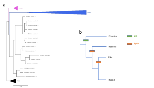
Figure 2: (a) Gene tree of Ly49 receptors across mammals, including main clades along with pig, horse, bison, sheep, and cow (abbreviated as PHACS). (b) Schematic of KIR/Ly49 expansion according to species tree.
Importantly, these pairwise statistical tests were unbiased in the sense that they included all significant gene family expansions, not only those pre-screened to be relevant to pregnancy. As such, for further analyses, I narrowed my field of search to revolve around genes already known to be relevant to placenta development and pregnancy, in this case natural killer cell receptors. Slodkowicz and Goldman (2020) showed that positive selection occurs more frequently in genes that carry immune functions. Additionally, uterine natural killer cells (uNKs) perform important functions throughout pregnancy, such as vascular remodelling, through MHC-interacting receptors. Though mammals share some uNK cell receptors, there are two main families of variable receptors: Ly49 receptors are expanded in rodents, while killer immunoglobulin-like receptors (KIR) are expanded in primates and other mammals (McQueen et al., 2002). The status of these receptors in lagomorphs, a group of mammals closely related to rodents that includes rabbits and hares, is not well studied. To detect expansions in lagomorphs, I used OrthoFinder (Emms and Kelly, 2019) to cluster genes by sequence, accounting for phylogenetic differences, screening for clusters of NK cell receptors. Figure 2(a) shows a phylogeny constructed of a cluster of Ly49 receptors; as expected, primates possess single copies of the receptor while rodents show a large expansion. However, two lagomorph species of pika show a late expansion of Ly49 receptors, while rabbits do not show the same. No compensatory expansion of KIRs was detected in rabbits either, an observation cross-referenced by searching the genomes of 3 closely related species (snowshoe hare, mountain hare, and brush rabbit). Instead, an expansion of leukocyte immunoglobulin-like receptors (LILRs) was detected. My project ended with testing de-novo genome annotation as a way of further elucidating what receptors rabbits and closely related species may be using.
Taken together, my time at the Crick generated data that provides various possible avenues for exploration, including investigating expansions that may underly interesting placental phenotypes, or better understanding immune genes during pregnancy in understudied species. Personally, I took home important skills in analyzing data, comparing methods, and substantially improved my working knowledge of programming languages like R and Python. I would like to thank my supervisor, Greg Slodkowicz, and Margarida Cardoso-Moreira for their friendly guidance throughout the project. I am extremely grateful to the Crick and the Rosa Beddington Fund for providing me with this unforgettable opportunity. I encourage all eligible students to apply for any future opportunities, as this has been an incredibly unique and gratifying experience.
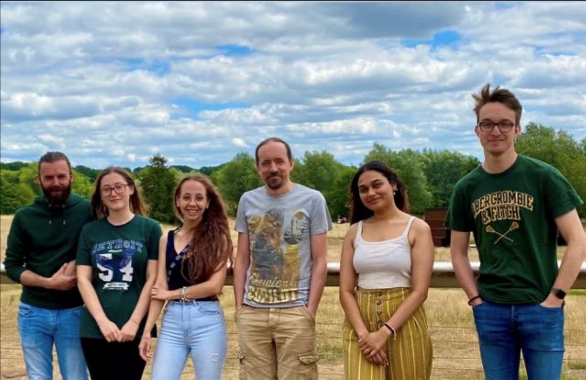
As a BSc Biomedicine Student at the University of East Anglia, I have had the pleasure of working in the Grocott Lab for eight weeks during my summer holidays. The Grocott Lab is part of the Norwich Developmental Biology and Stem Cell Network. It is researching the development of the eye, utilising chickens as a model organism. The team envisions the lab to work in three main areas: model organism work, computer modelling and human stem cell models. Figure 1 below shows the current Grocott Lab Team.
Figure 1: The team currently working in the Grocott Lab.
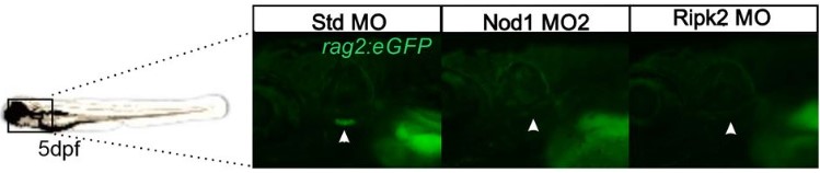
The title of my project was “Using Myc genes to search for optic vesicle progenitors”. Congenital malformations are a leading cause of infant death. Holoprosencephaly is a spectrum of related conditions in which the two sides of the brain are fused together. In extreme cases this results in a condition termed Cyclopia, where only one eye forms. The co-expression of c-Myc and N-Myc within the anterior neural folds may define paired growth fields that contribute to the outgrowth of optic vesicles. We hypothesised that the knockdown of one or both Myc genes in the anterior neural folds may result in impaired optic vesicle outgrowth and thus holoprosencephaly. This was inspired by reports that Myc genes regulate the growth of the neural retina from the optic cup lip at later stages of eye development.
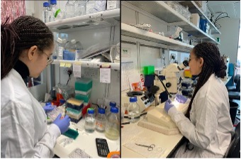
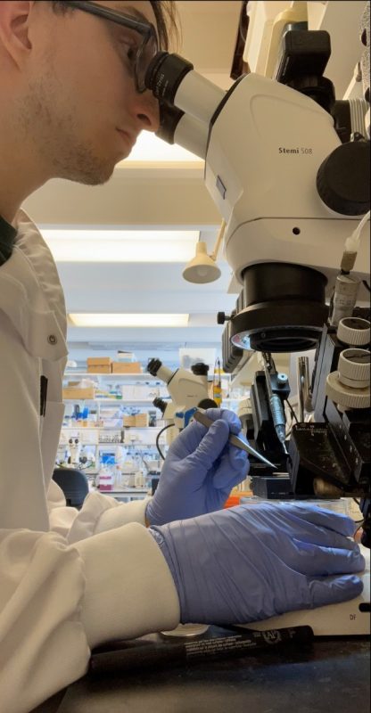
Using validated antisense morpholine oligonucleotides, I knocked down the N-Myc and C-Myc genes in developing chicken embryos to assess their impact on optic vesicle outgrowth and develop an understanding of the function of both of these genes. A typical day in the lab would include isolating embryos from eggs, preparing different chemical solutions needed to hydrate the chicken embryo, electroporation of the embryos with the morpholino (nucleic acid chains with a synthetic backbone, not recognised by cellular enzymes) mixed with DNA and incubating embryos overnight. I gathered a variety of skills during my eight weeks with the Grocott Lab. Under the close eye of Felicitas Ramirez (a postdoc in the lab and a wonderful mentor) I learnt techniques and skills necessary to culture a chicken embryo and master the art of electroporation. Electroporation is the process of introducing charged molecules, such as DNA or FITC labelled morpholinos into cells using a pulse of electricity to open pores in the cell membranes briefly.
Figure 2. Working at the microscope: electroporation.
The first hurdle was isolating the embryos from eggs and getting the correct stages of development. We found that many factors such as the humidity and temperature had an effect on the stage of growth we were isolating. Figure 2 shows me at my typical workspace, the microscope, electroporating the embryos that we had isolated in the morning.
Initially, I electroporated Hamilton & Hamburger stage 3-4 embryos and cultured these for approximately 12 hours. I expected embryos to grow to stage 10, in which the optic vesicles had formed, and I could observe a phenotype. After many rounds of electroporating and with a result showing no obvious phenotype we decided to change the technique and electroporate in ovo. We kept getting a negative result and saw nothing exciting happen to the chicken embryos. Several different methods of electroporation were attempted. The only technique we found to give promising results was electroporating older embryos (stage 7-8) using the traditional method.
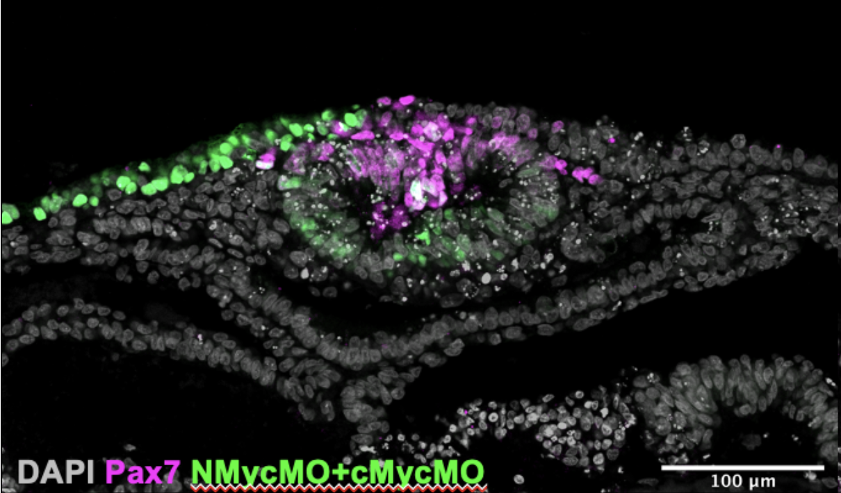
Once we had cultured the embryos and analysed them we further processed them for immuno- staining (another technique I learnt). This allowed us to look at the expression of four genes simultaneously and produced images like the one in Figure 3. We gained data that were interesting: The expression of Pax7 (a neural plate border marker) and the morpholino fluorescence was mutually exclusive. This confirms that Myc genes are important for the development of neural crest cell progenitors from the neural plate border.
Figure 3. A transverse section of the hindbrain of a stage 9/10 embryo stained for DAPI, Pax7 and N-Myc and C-Myc Morpholino.
From the results of our experiments we formed a new hypothesis: “The lack of obvious phenotype is because the electroporation is always mosaic and there are sufficient un-electroporated cells to compensate”. I am excited that I contributed to the project in a helpful way. Felicitas Ramirez is now continuing the project, examining the new hypothesis, and producing results which could lead to a Grocott Lab publication.
All of this would not have been possible without the support of the BSDB. I am very grateful for and appreciate the opportunity given to me. Additionally, I would like to thank Tim Grocott and his lab group for taking me in and allowing me to lend a helping hand in their research.
 (No Ratings Yet)
 (1 votes)