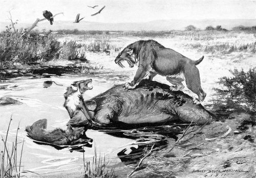
Smilodon californicus and Canis dirus fight over a Mammuthus columbi carcass in the La Brea Tar Pits. Public Domain, via Wikipedia
In the latest episode of Genetics Unzipped, Dr Kat Arney delves back into the ancient past, winding the clock back thousands of years to discover the stories of Denisovans and direwolves that researchers are now able to read in the fragments of DNA left in bones or even dirt.
One of the people who’s digging into the past through the use of ancient DNA to understand why a species might have vanished is Dr Kieren Mitchell from the University of Adelaide. While many may think that his species of choice – the direwolf – is fictional, they were definitely real, but the reasons they went extinct may come down to being too choosy about their meals and their mates.
Kat also speaks with Dr Benjamin Vernot – a researcher at the Max Planck Institute for Evolutionary Anthropology in Leipzig, Germany. Rather than studying bones, he’s been digging for DNA in more unlikely places in order to unearth the stories from our ancient ancestors.
If you enjoy the show, please do rate and review on Apple podcasts and help to spread the word on social media. And you can always send feedback and suggestions for future episodes and guests to podcast@geneticsunzipped.com Follow us on Twitter – @geneticsunzip
Welcome to our gallop through the preprints in developmental biology (and related subjects) published in July.
The preprints this month are hosted on bioRxiv, arXiv and preprints.org – use the links below to get to the section you want. Stand-out section for this month may be the Tools & Resources with some exciting new tools and methodologies.
PPARdelta signaling activation improves metabolic and contractile maturation of human pluripotent stem cell-derived cardiomyocytes Nadeera M. Wickramasinghe, David Sachs, Bhavana Shewale, David M. Gonzalez, Priyanka Dhanan-Krishnan, Denis Torre, Elizabeth LaMarca, Serena Raimo, Rafael Dariolli, Madhavika N. Serasinghe, Joshua Mayourian, Robert Sebra, Kristin Beaumont, Ravi Iyengar, Deborah L. French, Arne Hansen, Thomas Eschenhagen, Jerry E. Chipuk, Eric A. Sobie, Adam Jacobs, Schahram Akbarian, Harry Ischiropoulos, Avi Ma’ayan, Sander M. Houten, Kevin Costa, Nicole C. Dubois
The blood vasculature instructs lymphatics patterning in a SOX7 dependent manner Ivy Kim-Ni Chiang, Winnie Luu, Key Jiang, Nils Kirschnick, Mehdi Moustaqil, Tara Davidson, Emmanuelle Lesieur, Renae Skoczylas, Valerie Kouskoff, Jan Kazenwadel, Luis Arriola-Martinez, Emma Sierecki, Yann Gambin, Kari Alitalo, Friedmann Kiefer, Natasha L. Harvey, Mathias Francois
The Imprinted Igf2-Igf2r Axis is Critical for Matching Placental Microvasculature Expansion to Fetal Growth Ionel Sandovici, Aikaterini Georgopoulou, Vicente Pérez-García, Antonia Hufnagel, Jorge López-Tello, Brian Y.H. Lam, Samira N. Schiefer, Chelsea Gaudreau, Fátima Santos, Katharina Hoelle, Giles S.H. Yeo, Keith Burling, Moritz Reiterer, Abigail L. Fowden, Graham J. Burton, Cristina M. Branco, Amanda N. Sferruzzi-Perri, Miguel Constância
Retinal ganglion cell-specific genetic regulation in primary open angle glaucoma Maciej S. Daniszewski, Anne Senabouth, Helena H. Liang, Xikun Han, Grace E. Lidgerwood, Damián Hernández, Priyadharshini Sivakumaran, Jordan E. Clarke, Shiang Y. Lim, Jarmon G. Lees, Louise Rooney, Lerna Gulluyan, Emmanuelle Souzeau, Stuart L. Graham, Chia-Ling Chan, Uyen Nguyen, Nona Farbehi, Vikkitharan Gnanasambandapillai, Rachael A. McCloy, Linda Clarke, Lisa Kearns, David A Mackey, Jamie E. Craig, Stuart MacGregor, Joseph E. Powell, Alice Pébay, Alex W. Hewitt
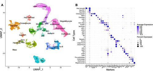
Liang, et al. use scRNAseq to identify the major cell types in the mouse liver
Downregulation of WT1 transcription factor gene expression is required to promote myocardial fate Ines J. Marques, Alexander Ernst, Prateek Arora, Andrej Vianin, Tanja Hetke, Andrés Sanz-Morejón, Uta Naumann, Adolfo Odriozola, Xavier Langa, Laura Andrés-Delgado, David Haberthür, Benoît Zuber, Carlos Torroja, Ruslan Hlushchuk, Marco Osterwalder, Filipa Simões, Christoph Englert, Nadia Mercader
Differential impact of a dyskeratosis congenita mutation in TPP1 on mouse hematopoiesis and germline Jacqueline V. Graniel, Kamlesh Bisht, Ann Friedman, James White, Eric Perkey, Ashley Vanderbeck, Alina Moroz, Léolène J. Carrington, Joshua D. Brandstadter, Frederick Allen, Adrienne Niederriter Shami, Peedikayil Thomas, Aniela Crayton, Mariel Manzor, Anna Mychalowych, Jennifer Chase, Saher S. Hammoud, Catherine E. Keegan, Ivan Maillard, Jayakrishnan Nandakumar
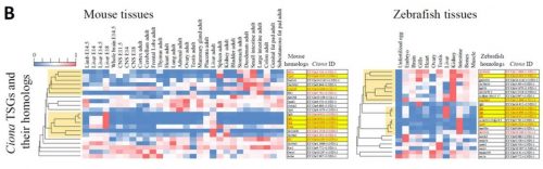
Evolution of a chordate-specific mechanism for myoblast fusion Haifeng Zhang, Renjie Shang, Kwantae Kim, Wei Zheng, Christopher J. Johnson, Lei Sun, Xiang Niu, Liang Liu, Theodore A. Uyeno, Jingqi Zhou, Lingshu Liu, Jimin Pei, Skye D. Fissette, Stephen A. Green, Sukhada P. Samudra, Junfei Wen, Jianli Zhang, Jonathan Eggenschwiler, Doug Menke, Marianne E. Bronner, Nick V. Grishin, Weiming Li, Kaixiong Ye, Yang Zhang, Alberto Stolfi, Pengpeng Bi
Comparing mice and zebrafish tissues from Matsubara, et al.
An open-access volume electron microscopy atlas of whole cells and tissues C. Shan Xu, Song Pang, Gleb Shtengel, Andreas Müller, Alex T. Ritter, Huxley K. Hoffman, Shin-ya Takemura, Zhiyuan Lu, H. Amalia Pasolli, Nirmala Iyer, Jeeyun Chung, Davis Bennett, Aubrey V. Weigel, Melanie Freeman, Schuyler B. van Engelenburg, Tobias C. Walther, Robert V. Farese Jr., Jennifer Lippincott-Schwartz, Ira Mellman, Michele Solimena, Harald F. Hess
A Comprehensive Overview of the Physical Health of the Adolescent Brain Cognitive Development Study (ABCD) Cohort at Baseline Clare E. Palmer, Chandni Sheth, Andrew T. Marshall, Shana Adise, Fiona C. Baker, Linda Chang, Duncan B. Clark, Rada K. Dagher, Gayathri J. Dowling, Marybel R. Gonzalez, Frank Haist, Megan M. Herting, Rebekah S. Huber, Terry L. Jernigan, Kimberly LeBlanc, Karen Lee, Krista M. Lisdahl, Gretchen Neigh, Megan W. Patterson, Perry Renshaw, Kyung E. Rhee, Susan Tapert, Wesley K. Thompson, Kristina Uban, Elizabeth R. Sowell, Deborah Yurgelun-Todd
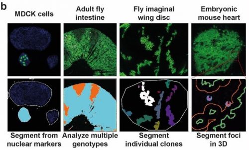
Spatial mapping of protein composition and tissue organization: a primer for multiplexed antibody-based imaging John W. Hickey, Elizabeth K. Neumann, Andrea J. Radtke, Jeannie M. Camarillo, Rebecca T. Beuschel, Alexandre Albanese, Elizabeth McDonough, Julia Hatler, Anne E. Wiblin, Jeremy Fisher, Josh Croteau, Eliza C. Small, Anup Sood, Richard M. Caprioli, R. Michael Angelo, Garry P. Nolan, Kwanghun Chung, Stephen M. Hewitt, Ronald N. Germain, Jeffrey M. Spraggins, Emma Lundberg, Michael P. Snyder, Neil L. Kelleher, Sinem K. Saka
My name is Helen Zenner and I am delighted to introduce myself as the new community manager of the Node. I started my scientific life as a cell biologist, specialising in membrane trafficking, but over the years I have found myself being drawn more and more towards developmental biology. Of course starting a postdoc in Daniel St Johnston’s lab really accelerated this progression, and although I spent most of the first two years claiming not to be a fly person, I have now fully embraced both the fly and developmental biology community!
As community manager at the Node I am excited to build on the excellent work of Aidan Maartens and the Development team. Fortunately Aidan has left me instructions on how to run the Node, and although it doesn’t have ‘Do Not Panic’ written in reassuring large letters, it looks to be an excellent guidebook and hopefully the change will be seamless. The transition has also been supported by Helen and Esperanza who run our sister sites, preLights and FocalPlane, so thanks to them as well.
Going forward, I hope to bring you some of the new features that you asked for in our recent community survey, as well as some additional ideas that we hope you will enjoy. But first and foremost, I would like to remind you that anyone within the community is welcome to post on the site. You can either post directly on the site once registered, or if you have any questions or would like support I would love to hear from you at thenode@biologists.com or helen.zenner@biologists.com
During vertebrate face development, bilateral streams of neural crest cells migrate from the neural tube to give rise to the facial prominences. A new study in Development combines high-resolution live imaging of chick facial development with a mathematical examination of cell behaviour to understand the dynamics of facial symmetry. We caught up with Adrian Danescu, Lisanne Rens and corresponding author Joy Richman (Professor and Director of the Pediatric Dentistry Graduate Program in the University of British Columbia in Vancouver, Canada) to find out more about the work.
Adrian, Lisanne and Joy (L to R)
Joy, can you give us your scientific biography and the questions your lab is trying to answer?
JR: I was originally trained as a dentist and then specialized in paediatric dentistry. It was while I was doing my specialty training that I first encountered the field of developmental biology. The tooth development project with Ed Kollar was so enjoyable that I went on to do a PhD in craniofacial development with Cheryl Tickle at UCL. She was an outstanding mentor and although your audience will be very familiar with her pioneering work on limb development, she also had three students that worked on the face and I was one of them. After that experience in London, the chicken was my main model organism until the last 15 years when I started also working on non-avian reptiles (lizards, snakes, turtles). My group has made discoveries concerning the molecular mechanisms of facial morphogenesis; however, all our previous work was done with static analysis. This is our first foray into time-lapse imaging to describe cellular behaviours in real time. We certainly did not anticipate the striking choreography of cell movements in the face way back when we started this project.
Adrian, how did you come to join Joy’s lab and what drives your research today?
AD: Like Joy, I also trained as a dentist, in Romania, and then came to Canada to take an MSc degree. I was interested in embryology and I decided to enrol in a PhD project in a lab that focused on craniofacial development – Joy’s lab at UBC was a perfect fit for me. My project involved facial birth defects, and their proper study required a suitable model. The lab was well versed in avian techniques and for me it was essential to learn quickly all the technical aspects necessary for the ongoing project. My entire PhD was a dynamic journey, with lots of opportunities for exciting projects and networking.
My main accomplishment in the lab was to develop a system to observe the movement of mesenchymal cells within the face during the early stages of development, prior to lip fusion, with high-resolution microscopy. This took several years of optimization and painstaking attention to subtle things such as finding a way to label individual cells. At the beginning of the project, I collaborated with an expert in lipid nanoparticles so I could deliver plasmids to the chicken face without electroporation. I now recognize the practical applications of these in vivo transfection methods after the implementation of the same technology to make COVID-19 mRNA vaccines. After gathering detailed tracking data, we started to collaborate with Leah Edelstein-Keshet and her postdoc Lisanne Rens from the department of Mathematics at UBC. They came at the data from a different angle and thanks to their insights the paper reached a deeper significance.
Lisanne, what is your research history and how did you come to be involved in this project?
LR: I am from the Netherlands, which is also where I obtained my PhD in mathematical and computational biology (from Leiden University). I typically use mathematical modelling techniques to describe processes in development, such as cell migration, angiogenesis, branching morphogenesis. At the start of this project, I was a postdoc at the mathematics department at UBC, working with my advisor Leah Edelstein-Keshet. I became involved in this project because Joy and her group were looking to quantify their data and understand it better. They got in contact with Leah, and I was very excited to be involved because I was already familiar with suitable methods, which I previously used to quantify simulated data. Just to be able to work with experimental biologists is a great opportunity. After our first meeting, Adrian sent me their data, and then it all started.
How has your research been affected by the COVID-19 pandemic?
AD & JR: I think we were fortunate enough that most of our experiments and analyses were completed before COVID-19 changed our way of working. There were only a few experiments left to finalize the paper and we planned them right after the restrictions were lifted. Communication via online platforms was convenient enough, especially for being able to bring other people from various places to participate in our discussions. However, the lab closure delayed the time we initially planned to finish the paper.
LR: For me, as a computational biologist, working at home is not as big of a deal compared to wet-lab biologists. However, like with any person, working from home comes with challenges. I find it lonelier, and it’s harder to keep being motivated. Also, with school closures, the kids are around.
What is the theory of developmental instability, and how does it relate to craniofacial abnormalities?
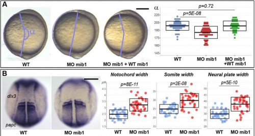
AD, LR & JR: Developmental instability refers to the range of fluctuations during development that can be usually compensated by various mechanisms to maintain normal development. There are both genetic and environmental factors at play, but our focus was to find evidence of instability at the cellular level. The theory is that all embryos will have some degree of developmental instability and, in the majority of cases, normal morphogenesis occurs. However, a slight increase in instability may be enough to lead to congenital malformations such as cleft lip. This instability is not only particular to the craniofacial area, but it may also concern all organs. One of the ways to measure developmental instability in other systems is to look at symmetry. Since we had dissected the midline of the face, we were able to compare the left versus right side. Through fruitful discussions with Lisanne and Leah Edelstein-Keshet, the idea to map the data back onto a grid was developed. In that way, we could compare directly each grid reference point in the equivalent anatomical location.
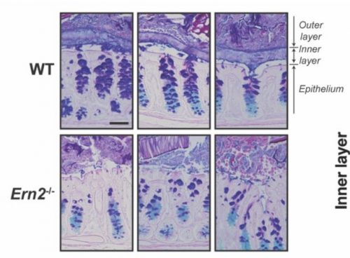
Composite figure of the key experimental data in the paper; for more details, see the full manuscript
Can you give us the key results of the paper in a paragraph?
AD, LR & JR: In this study, we wanted to understand the striking shape changes in early facial development prior to lip fusion. We turned to high-resolution live imaging in order to globally track hundreds of individual mesenchymal cells across the frontonasal mass. First, we discovered that mesenchymal cells moved; second, the movements alternated between states of order and disorder; third, clustering algorithms revealed that the movements are coordinated over large distances; and fourth, by interpolating the data we found symmetry that also fluctuates over time. We then used this interpolated data to map patterns of divergence and convergence that are again cyclical. We showed that all these cell behaviours are dependent on the actomyosin network. One of the most interesting observations made through mathematical modelling was the correlation between the switches in direction of movement with transitions from states of order to disorder.
What did the mathematical analysis of cell behaviours reveal that simple observation could not?
AD, LR & JR: First, the analysis confirmed some of the patterns we thought we were seeing. For instance, after quantification, the symmetry we noticed by eye became very clear, and the loss of symmetry was tremendous in the knockout tissue. We also noticed changes in the direction of the cells, but the pattern was not clear. This is where a divergence analysis helped. It revealed bands of convergence and divergence, and how they changed over time. Our velocity correlation analysis revealed the spatial distance across which cells are seemingly able to communicate. By clustering algorithms, we identified the spatial regions of coordinated motion, which was not possible to do by hand.
Furthermore, the order/disorder and the K-means clustering analyses revealed fluctuations of cell behaviour at a smaller scale, indicating developmental instability during normal midface development. Modelling also helped us discover the rapid switches of cell direction between divergence and convergence that happens within 20 minutes. The overall symmetry and periodicity during midface development were identified by the mathematical modelling as well.
The cell movements you observe are often symmetrical – what might explain this coordination of behaviour over such a long range?
AD, LR & JR: Several pathways, such as WNTs, BMPs, SHH, FGFs and so on, are at play during midface morphogenesis, and they may have a role in regional or more global coordination of cell movement. Furthermore, the forces generated through the extracellular matrix may contribute to these movements as well, due to cells being connected as a network. We identified one potential candidate to be WNT5A signal, with its expression overlapping the band of divergence and convergence that we identified. Our observations will pave the way for future investigations into the molecules that play an essential role in either buffering against instability or promoting the fluctuations. Ultimately, gene pathways associated with increased risk of clefting will be tested in this system.
When doing the research, did you have any particular result or eureka moment that has stuck with you?
AD: The first moment that stuck with me was observing how dissected faces grew and developed normally in a culture environment. I realized the advantage of this system, being suitable for direct observation under the microscope, in contrast with the side positioning that can be accessed in a developing embryo inside the egg. However, this was just the beginning of a long and laborious process to set up a series of methods to pursue my project.
LR: I was most surprised that we were able to link changes in divergence/convergence and coordinated/uncoordinated motion of the cells. Both of these quantities varied in time in a similar periodic fashion. This bears the question which one of those precedes the other, or how they are linked.
And what about the flipside: any moments of frustration or despair?
AD: There were countless moments of frustration, as always happens in research. Along the years, I learned to manage and embrace them as a way to grow. Learning the dissection techniques, optimizing the imaging setup for better clarity and stability of the culture during imaging, finding the right people to contribute to the project were all challenging, but what matters in the end is never, ever give up.
LR: For me, luckily not as many frustrations as with other projects I’ve done! I think the biggest challenge was how to interpret what the quantification of the data was telling us: what are the possible underlying mechanisms?
What next for you two after this paper?
AD: I graduated from both the PhD and the clinical program in Orthodontics last year. I have started to look for opportunities to find my way back into research. I intend to find the optimal way to work as a clinician and dedicate time for research. I expect this process to take longer as we are in the third wave of COVID-19 now in Canada, and there is still a lot of concern and uncertainty for the near future.
LR: I recently started a tenure track position at the TU Delft in my home country. I work in the mathematical physics group, continuing my line of research into mathematical modelling of cell and tissue biology.
Where will this story take the Richman lab?
JR: I would love to go further into the environmental influences that most affect developmental instability. I will expand the work to test specific pathways as mentioned above. In the current paper, we employed a global block on small GTPase signalling, but in the future we will be more specific.
I would love to go further into the environmental influences that most affect developmental instability.
Finally, let’s move outside the lab – what do you like to do in your spare time?
AD: I recently moved to Ontario after many great years in beautiful Vancouver. I generally try to allocate time for cooking, reading, travelling, doing outside activities, and spending time with my wife. I am learning that Ontario is fantastic for outdoor explorations with numerous lakes and parks. The summer is coming, and hopefully we will be able to enjoy a more normal summer after a tough year with so many lockdowns.
LR: Classical ballet is a big passion of mine, so during my time in Vancouver, I was practicing ballet downtown for two hours per week. Now in the Netherlands, I am back at my old ballet school. I also have two young kids who I spend most of my spare time with.
JR: I enjoy walking along the beach or in one of the many lovely urban forests near the UBC campus with my dog. I also am a passionate Masters swimmer and hope to return to club swimming soon. Right now, the pool is shut because of COVID-19. I also have really enjoyed spending lots of quality time with my two children in their twenties who came home to shelter during the pandemic. Oh yes, I have also kept my sour dough starter alive for a year now. Being a scientist helps!
Kat explores the world of transmissible tumours, looking at the history of contagious cancers in Tasmanian devils, dogs, clams and cannibal hamsters. Plus, the story of the man who caught cancer from his tapeworm.
If you enjoy the show, please do rate and review on Apple podcasts and help to spread the word on social media. And you can always send feedback and suggestions for future episodes and guests to podcast@geneticsunzipped.com Follow us on Twitter – @geneticsunzip
Where are you originally from, where do you work now, and what do you work on?
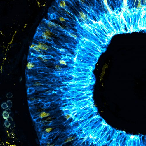
I am originally from Austria, where I also did my undergrad in biology. After that, I did a MSc in molecular biology in Austria and interned in various labs, including The European Molecular Biology Laboratory EMBL Heidelberg. For my PhD, I moved to Sheffield, to study cerebrovascular development. For my Postdoc, I am working at UCL, studying neurovascular unit formation in the zebrafish retina. To achieve this, I am combining experimental research with computational modelling to understand the biological processes that underpin development and disease.
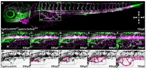
False-colour illustration of zebrafish retina neurons (blue) and glia progenitors (yellow) at 48 hours post fertilization, acquired with Zeiss AiryScan microscopy for her PostDoc. Image processing was conducted with Fiji software.
Has science always been an important part of your life?
I have always loved nature, plants, and animals. At home, we always had loads of books, stones, fossils, and other collections. A key element in my decision to go into STEM was my biology teacher in high school who loved his role and was a fantastic teacher. For his classes he used to bring in spiders, fish, and flowers for us to study. My love for STEM was reinforced over the years and it allowed me to learn many wonderful things, meet great people, and live in different countries.
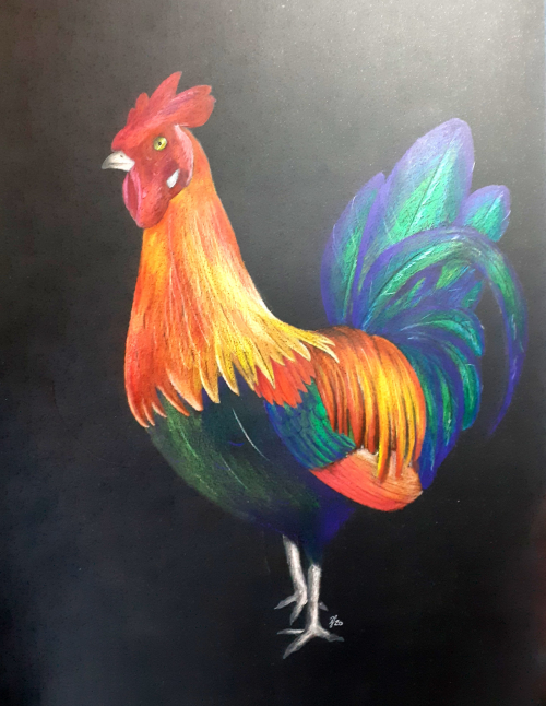
Realistic polychrome colour pencil study of a colourful rooster on black drawing paper. Similar to the parrot drawing, Elisabeth studied in this piece the effect of colour and contrast.
And what about art – have you always enjoyed drawing/painting/etc?
My mother would tell you that I was always covered in some form of paint or colour, before I could even stand! So yes, I always enjoyed experimenting with colours/art and I am extremely grateful that growing up my family embraced this and supported me.
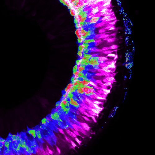
False-colour illustration of zebrafish retina neurons at 60 hours post fertilization, acquired with Zeiss AiryScan microscopy for her PostDoc. Image processing was conducted with Fiji software.
What or who are your artistic influences?
Nature is probably my biggest influence; be it through a microscope, camera, or most recently a telescope. To me there is nothing as inspiring as the natural things surrounding me.
“There is nothing as inspiring as the natural things surrounding me”
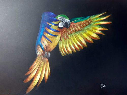
Realistic polychrome colour pencil study of a flying parrot on black drawing paper. This drawing is very much a study on natural movement as well as colours. Drawing with bold vibrant colours on black drawing paper allows particularly the study of colour contrast
How do you make your art?
For me, making art is the time where I do not have to be as rigorous as I am in my work life. So, I spend a lot of time experimenting rather than following one style/practice, e.g. currently I am exploring painting acrylic landscapes on canvas, drawing animals with colour pencils, and drawing monochrome objects with different perspectives. In 2020, I adopted a more structured approach, but I think the seemingly “unstructuredness” and chaos in itself is very enjoyable.
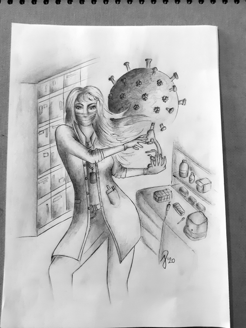
Pencil on drawing paper sketch portrait of a female scientist in the lab holding a microscope, with a Covid-19 virus in the back. The sketch illustrates the pivotal role of science in the Covid-19 pandemic.
Does your art influence your science at all, or are they separate worlds?
The experiments conducted in the lab are very data-driven with the need to adhere to protocols et cetera, so there is little room to be creative. However, communicating and presenting data is the other half of science; and for me this is where I can be creative. Working with images and microscopy data, the data themselves are highly inspirational to me.
Abstract false-colour polychrome colour pencil study of the head of a Scottish Highland cow on black drawing paper. The drawing was inspired by photography studies when walking in Yorkshire.
What are you thinking of working on next?
Currently I am working on a painting of the zebrafish brain vascular architecture colour-coded by depth and an autumn landscape on canvas. I constantly look at ways to improve my photography skills and just recently started to explore astrophotography.
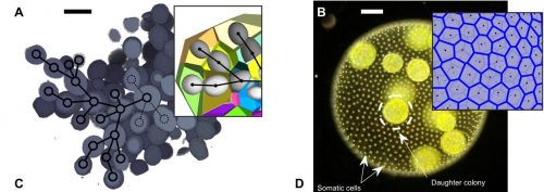
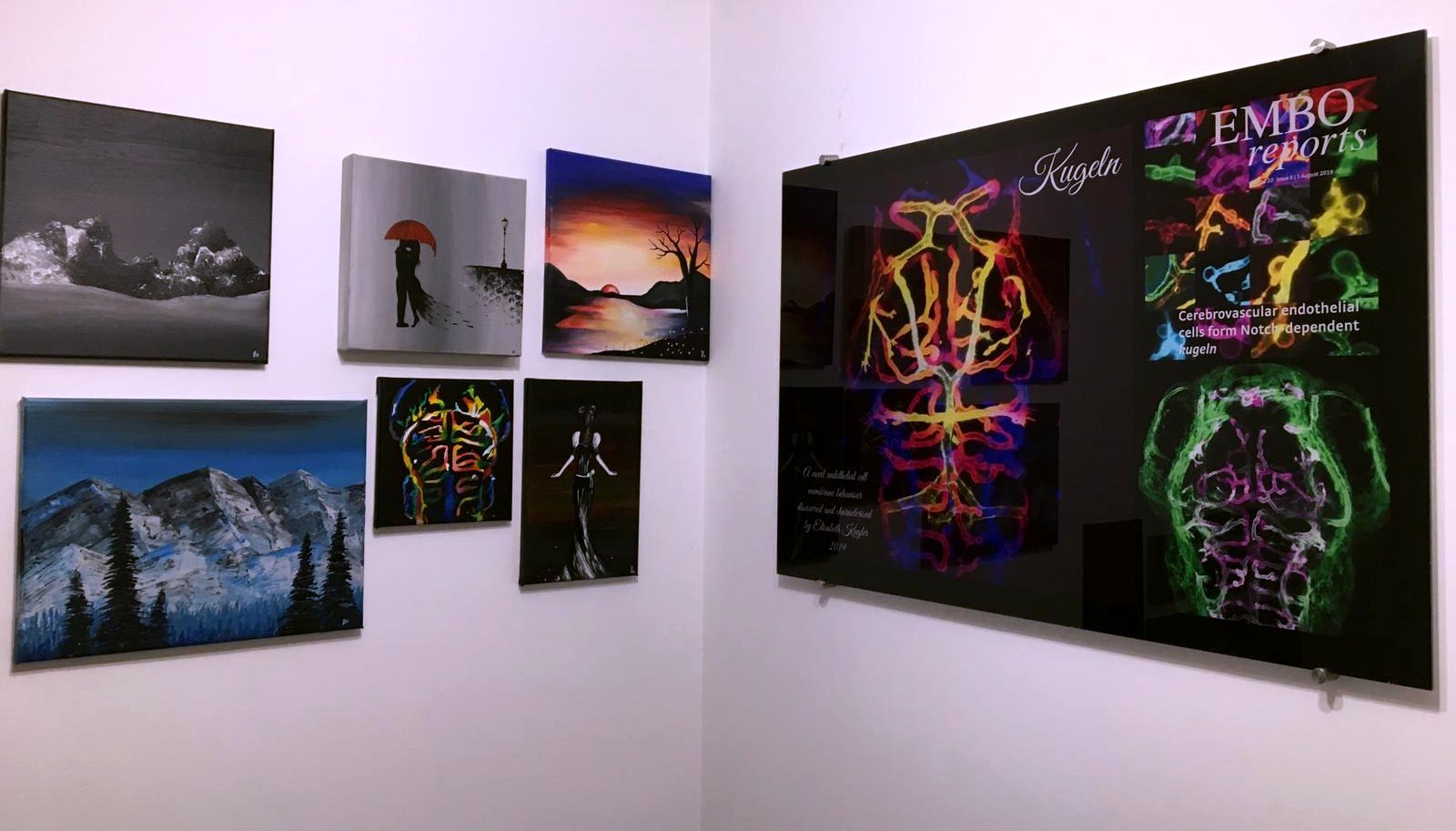
Part of Elisabeth’s personal gallery, showcasing art and science next to each other. Left, poly- and monochrome acrylic paintings on canvas. Right, collage of LSFM pictures showing the endothelial cell membrane behaviour kugeln, acquired during her PhD.
We’re looking for new people to feature in this series throughout the year – whatever kind of art you do, from sculpture to embroidery to music to drawing, if you want to share it with the community just email thenode@biologists.com (nominations are also welcome!).
Epithelial tubes perform crucial functions in various organs, providing routes for the transport of fluids and gases. A new paper in Development addresses the question of how epithelial tubes elongate during development, using a combination of mouse organ culture and mathematical modelling. To find out more about the work, we met four of its authors: PhD students Lisa Conrad and Steven Runser, senior scientist Roman Vetter, and their supervisor Dagmar Iber, Professor in the Department of Biosystems Science and Engineering at ETH Zurich.
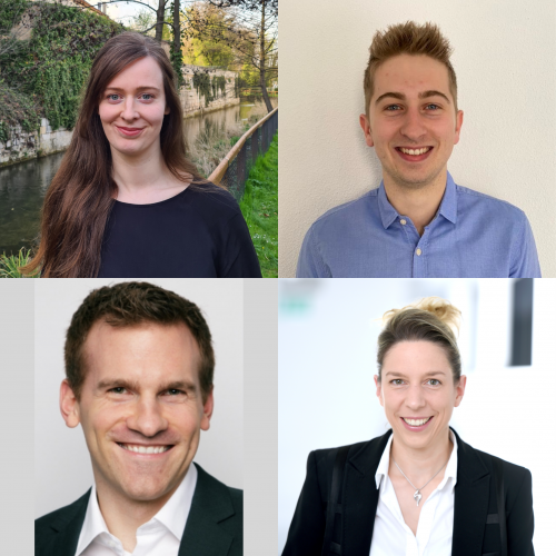
Lisa (top left), Steve (top right), Roman (bottom left) and Dagmar (bottom right).
Dagmar, can you give us your scientific biography and the questions your lab is trying to answer?
DI: I studied mathematics and biology, and did Masters degrees and PhDs in Cambridge and Oxford in each field. Since the very start, I have been interested in using mathematical modelling to uncover biological mechanisms. A Junior Research Fellowship at St. John’s College in Oxford gave me the freedom to pursue my own ideas and develop more precise, data-based models than the established conceptual models. Initially, I worked in immunology, but then switched to cell differentiation in bacteria, as the required quantitative data to build and validate models was available only for such simple organisms. These days, my group focuses on developmental mechanisms all the way up to human, and although we mostly collaborate with experimental groups, ETH allows me to also run a small wet lab to generate data and test ideas. The lab largely focuses on mouse lung and kidney development, but we maintain a rather broad interest in fundamental patterning mechanisms, and also collaborate with clinicians.
Roman, can you tell us a little about your research history and how you ended up working on developmental questions?
RV: I’m a computational physicist by training, and did my studies and PhD at ETH Zurich. With a few exceptions, my interest has always been in explaining nature’s wealth of emergent complex behaviour from simple, fundamental principles. During the earlier stages of my academic career, I found these problems mainly in the physical disciplines – from snow metamorphism to filament packing to crumpled shells and even fuel cell simulations. If you go through life with an open mind, open eyes and open ears, there’s an interesting question calling for a quantitative explanation virtually everywhere. My attention is drawn easiest by questions that combine geometrical shapes with mechanics and patterning. Lately, I have found such inspiration more and more in biology, where fundamental aspects of morphogenesis and development require taking new vantage points to advance further. I joined Dagmar’s group 2 years ago when she was looking for a senior biophysics modeller and I was looking to enter the field of computational biology to find new puzzles to solve – it was an instant match. I started as a postdoctoral researcher and recently became a senior scientist and lecturer in her group.
Lisa and Steve – how did you come to work in Dagmar’s lab and what drives your research today?
LC: I studied biology at the University of Freiburg and molecular medicine at Uppsala University. Developmental biology has fascinated me from the beginning; besides the interesting questions and methods in this field, the beauty of development is just so captivating! During a variety of lab projects, I noticed how much I enjoy projects that bring together expertise from different scientific backgrounds. By applying to the Life Science Zurich Graduate School, I found out about Dagmar’s group and got curious about their multidisciplinary approach to developmental biology. I started as the first PhD student with an experimental focus in the group’s then recently opened wet lab, eager to build on my experimental and research skills, while learning about new ways on how to tackle developmental questions from a different angle. It has been challenging to keep up with the aspects of the group’s research that are far from what I studied, but it’s also immensely rewarding when we can join forces to find new ways to better understand organ morphogenesis!
SR: I started my studies in cellular and molecular biology at the University of Strasbourg, but I quickly deviated towards more computational fields. I have always been interested in the design and application of mechanical simulations for the study of biological systems. To respond to this interest and to learn more about numerical approaches, I applied to Dagmar’s lab to do my master’s thesis. After completion of the thesis a little less than 2 years ago, Dagmar offered me the opportunity to continue in this field as a doctoral student. Since then, I have been developing and using different types of simulation models to study organ growth and morphogenesis.
How did you come to study tube elongation?
DI: The group had long been interested in lung and kidney branching morphogenesis. Although our ligand-receptor-based Turing mechanism could nicely predict where new branch points would form during lung and kidney branching morphogenesis, we noticed that the branch shapes that emerged in our simulations looked nothing like in the embryo because we were missing that bias in outgrowth that lets epithelial tubes of embryonic lungs and kidneys lengthen more than they widen.
Before this project, what mechanisms had been proposed for biased tube elongation?
DI: In lungs and kidneys, chemotactic movement towards a source of FGF10 or GDNF had long been noticed, and the extracellular matrix is thinner at the bud tips. In mammary glands, a constricting force had been proposed. In plants, hoop stress is a popular theory to explain their biased growth. We tested all those ideas, but none could explain the biased outgrowth of embryonic lung or kidney tubules. In fact, when checking the potential of hoop stress, we noticed that the lumen of the tubes is mostly very narrow, whereas the wall, the epithelium, is comparably thick. Although this is inconsistent with a role of hoop stress, it gave us the idea that fluid flow-induced shear stress may play a role.
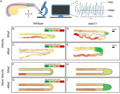
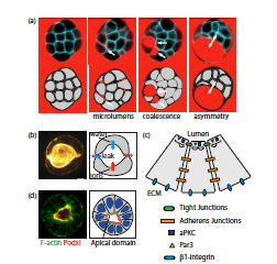
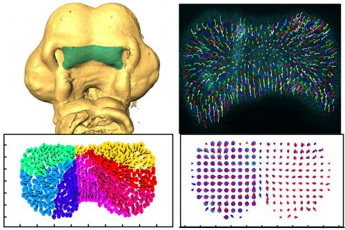
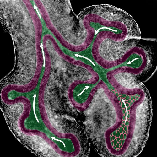
Fluid flow (arrows) from tip to base in the lumen (green) of developing lungs causes shear stress levels strong enough to be sensed by epithelial cells (magenta), giving them a direction in which to preferentially grow. This discovery provides a new explanation for the stereotypical directional bias in tube outgrowth observed during the development of branched organs such as lungs and kidneys.
Can you give us the key results of the paper in a paragraph?
SR: The key result of the paper is that the observed bias in epithelial tube outgrowth, the accompanying bias in the apical cell shape and the resulting biased orientation of cell divisions, can be explained with fluid-flow driven shear stress. After having ruled out all the previously proposed mechanisms, Roman used a Finite Element method to demonstrate that the collapse of the tubes in itself was very unlikely to result in a bias in outgrowth. Instead, the narrow luminal region meant that a fluid flow in the epithelial tubes might cause a significant level of shear stress on the cell walls. To prove the existence of a flow, Lisa injected micro-beads in the lung lumens and observed their movements over time. We simulated the effect that a flow with the measured velocity would have on the cell walls of a similar lung tube geometry. The shear stress levels thus calculated were well within the range of what epithelial cells can sense. Shear stress is well known to result in the elongation of endothelial cells along the flow direction in blood vessels. I used a cell-based vertex model to investigate the impact of such an elongation on epithelial tissues. Once parameterized based on quantitative data, the model was able to recapitulate all the measured features of the lung and kidney tube epithelium. The bias in cell division orientations was well in line with what had been measured, and similarly the bias in outgrowth of the tissue matched with the experimental observations.
How do you think lung and kidney cells sense shear stress, and how is this sensing translated into biased growth at the tissue level?
DI: Epithelial cells can sense shear stress with their cilium. How they translate this into a change in cell shape, and how the extent of the cell shape change relates to the shear stress level is not known. Microfluidic experiments may help to resolve this.
Do you have any ideas for what causes tube collapse in early development?
RV: Indeed we do have some ideas, and we’re working toward testing possible theoretical explanations with detailed computer simulations by Steve, and toward validating them with experimental data from our wet lab, together with Lisa and other group members. Tube shape and collapse is an exciting topic of ongoing research in our group, and we’re looking forward to telling you more once we have conclusive answers.
When doing the research, did you have any particular result or eureka moment that has stuck with you?
LC, SR: The project really picked up momentum when Harold Gómez, who is also shared first author, noticed, through careful examination of his beautiful lightsheet microscopy data, that the lumen is very narrow in many parts of the lung. But the most exciting moment was when we simulated the shear stress produced by the fluid flow, which we had finally succeeded in measuring experimentally, and realized that it was within the range cells can sense.
The most exciting moment was when we simulated the shear stress produced by the fluid flow.
And what about the flipside: any moments of frustration or despair?
DI: For me, certainly the first response by the referees. For years, I had asked experimental colleagues how to measure fluid flow in lung or kidney tubules and discussed strategies with my team. Yet, no one in my group felt they could pull this off. So, I decided to send the paper to Development, hoping that it would inspire some experimental lab to do that last experiment. After all, through a combination of (simple) experiments and simulations (that encompassed many different sophisticated techniques) we had excluded all previous proposals, suggested a new one, and provided convincing evidence for it. Yet, the referees would not have it. They not only insisted on that last experiment, but also saw little value in the paper as it was. I have seen it more than once that junior members got driven away from science by referee reports that had failed to recognise the value of their work. But not so Lisa: realising that those fluid-flow measurements were the make-or-break, she decided to just give it a try – despite all COVID restrictions. She remembered that Renato Paro had left an injector to our department when retiring, which no one used. She got it to work with the help of his former technician – and demonstrated fluid flow at the required level in the developing lungs.
LC: For me, the biggest source of frustration is when an experiment fails due to technical issues and the tissue samples that I harvested from an animal go to waste. Regarding the referee reports, Dagmar encouraged us to take on the challenge, and celebrating the small successes along the way kept spirits up! In the end, confirming our proposed mechanism was a really good experience.
How has your research been affected by the COVID-19 pandemic?
LC: Last year in March, ETH went into lockdown to help slow down the spread of the virus and our lab was closed for about 6 weeks. The reopening was done in several phases, so we had to work in shifts for a while and it took some time to start up experiments again. Working in shared facilities is still restricted, but most of the time we can figure that out by communicating with colleagues and working around each other’s schedules. In general, everything needs a bit more planning now. For the paper revisions, we had to set up a new experiment in the lab while COVID-19 restrictions were still in place, which of course provided an extra challenge. Having a colleague take a look at your (failed) experiment usually speeds up optimization and trouble-shooting and there are fewer opportunities for spontaneous exchange with other groups. Despite the additional work and stress brought by the pandemic, I feel like everyone at BSSE is supporting each other; especially Makiko Seimiya and Tom Lummen (BSSE Single Cell Facility), who have been awesome in sharing their advice and equipment, and helping me with many tries at the spinning disk confocal, which was a new microscopy system for me.
DI, RV, SR: As theoreticians, we have been forced into a home office for more than a year now. We miss the personal interactions, but remote work is otherwise straightforward for us.
What next for you two after this paper?
LC: I am currently finishing up a project that compares branching morphogenesis in lungs and kidneys. In a collaboration with Roman and other team members, we’re also looking at tube formation, where I’m using nephrogenesis in kidney organoids as a model.
SR: The 2D vertex model used in this paper offers many possibilities for the study of tightly packed cell sheets. However, numerous developmental events can only correctly be represented in three dimensions. With the advent of high performance computing, new computational frameworks representing cells more realistically in 3D can now be developed. I am currently developing such a model specifically designed for the simulation of epithelial tissues.
Where will this story take the Iber lab?
DI: The mechanism that defines the aspect ratio of tubes is one important puzzle piece to explain how complex organs are shaped and how organ-specific differences arise. There are many other fundamental questions concerning tubulogenesis, epithelial organisation, the physics of budding, the role of the mesenchyme and the final reorganisation of the lung epithelial tree into a fractal-like architecture. As a group, we are interested more generally in self-organisation during development.
Finally, let’s move outside the lab – what do you like to do in your spare time in Basel?
LC: I picked up diving a few years back and it’s so much fun! For a local spot, I would recommend the Bodensee; it’s really amazing to dive into a whole new world ‘at home’, now that travelling hasn’t really been an option. Basel has lots of relaxing greenery and I often go for walks along the Rhine. Since the start of the pandemic, I have also rediscovered the fun in crafting and caring for plants on my balcony.
SR: I enjoy a lot of different activities, which range from reading historical novels to playing football.
RV: Spare time comes and goes in phases. There are more things I’d enjoy doing than I could possibly fit into a full day – cycling across the country is one of many.
DI: I enjoy the Swiss mountains, swimming in the Rhine, the tennis court in front of my house – and the preparation for SoLa, the yearly running relay race, which we participate in with the entire group.
Hello all! I am Viktoria, the Sustainable Conferencing Officer of The Company of Biologists. I am here to quickly answer some questions about our Sustainable Conferencing Initiative.
What is the aim of the Initiative?
As biologists become increasingly aware of their environmental and social impact, questions about the environmental sustainability and social responsibility of our events have become more prominent. The Company of Biologists launched the Sustainable Conferencing Initiative in October 2020 to provide guidance and support on the sustainability of events. Furthermore, acknowledging the changes that the pandemic has brought on virtual communications, we are also here to offer insights into technologies that can be used for virtual and hybrid events. Our main aim is to facilitate the discussion of sustainability issues and innovative technologies in our community.
How will you share information?
We have our own website! You can visit sustainability.biologists and browse through our different pages. There is a Blog page where our team publishes pieces of information based on the questions coming from our community, and a Resources page that lists useful links regarding sustainability and innovative technologies. Also, in June our Forum became live. Its purpose is to offer a space to our community where ideas, best practices and questions can be shared, and we can all learn from and help each other.
Is there another form of support available?
Sustainable Conferencing Grants are available to fund innovative ideas that enable biologists to collaborate productively while minimising their impact on the environment. We accept applications from organisers of meetings, workshops, conferences, seminars, training events, and a wide range of activities in the fields covered by our journals. The events can be in-person, virtual or hybrid. Moreover, applicants for Scientific Meeting Grants may be awarded an additional £1,000 if they can demonstrate efforts have been made to reduce the environmental footprint of their event.
How can anyone join the discussion?
Visit our site to explore the information we share. If you register for an account, you will automatically gain access to our Forum where you can ask questions, offer advice, and engage with the community on sustainability and new technologies issues. You can also join our mailing list to receive our newsletters (we promise no spam emails).
Furthermore, you can find us on Twitter as @COB_Sustainable Come join us there and share ideas or questions by using our hashtag #Sustainable_Conferencing. Of course, you can always contact us via email at sustainability@biologists.com.
We are looking forward to connecting with our community and starting the discussion!
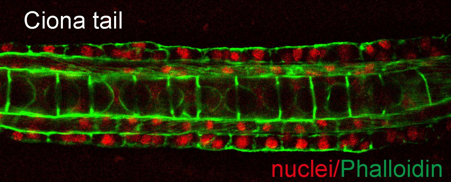
Over on The Company of Biologists WeChat channel, we’re enjoying getting to know the growing community. We recently published an interview with Dr Zhiyi Lv, a member of Professor Bo Dong’s group at Ocean University China in Qingdao, China. The Dong lab was established in 2014 and is interested in uncovering the cellular, mechanical and biochemical signalling networks that interact to drive the diverse morphogenetic processes during organ formation and tissue regression using marine ascidians and flies as models. You can find out more about Bo’s research in a ‘The people behind the papers’ interview published in Development last year and a recent Development presents… talk by Hongzhe Peng, a doctoral student in the lab.
Having gained his undergraduate and Master degrees at Northwestern A&F University in China, Dr Lv moved to Germany to obtain his PhD at the University of Göttingen. He remained there for his first postdoc position before relocating back to China to join Professor Bo Dong’s lab at Ocean University of China. Here, he tells us more about his scientific journey, including why he finds the dev bio field amazing and how labs differ between Europe and China.
When did you first become interested in science?
I don’t think there was a specific timepoint that where I thought, ‘aha, now I am interested in science!’ All kids are curious about the unknown world and they are always trying to explore the surroundings. In this way, scientists and children have a lot in common. Fortunately, I did not lose this curiosity as I grew up.
What attracted you to the field of developmental biology?
How a simple-structured (relatively) fertilised egg becomes a complex adult with head and legs attracted so many people since Aristotle’s time. Then, people realised that genes controlled the development. Now, increasing evidence suggests that mechanical forces contribute morphogenesis actively. It is amazing, isn’t it?
You gained your PhD at the University of Göttingen in Germany and stayed in the same lab for your postdoc but switched from biochemistry to biophysics of morphogenesis – can you tell us more?
I got my PhD under the supervision of Professor Grosshans. I worked on the regulatory mechanism of actin polymerisation. At that time, we identified that a F-BAR protein, Cip4, inhibits actin polymerisation by inactivating Diaphanous, which is an actin nucleator. We got very exciting data, which was published in Journal of Cell Science. Biophysics of embryogenesis has been an important topic in the Grosshans lab. I was impressed by my biophysical colleagues’ talks during our seminars. Professor Grosshans was very nice and always encouraged us to explore a new area. Two projects were running in the lab. One was mechano-transduction at cell-cell contact, and the other one was nuclear array self-organisation in a Drosophila syncytial embryo. I chose the second one for my postdoctoral project.
What are the differences in the lab between Europe and China?
The biggest difference is that experienced postdoc researchers are the main power in the biological labs in Europe. However, most bench work is done by the master and doctoral students. In this case, we need more time and effort to train the students.
Also, some labs in Europe are quite small – one PI tends to lead several postdocs and PhD students, although there are also big labs in Germany. In China, most labs, especially productive labs, are large!
How did you come to work in Professor Dong’s lab?
I met Professor Dong when we were in an EMBO symposium in Heidelberg, Germany in 2018. I was attracted by his work and also by his personality. We share similar scientific interests, and he asked whether I was willing to join to his group. Why not? It was a spontaneous decision.
You have been back in China for a couple of years now – what was it like coming home?
I have experienced ‘reverse culture shock’! For example, when we go to dinner with friends or colleagues, we do not split the bill in China. The leader or the senior person pays for all. As time passes, I will get used to Chinese culture again.
What question is your research currently trying to answer? The origin and the regulation of forces driving morphogenesis, and the crosstalk between genetic cascade and mechanical forces.
What are the main advantages and drawbacks of the model systems you work with, Drosophila and Ciona?
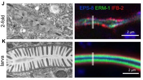
Some students in the lab often misunderstand that Drosophila is a user-friendly model compared to Ciona. The reason behind this might be that Drosophila is easier for genetic manipulation. But in my opinion, this is totally wrong – CRIPSR/Cas9 can also generate the mutant we want in Ciona. I think the advantage of Drosophila is that you can keep the stocks in the lab and you can do experiments whenever you want. The drawback of this model is that you have to take care the animals frequently. We need to collect Ciona from sea. So, the material is limited during early spring and late winter. We need to set up the inbred line in the lab. This is what we are currently doing. Ciona embryos and its larvae are smaller than Drosophila, which is a big advantage for imaging.
What is next for you?
I plan to focus more on Ciona embryogenesis research, and hope to involve myself in the Ciona community more actively.
Follow us on WeChat for exclusive interviews and research highlights written by the community, as well as useful resources to help navigate the publishing process.
Find out more about the Company’s efforts to engage with Chinese researchers
As the vertebrate body axis extends, HOX genes are sequentially activated in axial progenitors to specify their identity. A new paper in Development addresses what regulates the tempo of this HOX expression in human progenitors. To hear more about the story, we caught up with the paper’s two first authors, Vincent Mouilleau and Célia Vaslin, and their supervisor Stéphane Nedelec, Group Leader at the Institut du Fer à Moulin in Paris.
Vincent, Célia and Stéphane (L to R)
Stéphane, can you give us your scientific biography and the questions your lab is trying to answer?
SN: I studied Biology in Rennes and then Paris, where I did my PhD in the Department of Biology of the Ecole Normale Supérieure. I had the chance to work in the group of Alain Prochiantz under the supervision of Alain Trembleau, where I studied local protein synthesis in neurons. To follow up on this research I then joined Hynek Wichterle, who had recently started his lab at Columbia University. Hynek is a pioneer of in vitro differentiation of pluripotent stem cells (PSCs) to study development and diseases. It was very exciting to work in this environment in the early days of the field with great colleagues and so many things to explore, and this time at Columbia had a profound impact on my scientific career since I was still working on spinal cord development using in vitro approaches. I then moved back to France, working again in a very stimulating environment with Cécile Martinat at the I-STEM institute in Evry. There, I started projects aiming at studying human developmental biology using human PSCs (hPSCs). We developed a powerful approach to assess how extrinsic cues control cell fate and discovered pathways sufficient to convert hPSCs into distinct neuronal subtypes, including spinal motor neurons (MNs). Building on this work, I started a new lab in Paris, at the Institut du Fer à Moulin: a very dynamic and collaborative Neuroscience institute. The current story was largely developed there in collaboration with both the Hynek and Cécile groups, which was very satisfying.
We currently use spinal cord development as a model system to address two interrelated questions. First, the mechanisms by which a limited number of extrinsic factors control human spinal neuronal diversity and morphogenesis – in vitro differentiation of hPSCs is a unique model to approach this question. Second, the mechanisms by which mutations in ubiquitous genes perturb developmental programs to impair selective neuronal populations and cause MN diseases – here we take advantage of developmental studies to improve cell and tissue engineering.
Vincent and Célia – how did you come to work in Stéphane’s lab and what drives your research today?
VM: During my bachelor’s degree in Nantes I became fascinated by stem cells and neurodevelopment. I thus decided to join a Master’s program in Paris focusing on these two topics. The possibility of generating, in vitro, a specific subtype of human cells with the right ‘recipes’ fascinated me, and for my internship I joined the I-STEM institute and Stéphane’s lab. I then moved on to do a PhD, and helped Stéphane set up the new lab in Paris while continuing working between the two institutes. It was an intense but enriching experience.
CV: During my undergraduate studies at Sorbonne Université, I quickly became interested in developmental biology and neuroscience. During a first internship in the lab of Jean Livet in Paris, I studied neural lineages in the chick embryo spinal cord, which confirmed my interest in these two fields. This led me to join Stéphane’s lab, first as an intern and then a PhD student, to investigate molecular mechanisms controlling spinal cord development. The power of the in vitro approaches used in the lab allowed me to decipher signalling mechanisms controlling spinal neural diversification – a subject that fascinates me.
How has your research been affected by the COVID-19 pandemic?
VM, CV & SN: Our lab was completely shut down for 2 months last spring. Afterwards, we worked part-time on site to limit the number of people, which obviously significantly delayed the projects. However, working mostly with in vitro models helped, as it was easier to stop and restart the experiments. Also, the lockdown forced us to focus on writing and planning experiments, which was a positive side effect. Overall, this pandemic has most certainly delayed the progress of our research, but we were fortunate to return to the lab fairly quickly and be able to work in good conditions thanks to the heads of our institute, who did a fantastic job in dealing with the situation.
Before this project, what was understood about the relative influence of extrinsic and intrinsic factors on the pacing of the human HOX clock?
VM, CV & SN: We already knew a lot about the extrinsic and intrinsic mechanisms controlling the sequential induction of HOX genes during axial elongation. However, several aspects remain obscure; notably, the mechanisms pacing the clock within axial progenitors, in particular in humans. It was well established that cis-regulatory sequences within and outside the complexes are important for HOX gene sequential induction, and that progressive changes in chromatin structure along the complexes accompany the progression of the HOX clock. On the other hand, extrinsic factors such as retinoic acid (RA), Wnts, FGFs and GDF11 were shown to induce HOX gene collinear expression or modulate HOX gene expression patterns. However, whether these extrinsic factors were pacing the sequential activation within axial progenitors or were actuating an intrinsic timer was unclear.
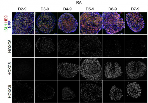
Effects of modulating the duration of retinoic acid exposure on HOX gene expression during in vitro motor neuron differentiation.
Can you give us the key results of the paper in a paragraph?
VM, CV & SN: We first characterized the expression profile of HOX transcription factors and MN subtype markers in the human embryonic spinal cord, so we could assess the functional consequences of HOX regulation in axial progenitors and properly define the identity of in vitro-generated human MNs. Using MN subtype as a readout, as well as transcriptional analysis of the axial progenitor stages, we showed that HOX genes undergo a temporal collinear activation in hPSC-derived axial progenitors that, upon differentiation, generate MN subtypes found in progressively more caudal regions of the spinal cord. Analysis of the transcriptomic data showed that the sequential activation of HOX genes was paralleled by an increase in FGF ligands and markers of active FGF signalling. This FGF activity was necessary for the HOX clock to proceed, and precociously increasing FGF levels hastened the expression of HOX genes expressed normally later on. The HOX clock was further accelerated with a rapid rise of the very caudal HOX10 genes when FGF was combined with GDF11, another extrinsic factor known to control the expression pattern of caudal thoracic and lumbar HOX genes in mouse and chick embryos. Slowing down or accelerating the clock in axial progenitors was always paralleled by a shift in MN subtype specification within the same time line of differentiation. These results demonstrated that the pace of HOX gene activation within axial progenitors is regulated by sequences of extrinsic factors. This observation argues against a solely intrinsic, chromatin-based, pacing mechanism. However, even in the most accelerating/caudalizing conditions, HOX genes are still expressed in a largely collinear sequence, which suggests that cell-intrinsic mechanisms likely ensure the order of expression. In addition, our work provides for the first time a method to efficiently generate well-defined MN subtypes for basic and translation approaches.
The pace of HOX gene activation within axial progenitors is regulated by sequences of extrinsic factors
Do you have any idea what controls the onset and duration of FGF signalling in hPSC cultures and in the embryo?
VM, CV & SN: The onset and duration of FGF signalling in axial progenitors are certainly controlled by extrinsic factors both in vivo and in vitro. Work from different labs has indicated that FGF and Wnt signals, provided in vivo by the primitive streak and the surrounding epiblast, and in vitro by addition of agonists in the medium, specify axial progenitors, which in turn induce different FGF and Wnt ligands. Thereby, a positive-feedback loop is generated, which likely contributes to an increase in FGF signalling overtime. Accordingly, in this study we observed a temporal induction of FGF ligands and of well-recognized downstream target genes in hPSC-derived axial progenitors. Then, in the neuronal lineage, the duration of FGF signalling in axial progenitors depends on the rate of their differentiation in neural progenitors. In vivo, neurogenesis-promoting RA from the abutting somites can repress FGF gene expression and pathway activity. In our study, the duration of FGF signalling is also likely controlled by the moment at which axial progenitors are exposed to RA. Of note, we also showed that FGF concentration can be integrated by axial progenitors so, in addition to the duration of FGF signalling, a progressive increase in FGF concentration might play a role in pacing the HOX clock. Whether this occurs in embryos is currently unclear.
The dynamics of intracellular signalling downstream of FGFs might also play an important role in rostro-caudal patterning. We showed that FGF activity on HOX genes requires activation of the MAPK pathway. In other models, this pathway adopts distinct signalling activity dynamics in response to variations in concentration or in duration of extrinsic factors. Whether changes in intracellular signalling dynamics downstream of FGFs play a role in HOX clock regulation is an interesting avenue to pursue.
How do you think your findings will impact clinical or bioengineering efforts?
VM, CV & SN: It’s another important aspect of the paper. Studying developmental principles using hPSC differentiation helps optimize differentiation strategies so specific cell or tissues types can then be used for disease modelling, drug screening or cell therapy approaches.
In our case, one consequence of the discovery of the HOX pacing mechanisms is the ability to efficiently and synchronously generate MN subtypes found at different positions in the human spinal cord. MN diseases, such as amyotrophic lateral sclerosis or spinal muscular atrophies, differentially impact MN subpopulations, but the basis of this differential vulnerability remains largely unknown. Providing the community with the ability to generate these MN subtypes might stimulate research on these currently incurable diseases.
Finally, considering the iterative use of HOX transcription factors to induce cell diversity in many lineages, it will be interesting to explore whether our strategy could help refine the production of other cell types, such as somite or neural crest derivatives.
When doing the research, did you have any particular result or eureka moment that has stuck with you?
VM: First seeing the efficient induction of caudal HOX genes while preserving MN induction was a particularly important moment. I’m also happy that we characterized HOX expression patterns and MN subtype markers in human embryos in collaboration with Gist and Mackenzie, who initiated that at Columbia. I think it will be an important resource.
CV: I tested different concentrations of Wnt agonist and I was particularly thrilled when I finally understood why a specific cell line required higher concentration of this agonist to generate caudal MNs. It’s a result that is a bit hidden in the manuscript but might have important consequences when people want to use these protocols with their favourite cell line. I also remember when I assembled images and graphs for the first time to organize the figures, I realized the accomplished work, even if much remained to be done at this time, and I was very proud of all the work we did together with Vincent.
And what about the flipside: any moments of frustration or despair?
CV: Experimentally, it has not always been smooth and easy: hPSCs can be tricky to deal with and are always demanding. However, I had the chance to work very closely with Vincent and constantly support each other, which helped a lot, both scientifically and personally. And finally, discovering such interesting results always cheered us up.
VM: As Celia said, working with hPSCs has its pros and cons as cells need to be taken care of almost every day, and all products and reagents need to be carefully calibrated. We had coating issues at some points and still unexplained cell death at another. These periods were very frustrating.
What next for you two after this paper?
CV: I am currently exploring the signalling mechanisms downstream FGF using reporters of signalling pathway activities. I will defend my thesis in a few weeks. Then I’m favouring a career in a biotech or industry.
VM: After defending my thesis I wanted to look for jobs abroad but the pandemic delayed this project. In the meantime, I’m helping with the national effort to test and track Covid patients while exploring future plans.
Where will this story take the Nedelec lab?
SN: On one hand, as we always try to combine developmental biology with cell engineering, we are exploring what’s downstream of the extrinsic factors that pace the HOX clock, the mechanisms by which they signal to the genome to induce distinct cell fates, and how they control spinal cord morphogenesis (using a new type of organoid model). 3D in vitro differentiation provides an experimentally accessible model for fine modulations of signalling pathways that can be coupled to genomic analysis while tracking consequences on cell fate and tissue shape. In collaboration, we are implementing optogenetic approaches and genomic approaches to address these questions.
On the other hand, we use the products of these developmental studies to study the basis of the differential vulnerability of MN subtypes in different forms of paediatric MN diseases called spinal muscular atrophies. For that, we have created a very stimulating network of collaborators, including clinicians and cell biologists.
Finally, let’s move outside the lab – what do you like to do in your spare time in Paris?
CV: A long time ago, before the pandemic, I really enjoyed living in Paris and often went to the cinema or museum, and I loved to discover new restaurants. But nowadays, it’s more biking or walking in the city and around, when the weather is nice!
VM: I like to walk randomly and get lost in the maze of Paris, discovering new streets and monuments randomly. When it was still possible, I particularly enjoyed waking up early on weekends to go to the Louvre to walk around the museum with almost nobody around. I also really enjoyed going to bars with co-workers to share problems and discuss projects. While I reduced this activity during my PhD, I’m also a big fan of Aikido.
SN: As Célia and Vincent mentioned, Paris will, hopefully, soon be Paris again, so we can enjoy the theatres, museums and the terraces. I also like rock climbing and, while it might sound surprising, Paris is not such a bad place for it. The nearby forest of Fontainebleau is a fantastic bouldering spot with endless possibilities. As my daughter has started to really enjoy it as well, I try to go as much as I can.
 (No Ratings Yet)
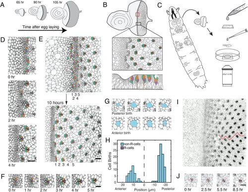


.jpg)