Some of you may have been so fortunate as to receive gift cards for Amazon.com or local bookstores in your Christmas stockings. While I wouldn’t think of dissuading you from purchasing the latest Louise Perry mystery or the memoirs of pre-eminent singers and chefs, I would recommend that you consider a new intellectual thriller, Evolution Evolving.
Imagine if two outstanding evolutionary biologists realized that evolutionary theory cannot explain adaptation and biodiversity without incorporating developmental biology. Imagine them inviting three developmental biologists to work on a book with them to construct the foundations of a more complete evolutionary theory. This book will become Evolution Evolving: The Developmental Origins of Adaptation and Biodiversity, a volume co-authored by evolutionary biologists Kevin Lala and Marcus Feldman, together with evolutionary developmental biologists Tobias Uller, Nathalie Feiner, and me.
This is not a textbook. It is a symposium, a working out of ideas, such that the reader is in dialogue with the book. The book presents evidence for certain views — that plasticity is universal and fundamental for evolution; that organisms are multigenomic holobionts whose symbionts can create new phenotypes and reproductive isolation in the animals they co-create; that there are multiple pathways of inheritance, including symbionts, epialleles, culture, and parental effects, and that some of these modes of inheritance allow the transmission of environmentally induced traits. Most of us had trained to see evolution as changes in gene frequency and development as changes in gene expression. This book organizes evidence that these genocentric explanatory mechanisms are inadequate to explain adaptations or the diversity of life.
Reading the book should make one question and refine one’s own ideas, to question one’s assumptions. Yes, these developmental phenomena happen; but are these differences important enough to change the way you think about evolution, organisms, development, and science? This book presents evidence that these phenomena — developmental plasticity, developmental symbiosis, and epigenetic inheritance systems — are critically important and that evolutionary biology gains enormous explanatory power only if it fully incorporates them. Some people have agreed with us. Marc Kirschner has called the book “a tour de force,” and Jessica Riskin has nominated the volume as a Scholarly Book of the Year, calling it “exhilarating reading. It is not just a book but an intellectual revolution.” Some people have disagreed. Evolutionary biologist David Houle doesn’t think these phenomena are important enough to change the way we think about evolution; moreover, “they are difficult to study.”
Twenty-five years ago, I predicted that evo-devo would cease to exist because it would become part of normative evolutionary biology. This is now happening. Look at the recent articles in PNAS about the genes responsible for the cryptic and mimetic pigmentation of insect wings. They are not classified under “developmental biology,” or even as “evolutionary developmental biology.” Rather, they are listed as “evolution.” Similarly, an evo-devo paper on the rates of prehistoric human teeth and brain development is listed in the “evolution” category, not as “development.” It seems that evolutionary developmental biology is becoming part of evolutionary biology. This book shows the many ways in which these fields can be merged.
Evolution is undergoing a metamorphosis, retaining some features, while jettisoning and repurposing others. It is evolution, but not as we knew it. It is an evolution where proximate and ultimate causes co-mingle, and where developmental mechanisms can bias the directions of evolutionary change. It is an evolutionary biology where the environment not only selects the phenotype but helps construct it. Evolution Evolving is an evolutionary biology book where developmental mechanisms are major players in the evolutionary processes that create adaptations and biodiversity.
The application deadline for the workshop is the 24th of January.
This is a residential workshop at Chicheley Hall on 2nd- 4th April 2025.
An important part of science is getting your results and ideas across to others, through papers, presentations, theses, grant proposals, conversations and interviews. Your audience may include specialists in the field, those from other disciplines, industry, or the general public.
How can you best communicate your science?
This workshop brings together experts in different fields to help you explore and develop your communication skills.
Working together with others on the course you will learn how to structure stories, bridge disciplines, simplify concepts and communicate effectively with a range of audiences. You will also get in-depth tutoring and practice in storytelling and public talks, developing hands-on demonstrations and multimedia (podcasts/YouTube/TikTok).
The Genetics Society will cover travel (within the UK only), accommodation and meals for successful applicants.
Tutors will include: Helen Keen (Award winning comedy writer and performer; author of the Radio 4 series, “It Is Rocket Science!”) First Create the Media (Led by award-winning writer and broadcaster Kat Arney) Alison Woollard (Presenter of the 2013 Royal Institution Christmas Lectures and Lecturer at University of Oxford)
Organiser: Jonathan Pettitt (Professor in Genetics, University of Aberdeen; Winner of the 2020 Genetics Society JBS Haldane Lecture) Cristina Fonseca (Science Communicator)
Who can attend?
The course is open to PhD students and postdoctoral researchers working in genetics and related areas.
Carer’s Award. In recognition of carer’s responsibilities, an award of (up to) £60/day will be made available to enable participants with carer responsibilities to attend this workshop. Awardees can spend this money as they think will best support their attendance.
With almost 200 posts published on the Node in 2024, below are just a few of our highlights.
Thank you to everyone who has contributed to the Node in the past year.
Have you read a Node post that you really enjoyed this year? Let us know in the comment section!
Behind the paper stories
Every paper has a story behind it. In these posts, we discover the highs and lows, the unexpected turns, and the fascinating discoveries from the breadth of developmental and stem cell biology.
Using an image or a video as a hook, these short posts bring people’s attention to a paper, a technique or a location that is of interest to the developmental and stem cell biology community.
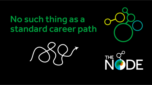
“No such thing as a standard career path” interview series
In this new series, we chatted to several developmental biologists who have had vastly different career trajectories. Check out all the interviews in this series so far.
The Node correspondents
Correspondents are researchers who are also interested in science communication. They work with the Node team to develop and create content on a broad range of topics. Here are a few highlights of posts produced by the correspondents:
Do you want to broaden your science communication experience alongside your research? We are looking for new correspondents for the Node. Find out more and apply by 20 January 2025!
Even though we have grouped posts into different series, we always welcome posts that don’t necessarily fit into any of our existing blog series.
Remember, the Node is your site: once you’ve registered, you can freely share your blog post, job advert or event notice with the community. If you have any questions, just get in touch.
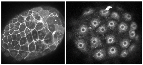
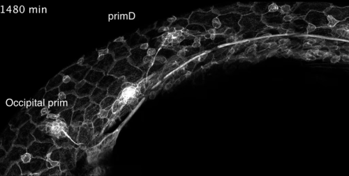
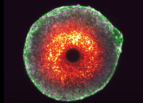
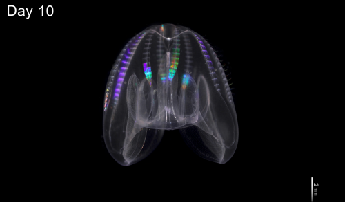
Over the past 12 months, Development has featured 24 cover images. Now, it’s your chance to pick your favourite!
To find out more about each cover image, you can visit Development’s 2024 issue archive. Thank you to everyone who’s contributed to this collection of wonderful images.
*The poll is now closed. Thank you to everyone who voted!*
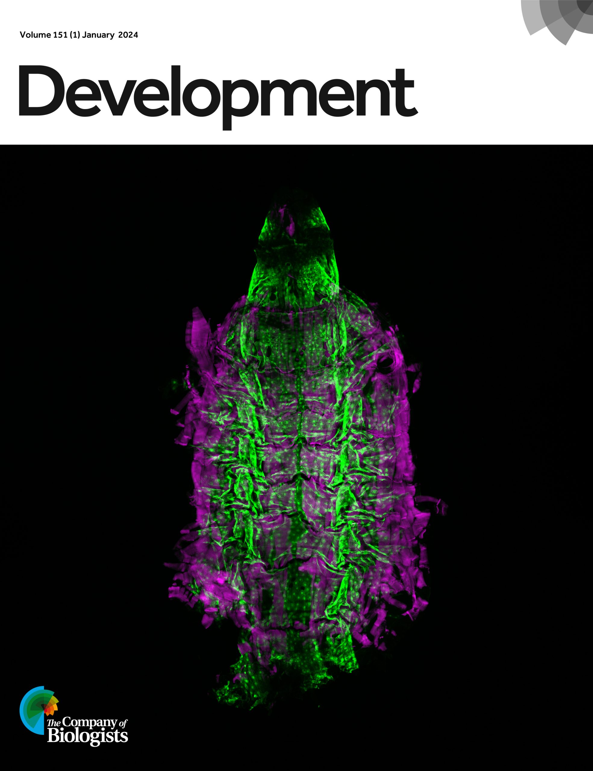
Issue 1
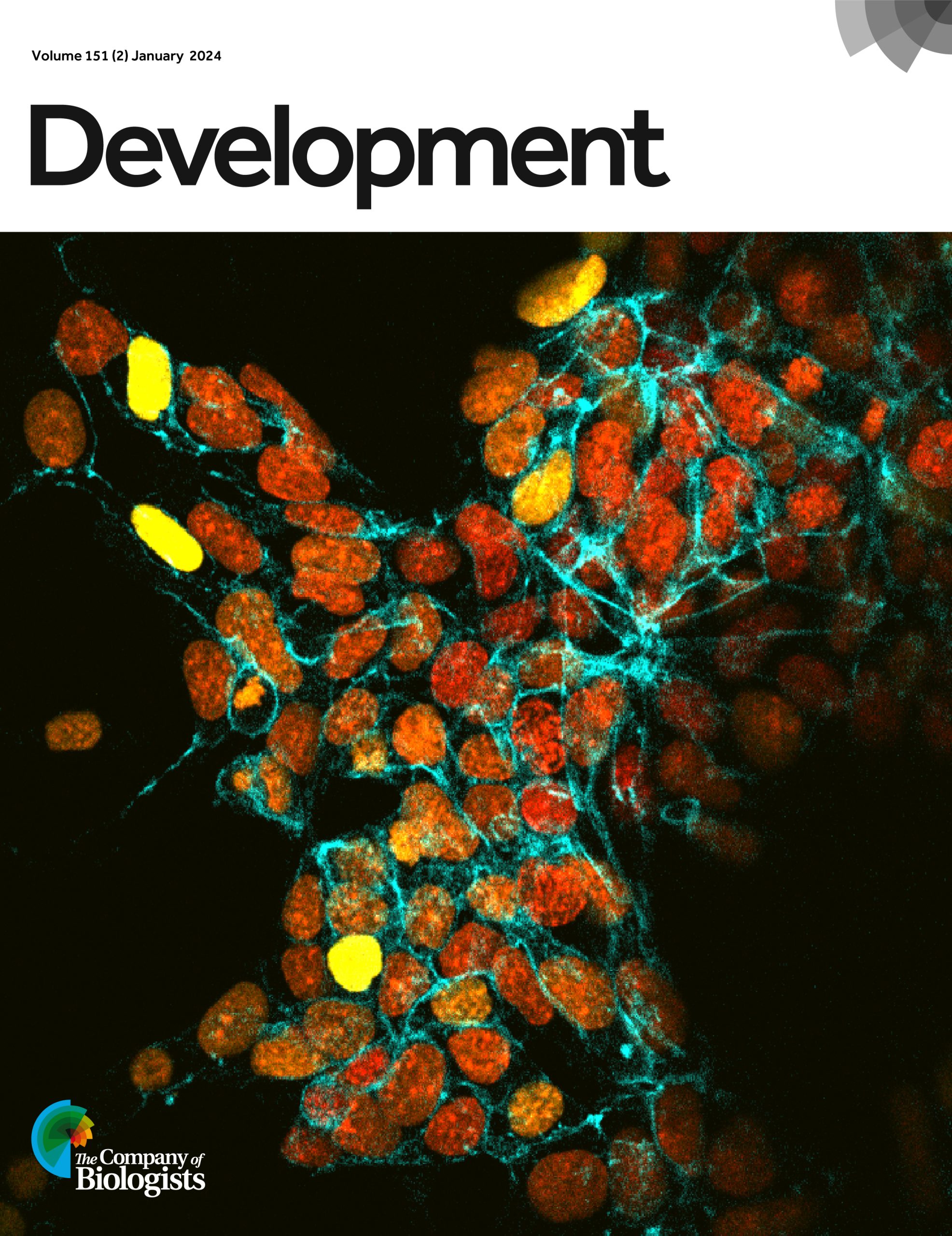
Issue 2
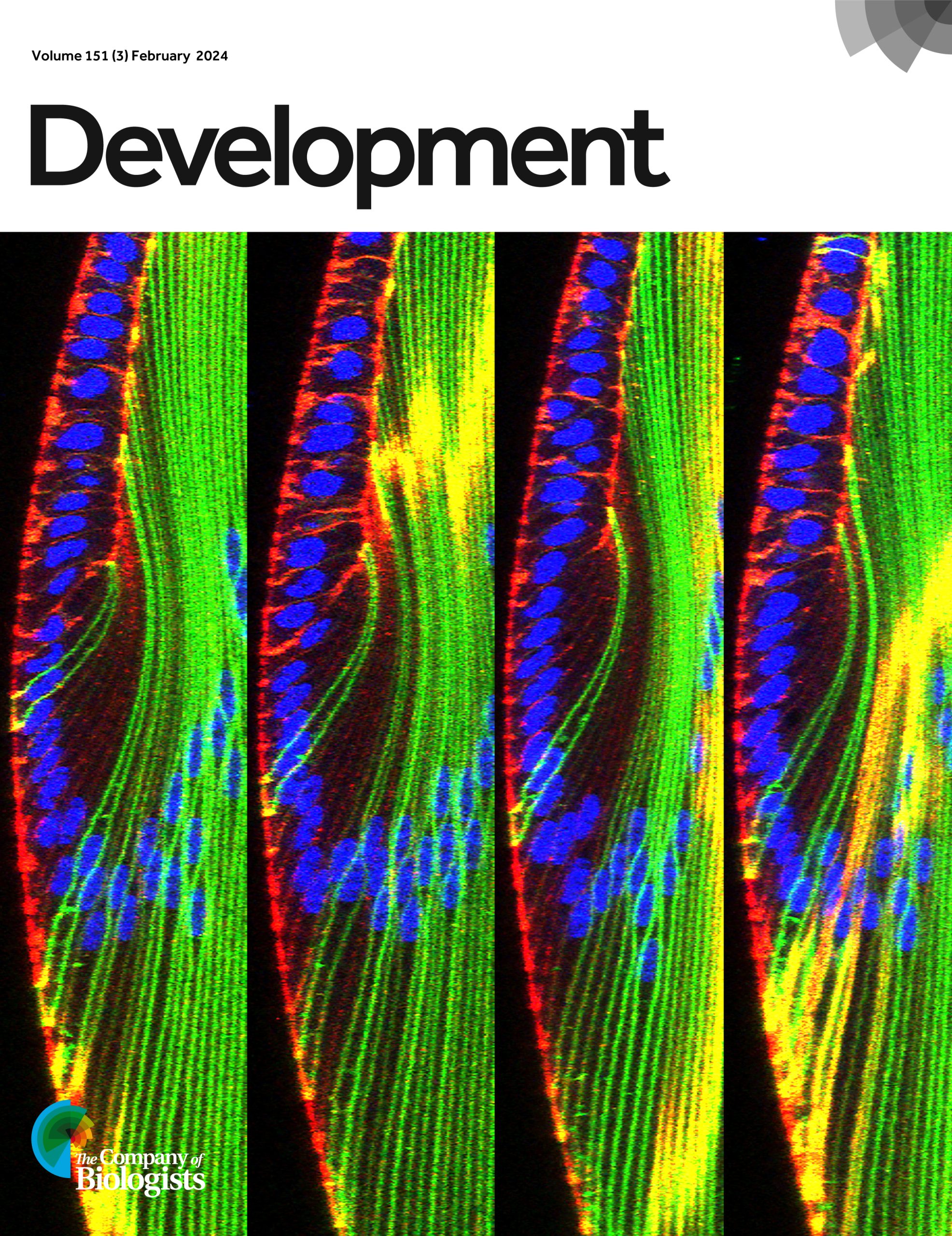
Issue 3
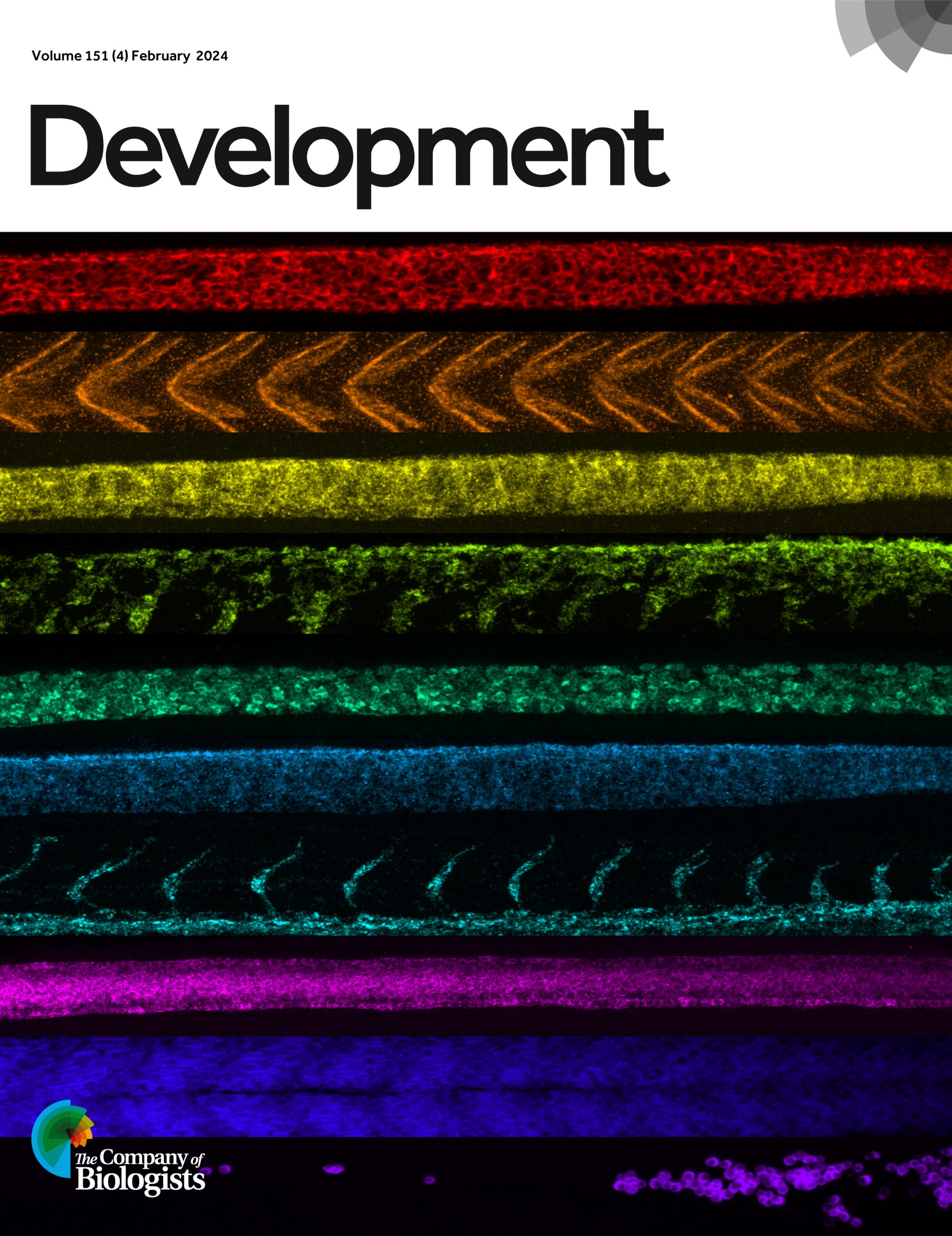
Issue 4
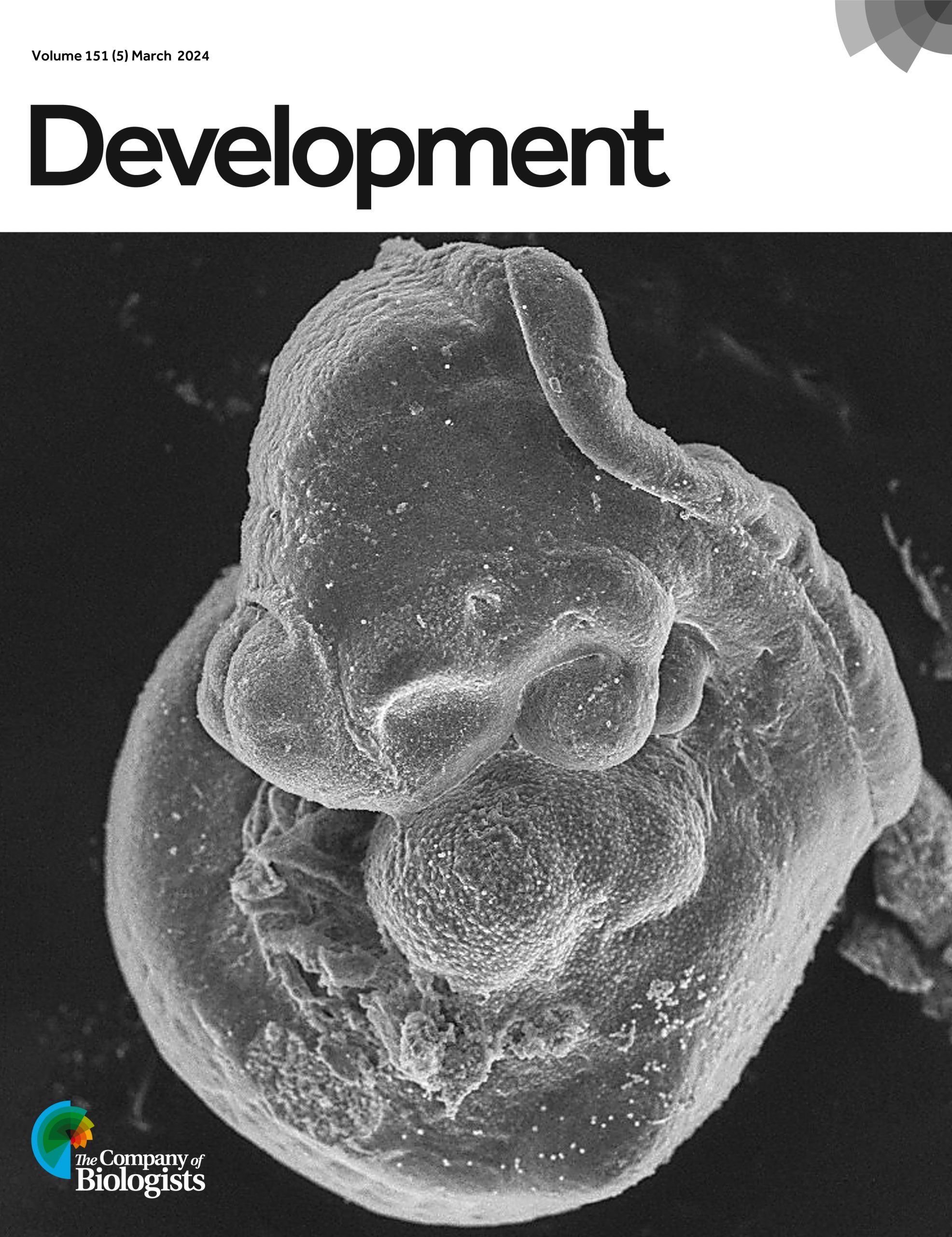
Issue 5
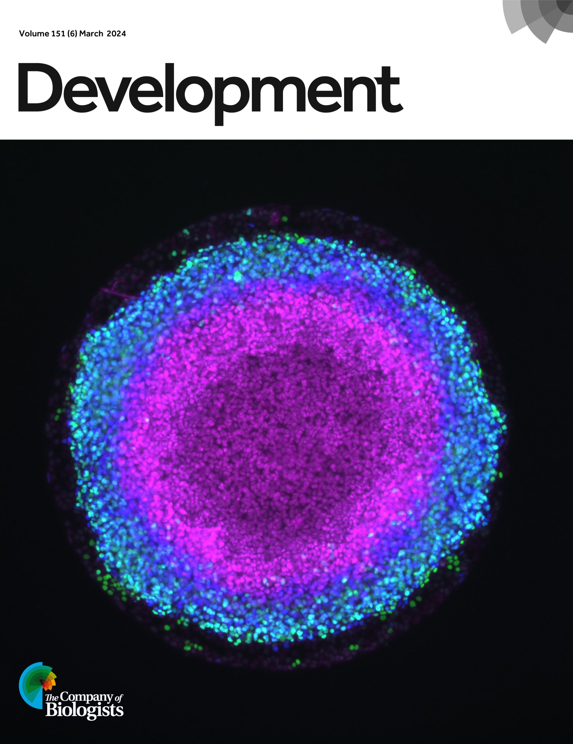
Issue 6
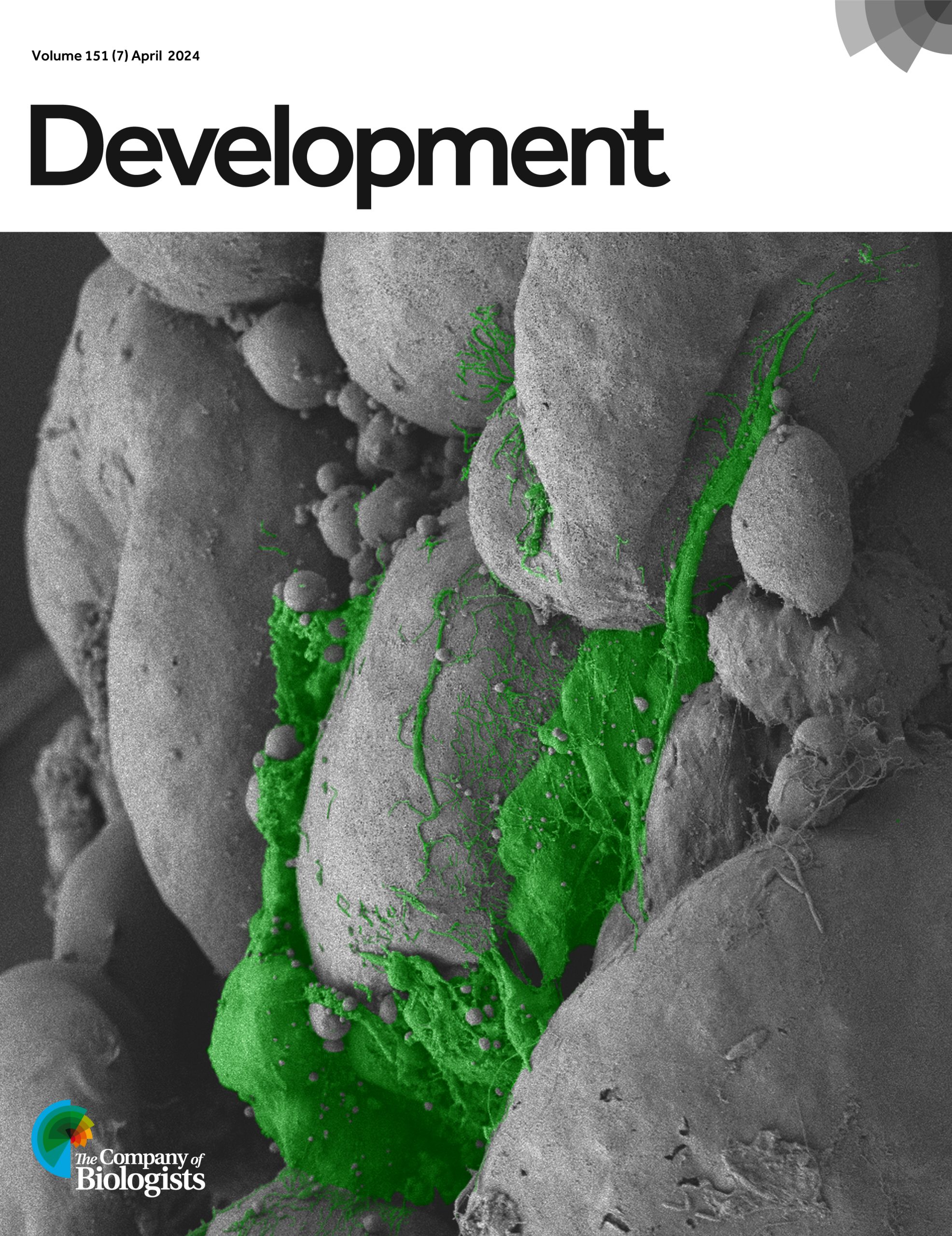
Issue 7
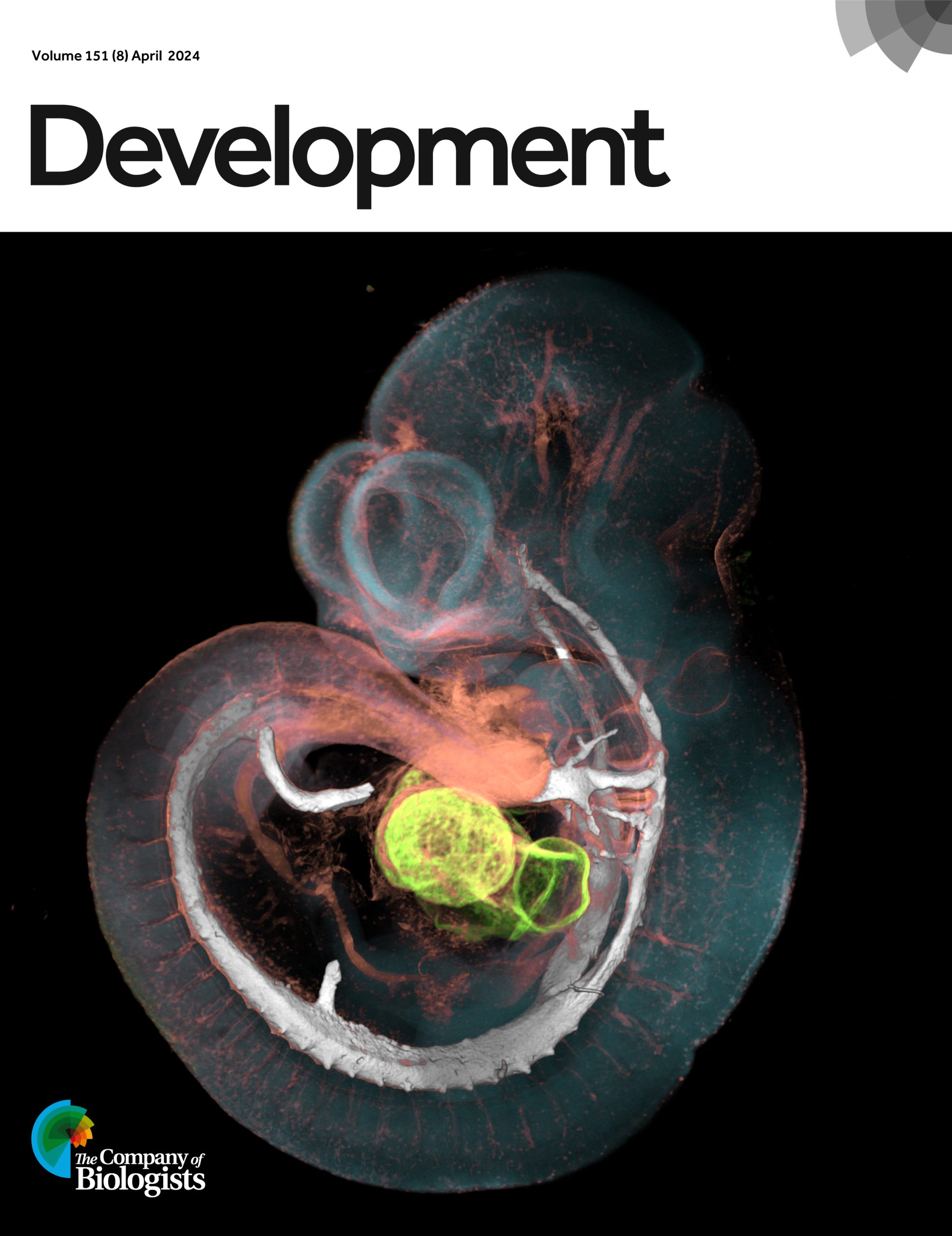
Issue 8
Issue 9
Issue 10
Issue 11
Issue 12
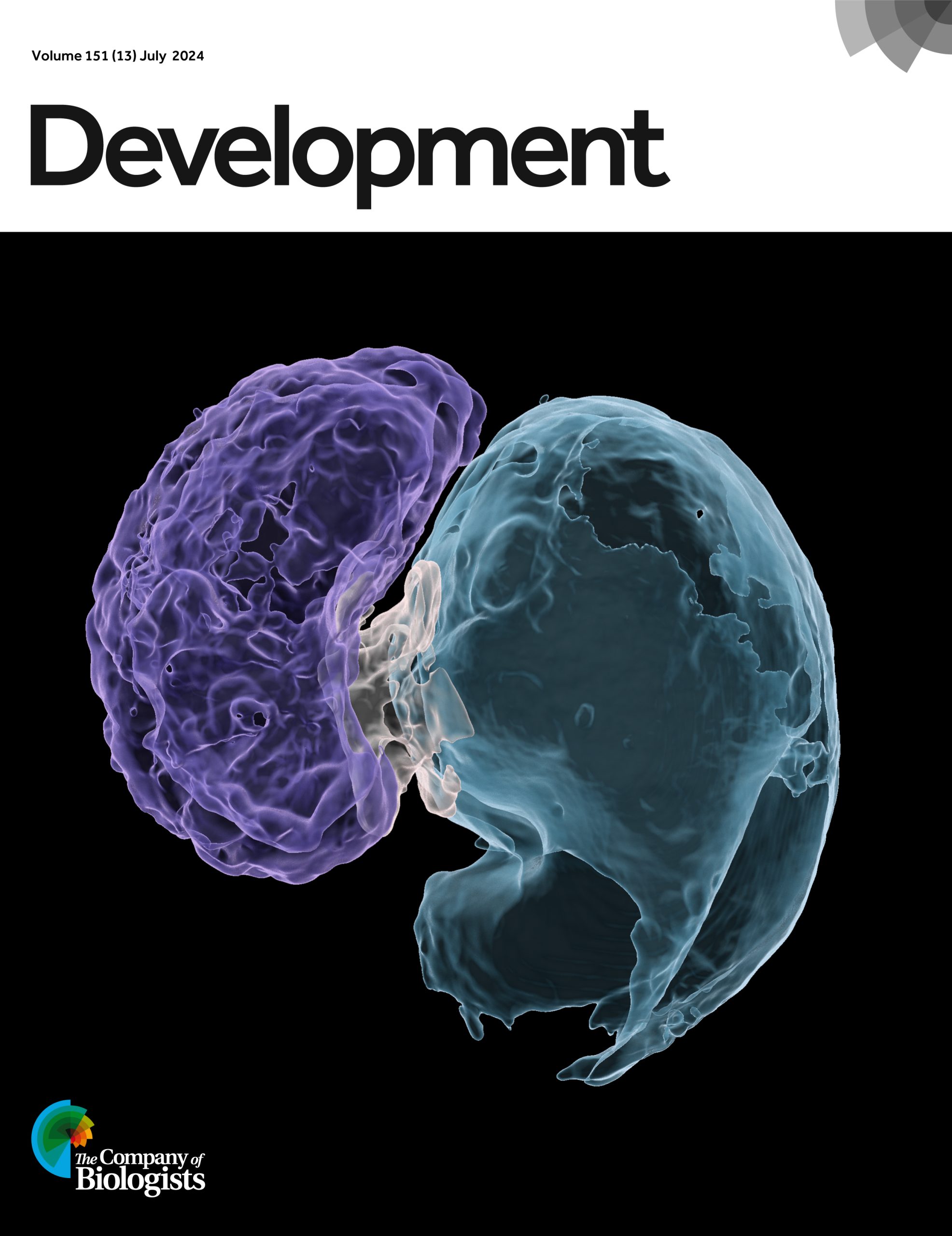
Issue 13
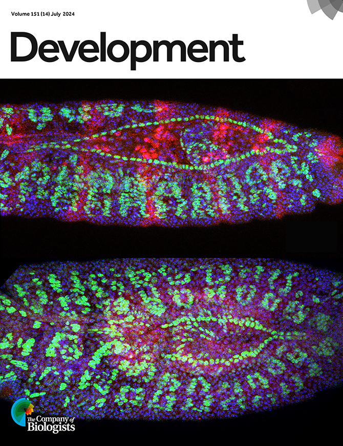
Issue 14
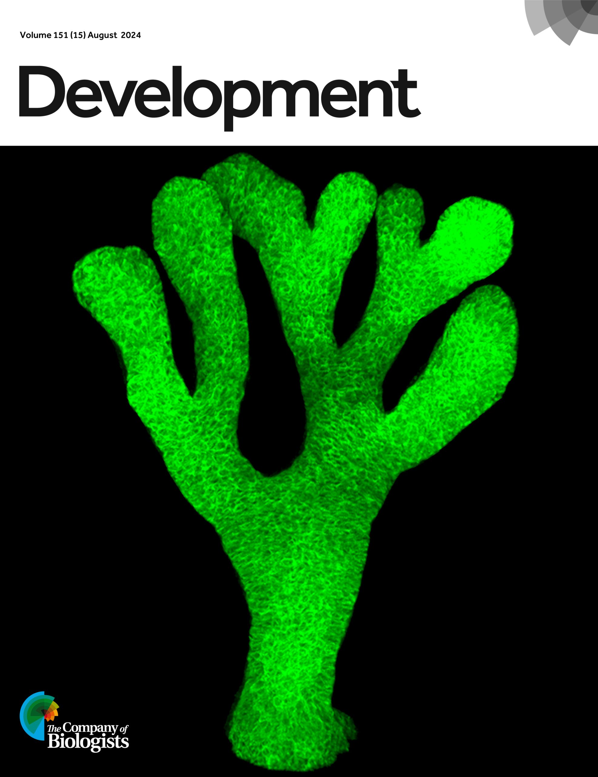
Issue 15
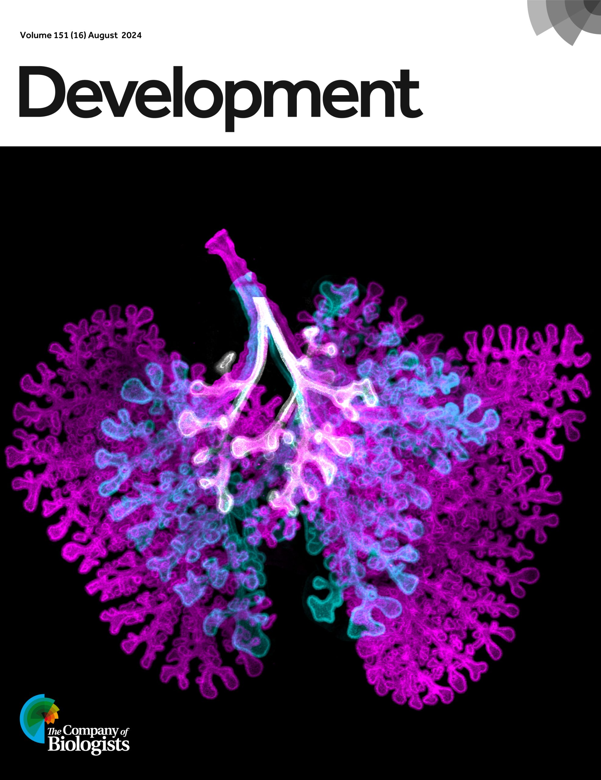
Issue 16
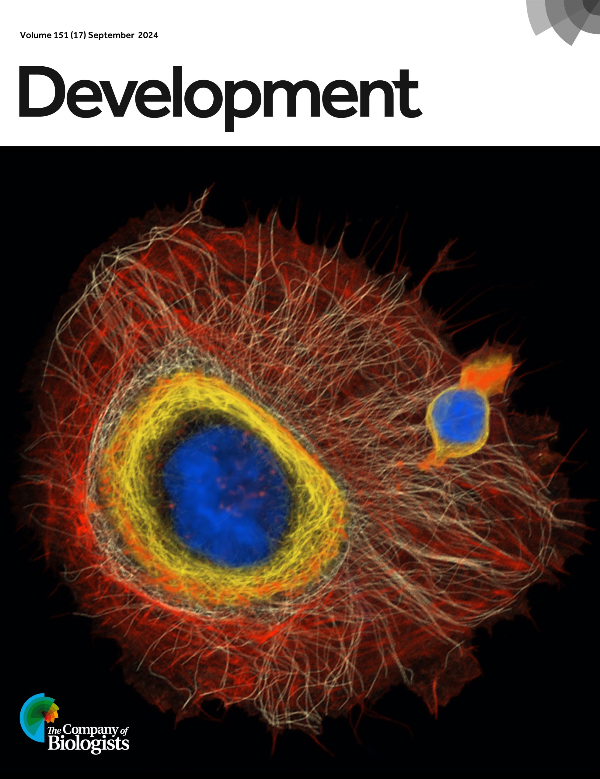
Issue 17
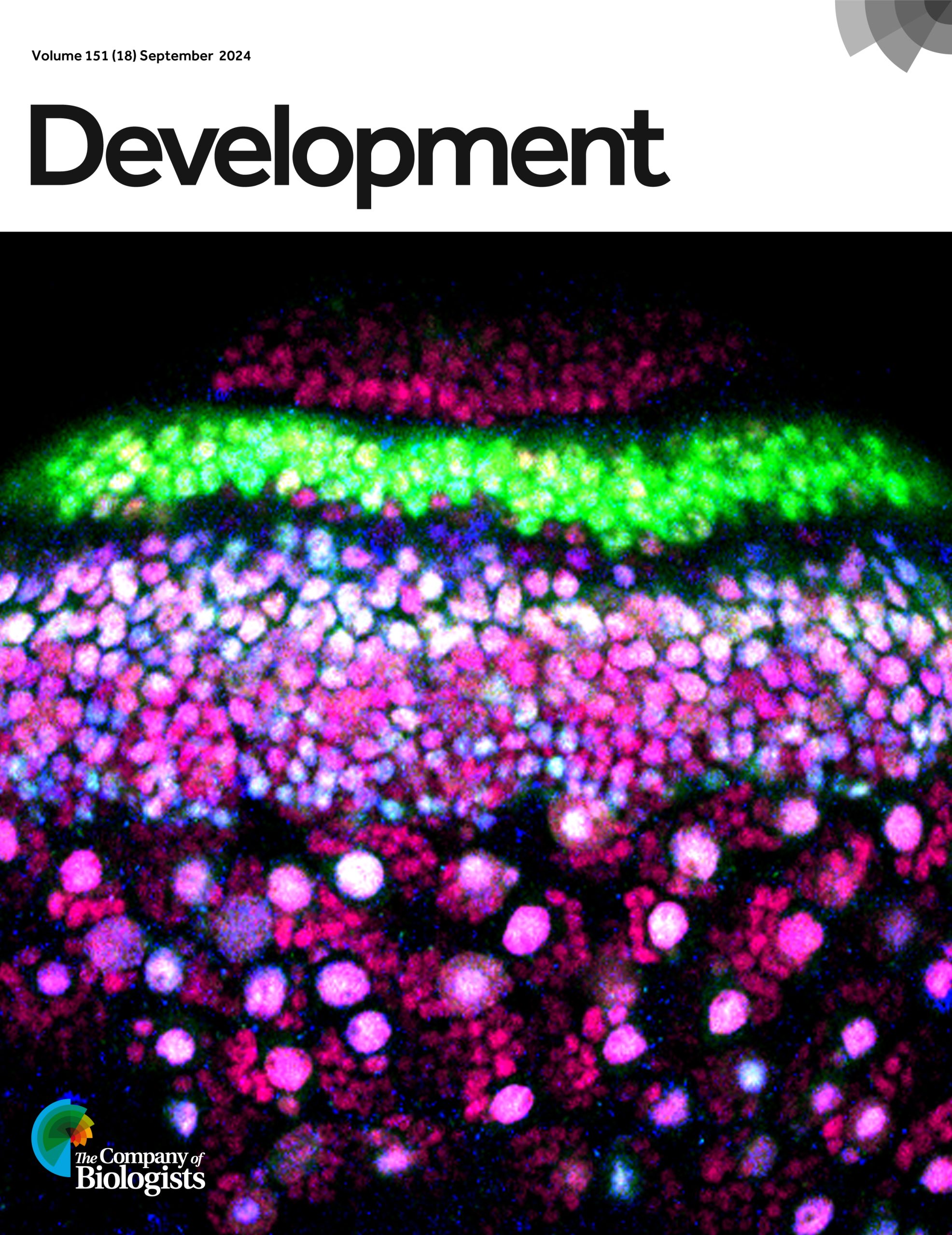
Issue 18
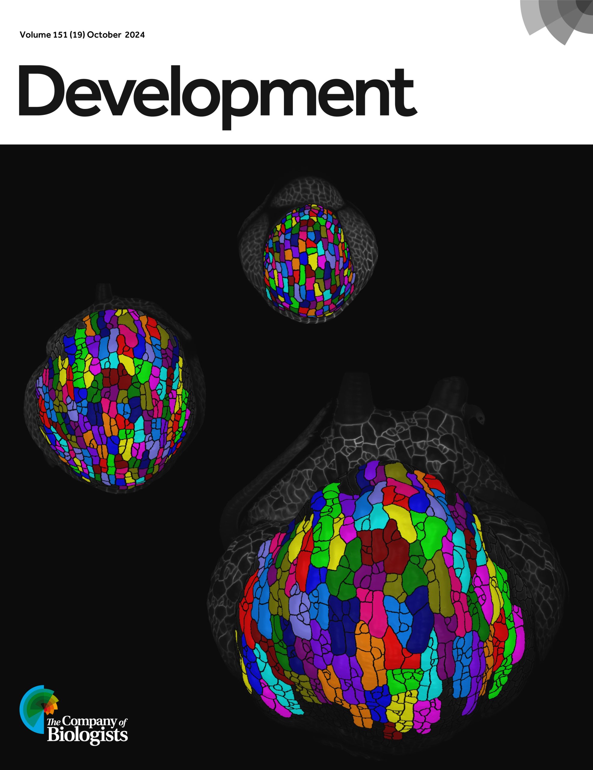
Issue 19
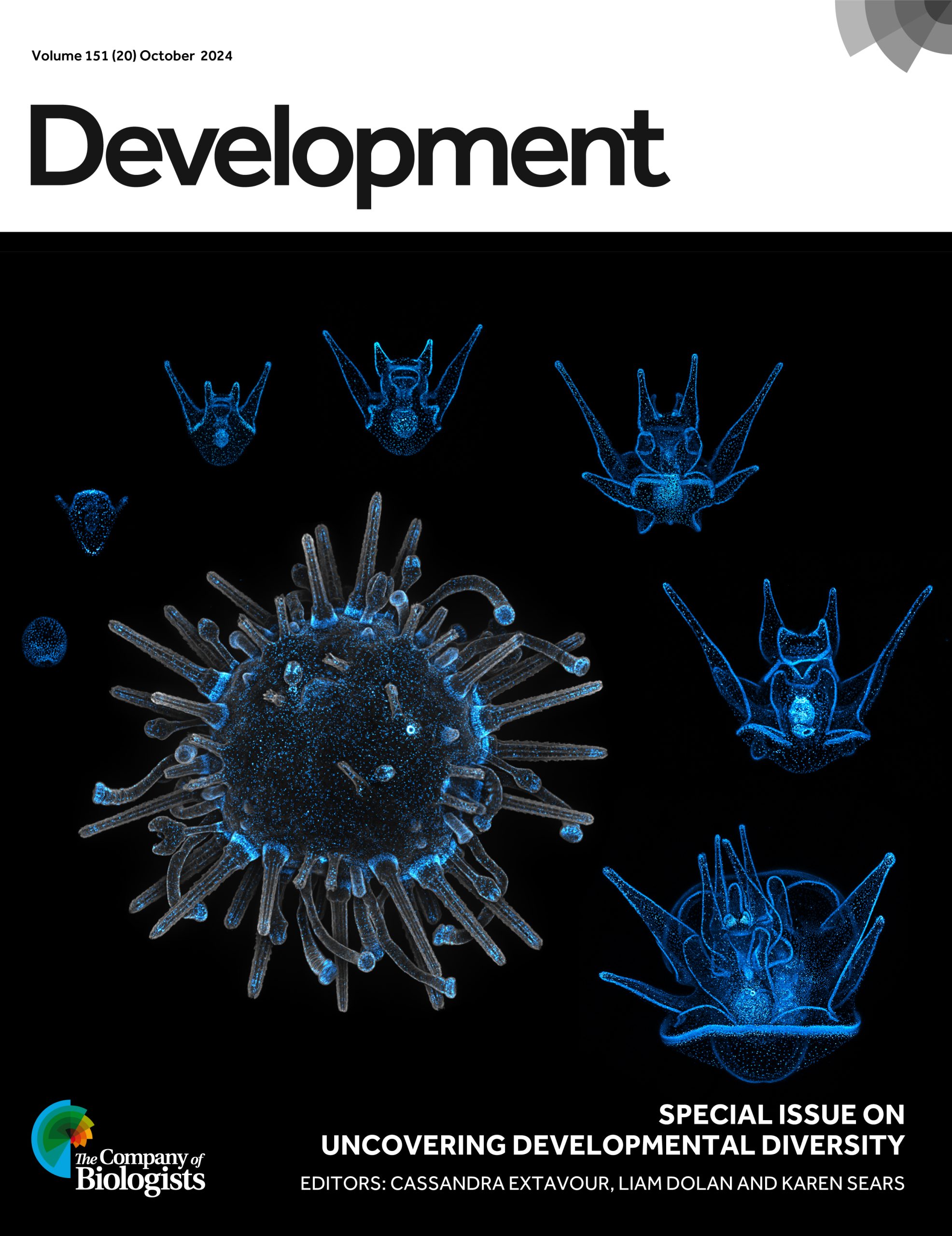
Issue 20
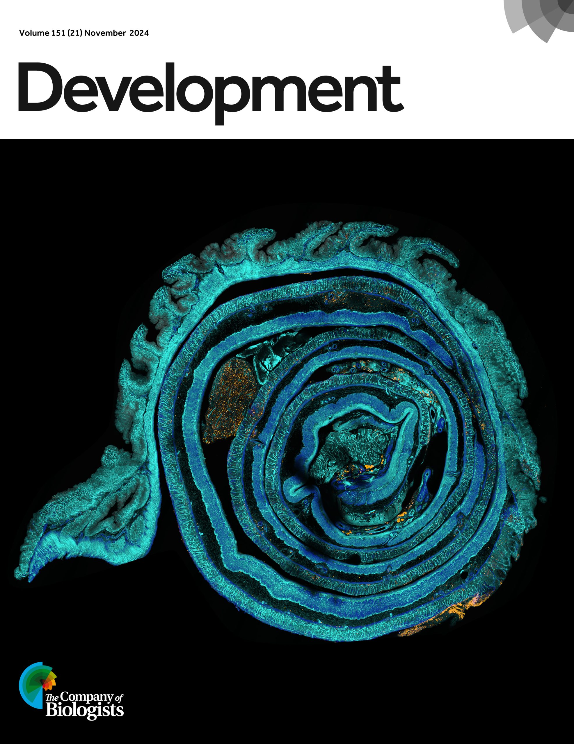
Issue 21
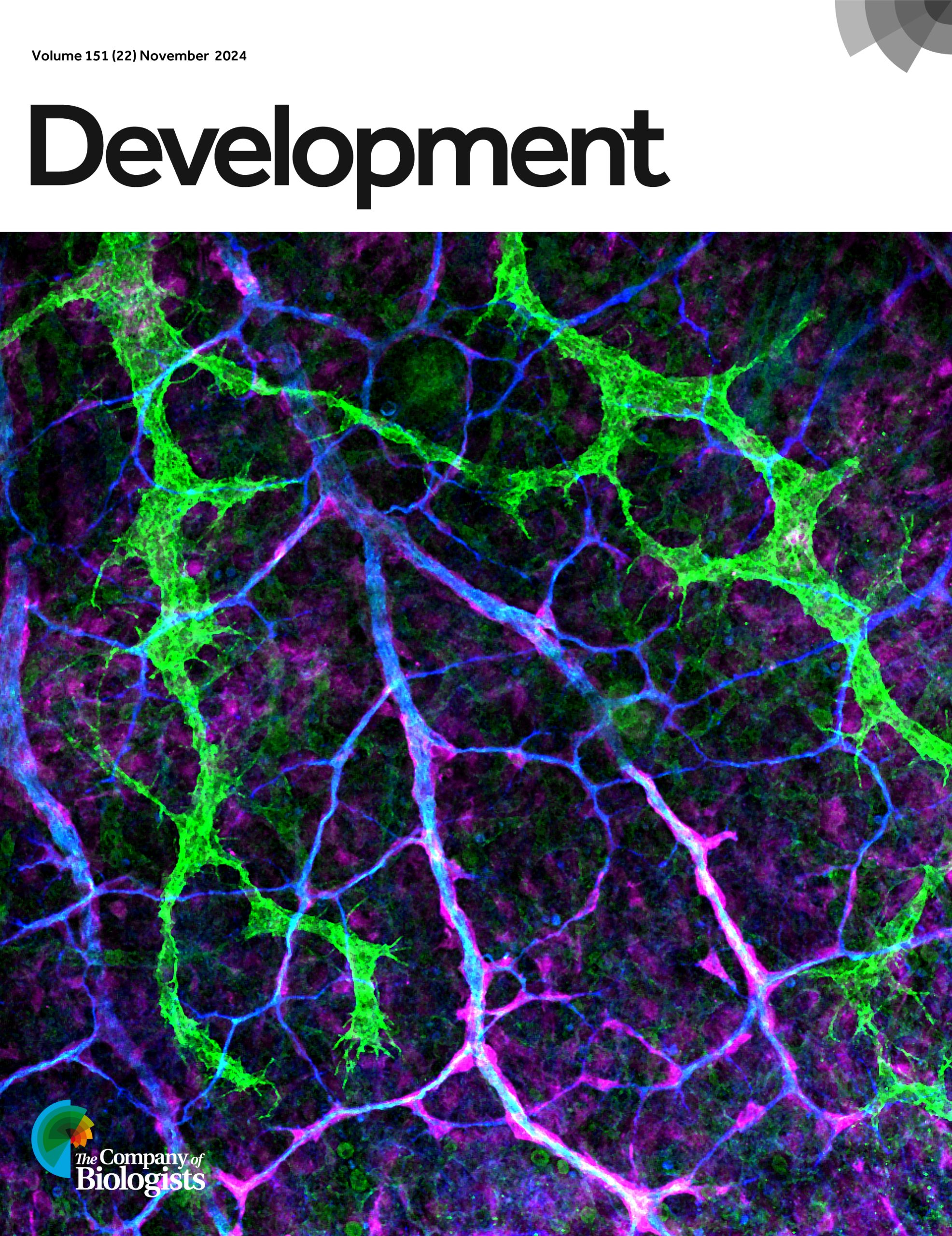
Issue 22
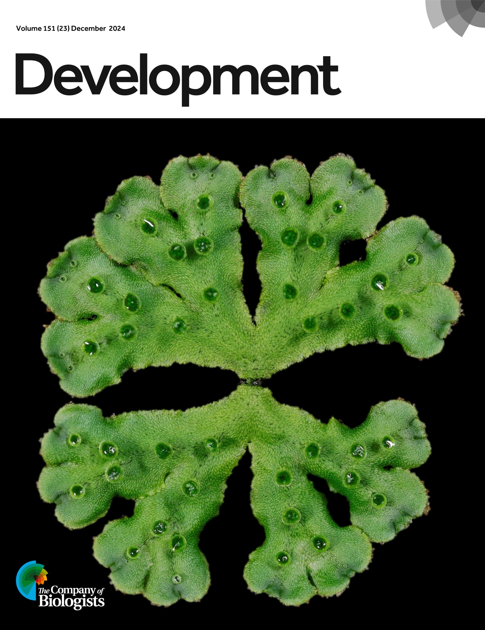
Issue 23
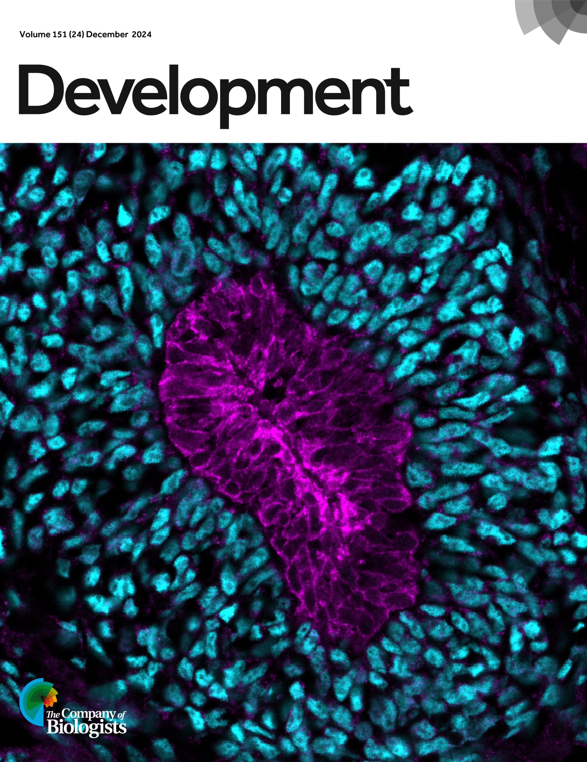
Issue 24
Visit Development’s 2024 issue archive to find out more about each cover image.
Vote for the 2024 Development cover image of the year
*The poll is now closed. Thank you to everyone who voted!*
The final webinar of 2024 featured two early-career researchers working on gene regulation and will be chaired by Development’s Senior Editor, Alex Eve.
PRESS RELEASE: Millions of people around the world are affected by retinal degenerative diseases. In most cases, loss of vision is caused by damage to the macula, a region in the centre of the retina. The macula is rich in cone photoreceptors – cells important for perceiving colour and seeing finer details. Currently, there are no approved treatments to replace the damaged macula, despite its huge impact on the quality of life. Now, a team of researchers from the University of Montreal, led by Professor Gilbert Bernier, found that blind minipigs receiving retinal transplants made from stem cells showed signs of restored vision. They published their study in the journal Development on 5 December 2024.
In this study, Professor Bernier’s team developed a method to coax stem cells into forming sheets of cells that recapitulate the structure of the human retina. The type of stem cells they used are called human induced pluripotent stem cells – immature cells ‘reprogrammed’ from an adult (mature) cell that can differentiate into any type of cells in the body. Using the stem cells, the researchers made ‘retinal sheets’ that are enriched in immature versions of the cone photoreceptor cells, which could become mature cone cells when cultured in the lab.
After successfully creating the retinal sheets in a dish, the researchers tackled the next challenge: transplanting these sheets into minipigs with damaged macula. Professor Bernier explains, “To get as close as possible to human clinical application, we have chosen minipigs because the size of their eyes is near that of humans and the animals are about the same weight as humans. Hence, all surgeries in our study could be performed by a retinal surgeon.”
Upon transplantation, the researchers found that the retinal grafts were able to integrate into the minipig’s damaged retinal tissue. Encouragingly, the minipigs showed signs of restored vision: new neural connections were formed between the grafted photoreceptor cells and the minipigs’ neural cells, and the scientists could detect neural activity of the photoreceptors at the grafted area when the minipigs were placed in a well-lit room.
Given the pressing need to develop therapeutic interventions against vision loss, researchers around the world are testing different ways to repair damaged macula. “Some approaches use dissociated photoreceptor cells; others create micro-dissected retinal organoids, which are lab-grown ‘mini-organs’ in a dish,” says Professor Bernier. “In contrast, our method allows the spontaneous formation of a flat retinal tissue that is already polarised and organised, as in the human embryonic retina.” He adds that their method can generate large yields of retinal tissue for transplantation.
A limitation in this method lies in the difficulty of controlling the placement and orientation of the grafts during surgery. The macula is only 4mm in diameter – about the length of a grain of rice. “To properly orient, place and stabilise the graft in the retina remains a big surgical challenge,” says Professor Bernier. His team are now working to improve the transplantation success rate. They are validating an experimental retinal surgery device to ensure proper orientation and implantation of the graft at the correct retinal disease site. Although many challenges remain, this study demonstrates the potential of retinal sheet transplantation for treating retinal degenerative diseases.
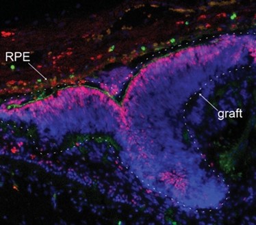
Integration of a human retinal sheet (graft-dashed line) transplanted into a degenerated minipig retina, showing expression of the photoreceptor-specific markers CRX (in red) and PNA (in green) within the graft. Note the graft polarization and close association with the pig’s eye retinal pigment epithelium (RPE). Image Credit: Dr. Andrea Barabino
Enthusiastic about science communication and looking for a chance to broaden your writing experience alongside your research activities? The Node, the community site for developmental and stem cell biologists, is looking to appoint three correspondents who will play a key role in developing and writing content over the coming year.
In 2024, we have been working with Alex Neaverson, who’s used her artistic talents to create illustrations for the Node. You can check out Alex’s illustrations about Rita-Levi Montalcini’s extraordinary life, and the post about the work of Millie Race, winner of the Young Embryologist Network Sammy Lee Award. Thank you Alex for making the Node more colourful!
As a correspondent, you will be expected to contribute around six posts over the course of the year – this could involve creating your own blog series around a theme of your choice, reporting on the latest exciting developments in developmental and stem cell biology, interviewing inspiring scientists, or writing about conferences and other events. We are also open to any other ideas you might have as we would like to shape a programme that both appeals to your interests and benefits the research community.
You will also gain insight into the publishing industry through meetings with the Community Managers and receive regular feedback on your writing. We will help raise your profile as a researcher and science communicator and are also happy to support you by contributing towards conference attendance costs relating to the role, providing reference letters, or in other ways.
Please note, we are also recruiting correspondents for FocalPlane, so when applying you will have the option of choosing to apply for the Node, FocalPlane or both.
We encourage applications from all individuals regardless of sexual orientation, gender identity or expression, religion, ethnicity, age, neurodiversity or disability status. We also welcome applicants from a range of geographic locations.
Please get in touch with us if you have any questions about the programme at thenode@biologists.com
Hopefully some of you will have seen the recent editorial in Development on our approach to peer review. If you haven’t read it yet, please do take a look. In it, James Briscoe (the journal’s Editor-in-Chief) and I discuss some of the initiatives that the journal has taken to try and support authors through the peer review process – including, most recently, encouraging authors to include a ‘Limitations’ section in the discussion of their article, giving you an opportunity to lay out explicitly the scope and extent of your study and, where appropriate, to respond to referee concerns by acknowledging them rather than addressing them experimentally.
Off the back of this editorial, James has also written a blog post that I’d really encourage you to read. Entitled ‘In Praise of Peer Review‘, James sets out why he believes that peer review (in some form) is an invaluable and irreplaceable part of scholarly communication. Alongside the debate that’s been going on around eLife’s exclusion from Web of Science (and subsequent decision to send a partial feed of articles for indexing, the piece has generated some discussion on social media both around whether peer review actually works to guard against publication of fraudulent, sloppy or otherwise dubious papers, and around the degree to which it actually helps to improve papers. I think James has done a great job of setting out the ‘why’ of peer review, but here I thought I’d give my view on the ‘what’: what should a peer review report comprise?
But before I start, let’s remember that – in the majority of cases at least – peer reviewers are both 1) highly knowledgeable in the field of the paper they’ve agreed to review and 2) well-meaning. Yes we all know of cases where papers have been sent to referees that weren’t sufficiently expert or who set out to block publication for political or petty reasons. But these are in the minority – most reviewers are competent to do the job they’ve been asked to, and they want to do it well. And they do it for little or no reward, because they believe that it’s an important part of their responsibility as a member of the academic community. If or how they should be rewarded is a whole other topic that I won’t get into now, but I am incredibly grateful for their dedication.
So, what do I want a referee to do?
Firstly, I want a referee to be respectful. Remember that there are people behind the data and – before hitting the ‘submit’ button on their report – pause to consider the potential impact of your words on the authors, particularly the students and postdocs who’ve actually done the work. At Development, we’re very fortunate that the vast majority of referees do abide by this guidance, but that’s not to say that I’ve not come across the odd report that felt overly combative or dismissive in tone – and that’s not OK.
Secondly, I want the report to be reasonable in terms of the amount of additional work requested. Think about the amount of time (and money!) that might be involved in addressing any particular point and ask how important that point really is to the main story of the paper. Which leads me on to:
Thirdly, I’d ask the referee to focus primarily on addressing the question ‘do the data support the conclusions?’ and not ‘what could the authors do to make the conclusions more interesting?’. While it’s very useful to get expert opinion on how important/relevant/useful/important the paper will be for the community, it’s primarily the editor’s job to decide on whether the paper is – in principle – appropriate for the journal in question.
And finally, I want the referee to be honest about what aspects of the paper they can and can’t (or even did and didn’t) assess. Are you able to judge if the authors have used appropriate statistical analyses? (And if so, did you actually check?!) If the paper contains computational work, do you have the expertise to assess it fully? If the authors deposited data, did you look at it? We fully appreciate that referees can’t always be experts in every area of a paper – particularly an interdisciplinary one – and we try to recruit referees with complementary expertise, but it’s really useful to know what you did and didn’t review.
Most reports I read (and I read a lot!) do largely follow these guidelines, but there is still a definite tendency for a referee report to read a bit like a shopping list of potential experiments and textual revisions. Experienced authors can often read the nuance to decide which points to tackle experimentally, and good editors will (either pro-actively or in response to author queries) help to navigate the revision process. But referees can also do their bit to shepherd papers through the often all-too-painful process of publishing by remembering that there’s both a financial and a temporal limit to how much a group of authors can (and should) do to revise a paper, that a single paper can’t solve a whole research question, and that their opinion isn’t necessarily any more valid than that of the authors (or, for that matter, the other referees).
We could discuss ad nauseam the benefits and problems of pre-publication peer review in its current form (and I frequently do!), and alternative models are beginning to emerge that can act in parallel to, or even replace, our current system. But let’s also think about the little steps that we can take to make the current system less onerous and more constructive – thus easing the path to publication.
The massive presence of disorder and variability challenges the traditional metaphor of the developmental process as a perfectly executed program leading to precise mechanisms at every level [1,2]. Yet, the final outcome —the organism— remains both astonishingly complex and remarkably reproducible. This paradox piqued the interest of Dimitri Fabrèges and Takashi Hiiragi. Back then, around 2017, Takashi was research group leader at the EMBL Heidelberg, and Dimitri a postdoc in his group. They began to explore the idea of disorder and variability from a provoking viewpoint: instead of undermining the precision of the developmental process, randomness and variability might actually act as driving forces that ensure precision and reproducibility.
Motivated by this hypothesis, the researchers focused on the early stages of mammalian development; particularly, on the initial cleavage process of mouse, rabbit and monkey embryo, encompassing the first cell divisions post-fertilization up to the 16-cell stage. A first analysis showed that the division times of different cells in the same embryo were progressively being desynchronized. A pivotal moment in this sequence is the 8-cell stage —that is, after 3 division rounds— a moment in which cells already divided in a quite disorganized way. Due to this high variability, researchers found that the beginning of this stage was characterized by a highly heterogeneous set of cell packing configurations. However, such initial variability is smoothly but steadily reduced along the so-called compaction process, leading to a seemingly common, spherical-like structure at the end of the stage. Such a structure guarantees that, in the next round of divisions —i.e., at the 16-cell stage— there will be a suitable proportion of inner and outer cells. Achieving this correct proportion is essential: inner cells will lead to the organism itself, while outer cells form the placenta and extra-embryonic material. This observation raised a challenging question, namely, how can one support the intuitive claim that embryos begin highly heterogeneous but become remarkably similar with a more rigorous foundation.
This was the perfect challenge for Virginie Uhlmann, an expert on biological image processing who, at the time, had just started as research group leader at the EMBL-EBI, in Cambridge. She tackled this question by developing an advanced computational framework able to analyze and track the geometric changes in the embryonic shape in high detail. This approach conceptualized an embryo’s developmental path as a trajectory within a high-dimensional space whose coordinates captured relevant geometrical properties [3]. The key result was that, indeed, the trajectories exhibited significant initial disparity but converged surprisingly by the end of the compaction process in a particular region of the abstract space that characterized the embryo geometry.
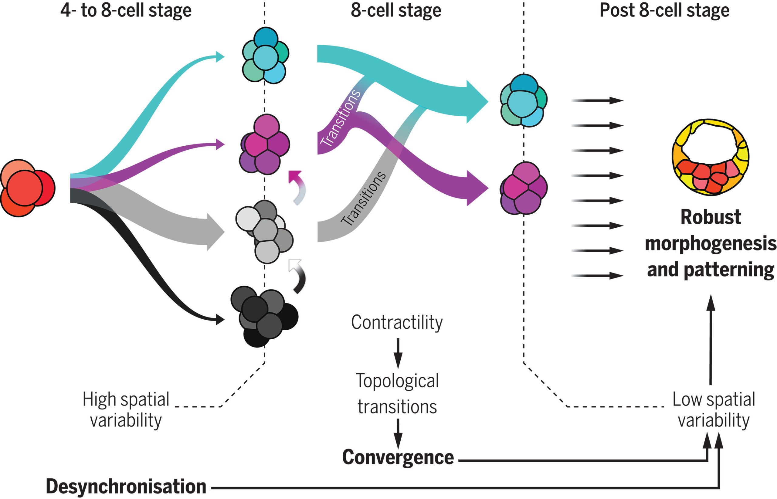
Which structure was represented in this region? Why did this particular structure seem to act as the developmental “target”? At the beginning of the winter of 2018, Takashi’s research group organized a retreat in the Catalan coastal town of Sitges, gathering several groups from the ISTA. Among the attendees were Edouard Hannezo, who just opened his research group as PI, and his first postdoc, Bernat Corominas-Murtra —both physicists working on biological problems. The evening was windy and stormy, and a little café was the refuge where they largely discussed with Dimitri and Takashi about the challenge of identifying and explaining the emergent structure. Although no immediate solution came out, Edouard and Bernat concluded that a deeper and simpler structural characterization was necessary —that is, complementing the geometric analysis with a topological one, stripping out all details but the raw structure. Weeks after, they stumbled upon a relatively recent publication showing a key mathematical finding: there are exactly different 13 ways to pack 8 spheres such that none of them exhibit independent movement. This result provided the key to define a classification scheme: either the embryos conform approximately to one of these 13 packing configurations, or they are floppy, meaning that some cells retain independent movement [4]. By establishing a suitable notion of “distance” among sphere packings, the researchers could classify embryos at various developmental time points. Their analysis revealed that, although variability was very high at the onset of the 8-cell stage, as the compaction process progressed, embryos consistently converged towards these similar packing structures along similar developmental pathways. At that point, the target structure was identified: the D2d packing of 8 spheres, in the Schoenflies notation.
Fig 1: 4-cell stage embryos give rise to many shapes at the beginning of the 8-cell stage, during which cell contractility triggers topological transitions. Ultimately, embryos are driven toward the most optimal packing (cyan). In parallel, the cell-autonomous desynchronization progressively increases temporal variability and helps to maintain topological optimality through generations, lowering spatial variability and promoting robustness. Picture taken from (Fabreges et al. 2024).
How does the embryo, without any external help, solve this kind of Rubik’s cube, i.e., transition from an arbitrary cell configuration to a specific optimal only one through successive cell rearrangements? Looking at the empirical data, one observable stood out above the other due to its clear trend: Adhesion was increasing along the compaction. Edouard suggested to challenge the simple hypothesis whether this slight change in the cell adhesion was enough to trigger all the topological rearrangements. The hypothesis has deep consequences. It implies that an increase on the cell adhesion could not only trigger deformations within the cells (i.e., increasing the contact surface, for example), but also qualitative reorganizations of the whole embryonic cell mass in a reproducible way. Computer simulations showed that such a genetically encoded slight increase in cell adhesion, coupled with significant random fluctuations in cell positions —disorder— was paradoxically facilitating the transition from any arbitrary packing of cells to the single optimal configuration. This hypothesis stands out as the simplest and, in the case of the mouse, it enabled even to reproduce in-silico the developmental trajectories of real embryos. In the case of rabbit and monkey, the role of other agents, like the zona pellucida —an external membrane that may exert a compressing force to the cell packing— could not be fully discarded.
At this point, the puzzle of the convergence towards a common, suitable embryo configuration was solved. However, the role of the temporal variability, which was experimentally observed at the starting point of the whole project and inspired it all, remained to be understood. Using several genetic perturbations, the results were surprisingly concluding that initial variability was actually required to achieve precise convergence. In particular, embryos in which cell divisions occurred more synchronously than in the wild-type ones showed a poorer convergence at the end of the compaction process, thereby hampering the further development of the embryo. The provoking hypothesis of Takashi and Dimitri on the role of stochasticity was thus proven to be fully consistent.
The researched path was not easy: Big part of the project was carried out during the COVID-19 pandemic. In turn, during the project, Dimitri and Takashi moved to Utrecht, to the Hubrecht Institute, Virginie to the University of Zurich, and Bernat to the University of Graz. Researchers from several institutions1 provided their bits of knowledge in the multiple challenges that paved the achievement of the results, and, as in living organisms, the sum of different expertises —biology, physics, mathematics and computer science— ended up in something that was much more than the sum of its parts. As in any adventurous interdisciplinary research, moments of joy and concern alternated, sometimes without pause in between… All in all, this research provides a new, constructive interpretation of the striking amount of disorder observed along developmental stages: When coupled to the changes in cell mechanics, the interplay among them can lead to significant and precise reorganization events within embryos, paving the way for a new understanding on how complex geometries and, in general, organization patterns arise in living beings. Disorder, therefore, far from being a problem the system has to deal with, may be one of the leading forces driving the precision of organism development.
Publication:
Dimitri Fabrèges et al. Temporal variability and cell mechanics control robustness in mammalian embryogenesis. Science 386, eadh1145 (2024)
References:
[1] M. Carlson, W. Reeves, M. Veeman, Stochasticity and stereotypy in the Ciona notochord. Dev. Biol.397, 248–256 (2015).
[2] R. Dumollard, N. Minc, G. Salez, S. B. Aicha, F. Bekkouche, C. Hebras, L. Besnardeau, A. McDougall, The invariant cleavage pattern displayed by ascidian embryos depends on spindle positioning along the cell’s longest axis in the apical plane and relies on asynchronous cell divisions.eLife6, 1–23 (2017).
[3] R. Delgado-Gonzalo, N. Chenouard, M. Unser, Spline-based deforming ellipsoids for interactive 3D bioimage segmentation. IEEE Trans. Image Process.22, 3926–3940 (2013).
[4] N. Arkus, V. N. Manoharan, M. P. Brenner, Minimal energy clusters of hard spheres with short range attractions. Phys. Rev. Lett.103, 118303 (2009).
1Other institutions involved:
Institute for the Advanced Study of Human Biology (WPI-ASHBi), Kyoto University, Kyoto, Japan.
Department of Developmental Biology, Graduate School of Medicine, Kyoto University, Kyoto, Japan.
Research Center for Animal Life Science, Shiga University of Medical Science, Shiga, Japan.
INRAE, BREED, Paris-Saclay University, Jouy-en-Josas, France.
École Nationale Vétérinaire d’Alfort, BREED, Maisons-Alfort, France.
PhD position in the Denholm lab at the University of Edinburgh, UK
You will use modern techniques to study the development and/or physiology of one of the most powerful water-conserving systems in nature – the beetle cryptonephridial (or ‘buried kidney’) complex.
Insects can live and thrive in some of the most inhospitable environments on earth, including extremely desiccating conditions such as deserts. Many species possess a powerful water-conserving system called the cryptonephridial (or ‘buried kidney’) complex (CNC), which recovers water from the rectum and recycles it back to the body. This remarkable system even allows water vapour absorption from moist air, providing a novel physiological mechanism for water uptake. It is estimated that >400,000 insect species have a CNC, with CNCs being particularly common in beetles. The broad principles underpinning CNC physiology were laid down half a century ago, and the system has since become a staple textbook example of a countercurrent exchange system. Despite this, next to nothing is known about CNC development, molecular physiology, endocrinological regulation or evolution.
In this project you will use the model beetle species Tribolium and exploit enabling technologies including genomics, single-nuclei RNAseq, informatics, imaging and in-vivo analysis to identify how this system develops and functions. We have catalogued gene expression profiles (using snRNAseq) from the CNC of this species (in both embryo and adult), providing a window into its embryonic development and the molecular players involved in its physiological function at single-cell resolution.
The techniques you will use and be trained in include: (1) Bioinformatics. You will use this to prioritise key genes involved in the development and function of the system. (2) Hybridisation chain reaction fluorescent in situ hybridisation. You will use this to map expression of candidate genes in embryonic, larval and adult CNCs. (3) Gene knock-down: you will use RNAi to knock-down gene activity for each candidate and, use (4) Microscopy (both fluorescence confocal and electron microscopy) and simple physiological assays to establish roles for these genes in CNC development and function.
Results from this project will significantly expand our understanding of one of the most powerful water-conserving systems in nature, one that is fundamental to insect physiology, ecology and evolutionary success.
 (No Ratings Yet)

 (1 votes)