August in preprints
Posted by the Node, on 4 September 2024
Welcome to our monthly trawl for developmental and stem cell biology (and related) preprints.
The preprints this month are hosted on bioRxiv and arXiv – use these links below to get to the section you want:
- Patterning & signalling
- Morphogenesis & mechanics
- Genes & genomes
- Stem cells, regeneration & disease modelling
- Plant development
- Evo-devo
Developmental biology
| Patterning & signalling
Multiple Notch ligands in the synchronization of the segmentation clock
Marcos Wappner, Koichiro Uriu, Andrew C. Oates, Luis G. Morelli
Tess A. Leathers, Raneesh Ramarapu, Crystal D. Rogers
The multi-level effect of chlorpyrifos during clownfish metamorphosis
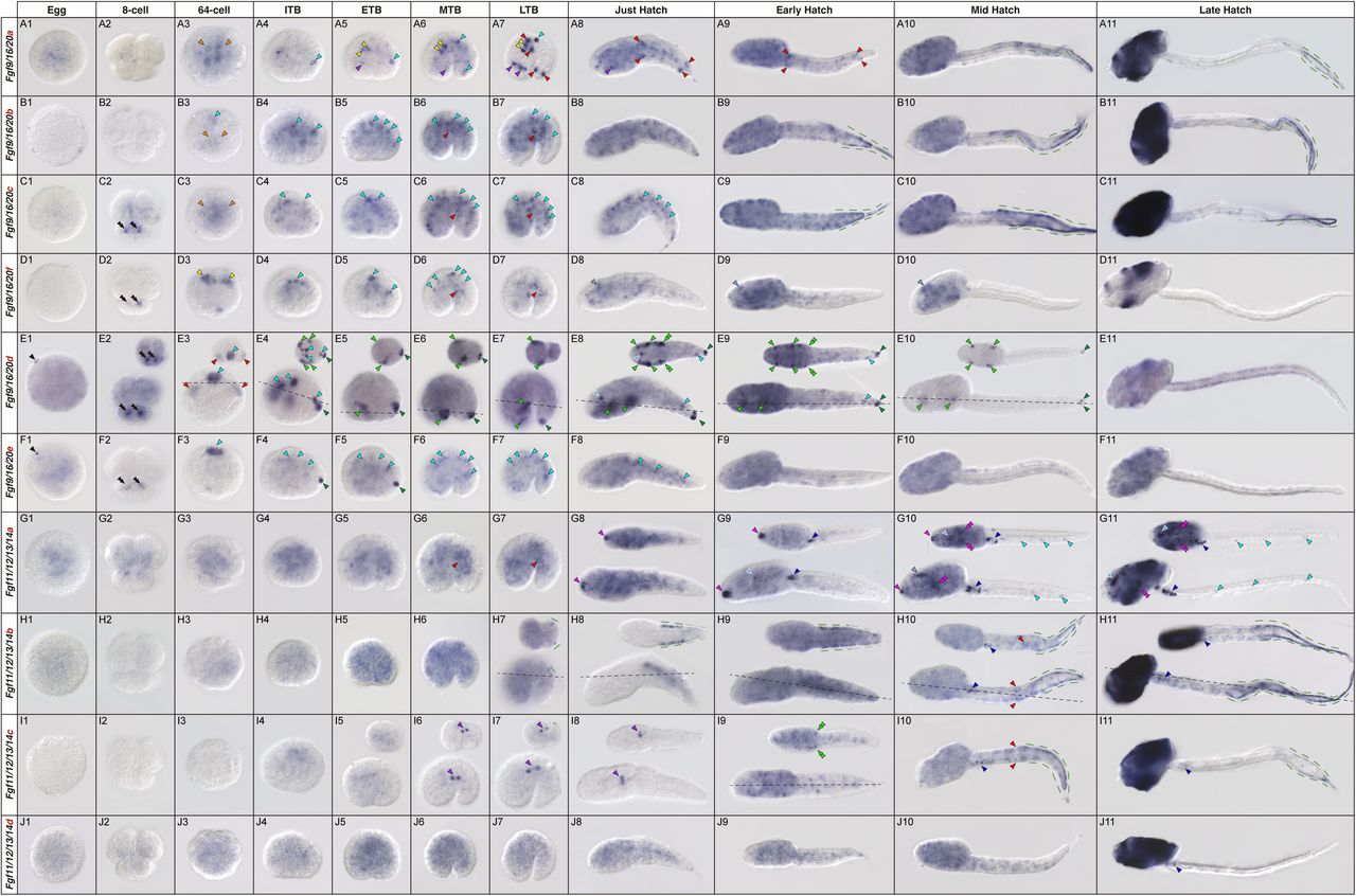
Mathieu Reynaud, Stefano Vianello, Shu-Hua Lee, Pauline Salis, Mélanie Dusseune, Kai Wu, Bruno Frederich, David Lecchini, Laurence Besseau, Natacha Roux, Vincent Laudet
Timothy J. Duerr, Melissa Miller, Sage Kumar, Dareen Bakr, Jackson R. Griffiths, Aditya K. Gautham, Danielle Douglas, S. Randal Voss, James R. Monaghan
Navyashree A Ramesh, Allison M. Box, Laura Buttitta
Bart Theeuwes, Luke TG Harland, Alexandra Bisia, Ita Costello, Mai-Linh Ton, Tim Lohoff, Stephen J Clark, Ricard Argelaguet, Nicola K Wilson, Wolf Reik, Elizabeth Bikoff, Elizabeth J Robertson, Berthold Gottgens
ETS TRANSCRIPTION FACTOR POINTED CONTROLS GERMLINE SURVIVAL IN DROSOPHILA
Alicia E. Rosales-Nieves, Miriam Marín-Menguiano, Lourdes López-Onieva, Juan Garrido-Maraver, Acaimo González-Reyes
Hinako Maeda, Hiroshi Sasaki
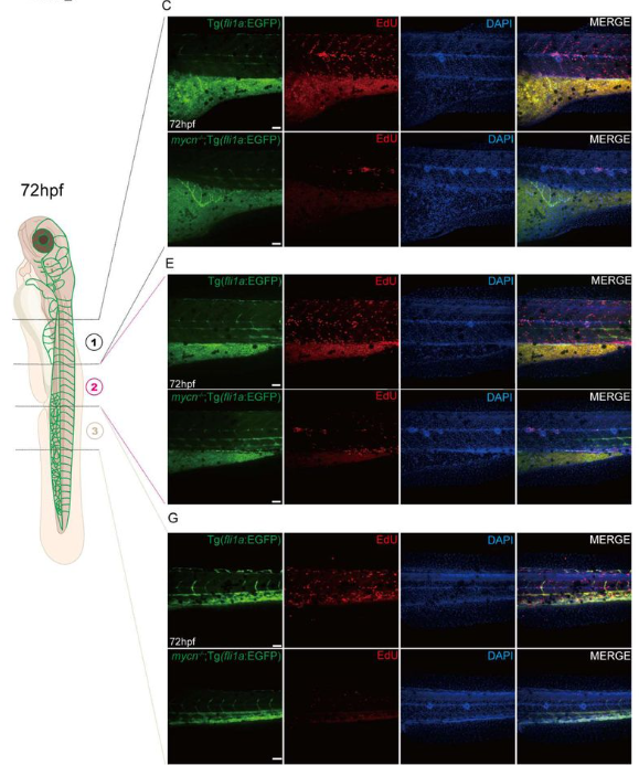
Mycn regulates vascular development through PI3K signaling pathway in zebrafish
Guo-Qin Zhao, Tao Cheng, Peng-Yun Wang, Jing Mo, Feng Yu, Yang Dong, Yun-Fei Li, Yu Feng, Peng-Fei Xu, Li-Ping Shu
NOTCH-driven oscillations control cell fate decisions during intestinal homeostasis
View ORCID ProfileSonja D. C. Weterings, Hiromune Eto, Jan-Daniël de Leede, Amir Giladi, Mirjam E. Hoekstra, Wouter F. Beijk, Esther J. M. Liefting, Karen B. van den Anker, Jacco van Rheenen, Alexander van Oudenaarden, Katharina F. Sonnen
View ORCID ProfileYara Fadaili, Hui-Chun Lu, Hyung Chul Lee, Amra Ryazapova, Claudio D. Stern
Effects Of Aryl Hydrocarbon Receptor Ligand TCDD On Human Trophoblast Cell Development
View ORCID ProfileVinay Shukla, Khursheed Iqbal, Hiroaki Okae, Takahiro Arima, Michael J. Soares
View ORCID ProfileRebecca Garrett Jaszczak, Jay W Zussman, Daniel E Wagner, Diana J Laird
| Morphogenesis & mechanics
Material Properties Of The Embryonic Small Intestine During Buckling Morphogenesis
Jenny Gao, Lucia Martin, Elise A. Loffet, John F. Durel, Panagiotis Oikonomou, Nandan L. Nerurkar
Bitan Saha, Harsha Mahabaleshwar, Charmaine Ho Min, Leslie Boon Haw Leong, Levene Wenqian Chua, Samuel Kwok, Tom J Carney
Morphogenic, molecular, and cellular adaptations for unidirectional airflow in the chicken lung
Kamryn N Gerner-Mauro, Lisandra Vila Ellis, Guolun Wang, Richa Nayak, Peter Y Lwigale, Ross A Poché, Jichao Chen
Mll4 regulates tooth enamel development
Jung-Mi Lee, Hunmin Jung, Qinghuang Tang, Woojung An, Soo-Kyung Lee, Jae W. Lee, Yungki Park, Hyuk-Jae Edward Kwon
Developmental control of E-cadherin junctions by mechanical contractility in Drosophila embryos
Na Zhang, Wangfang Liu, Huiying Lu, Matthias Haering, Fred Wolf, Joerg Grosshans, Zhiyi Lv, Deqing Kong
Mast cells are not essential for pubertal mammary gland branching.
Simran Kapoor, Jimmy Marsden, Clara M Munz, Cyril Carvalho, Marlene Magalhaes Pinto, Bert Malengier-Devlies, Solvig Becker, Guillaume Seuzaret, Katelyn Patatsos, Ramazan Akyol, Marc DALOD, Amy B Pedersen, Gillian J Wilson, Rebecca Gentek
| Genes & genomes
Anna Fleming, Elena V. Knatko, Xiang Li, Ansgar Zoch, Zoe Heckhausen, Stephanie Stransky, Alejandro J. Brenes, Simone Sidoli, Petra Hajkova, Dónal O’Carroll, Kasper D. Rasmussen
Members of an array of zinc finger proteins specify distinct Hox chromatin boundaries
Havva Ortabozkoyun, Pin-Yao Huang, Edgar Gonzalez-Buendia, Hyein Cho, Sang Y. Kim, Aristotelis Tsirigos, Esteban O. Mazzoni, Danny Reinberg
Shreeta Chakraborty, Nina Wenzlitschke, Matthew J. Anderson, Ariel Eraso, Manon Baudic, Joyce J. Thompson, Alicia A. Evans, Lilly M. Shatford‑Adams, Raj Chari, Parirokh Awasthi, Ryan K. Dale, Mark Lewandoski, Timothy J. Petros, Pedro P. Rocha
Temporally restricted activities of En1 regulatory elements underlie distinct limb malformations
Alessa R. Ringel, Andreas Magg, Natalia Benetti, Robert Schöpflin, Mira Kühnlein, Asita Carola Stiege, Ute Fischer, Lars Wittler, Stephan Lorenz, Stefan Mundlos, Lila Allou
Mechanistic basis of lineage restriction
Bohou Wu, Jae Hyun Lee, Kara M. Foshay, Li Zhang, Croydon J. Fernandes, Boyang Gao, Xiaoyang Dou, Chris Z. Zhang, Guoping Fan, Becky X. Xiao, Bruce T. Lahn
Simeiyun Liu, Andrew D. Holmes, Sol Katzman, Upasna Sharma
Yasuhisa Munakata, Mengwen Hu, Yuka Kitamura, Adam L Bynder, Amelia S Fritz, Richard M Schultz, Satoshi H Namekawa
Sharvani Mahadevaraju, Soumitra Pal, Pradeep Bhaskar, Brennan D. McDonald, Leif Benner, Luca Denti, Davide Cozzi, Paola Bonizzoni, Teresa M. Przytycka, Brian Oliver
A newly evolved gene is essential for efficient sperm entry into eggs in Drosophila melanogaster
Sara Y. Guay, Prajal H. Patel, Jonathon M. Thomalla, Kerry L. McDermott, Jillian M. O’Toole, Sarah E. Arnold, Sarah J. Obrycki, Mariana F. Wolfner, Geoffrey D. Findlay
Mechanistic origin and preimplantation development of uniparental and polyploid blastomeres
Yan Zhao, Andrea Fernández-Montoro, Greet Peeters, Tatjana Jatsenko, Tine De Coster, Daniel Angel-Velez, Thomas Lefevre, Thierry Voet, Olga Tšuiko, Ants Kurg, Katrien Smits, Ann Van Soom, Joris Robert Vermeesch
Suad Hassan Alsukari, Huei Teng Ng, Lilly Lang, Sharna Lunn, Shanthi Beglinger, Lauren Carr, Michael Boyes, David Andrew Turner, Bettina Wilm
Huishan Wang, Xingyan Liu, Yamin Liu, Chencheng Yang, Yaxin Ye, Xiaomei Yu, Nengyin Sheng, Shihua Zhang, Bingyu Mao, Pengcheng Ma
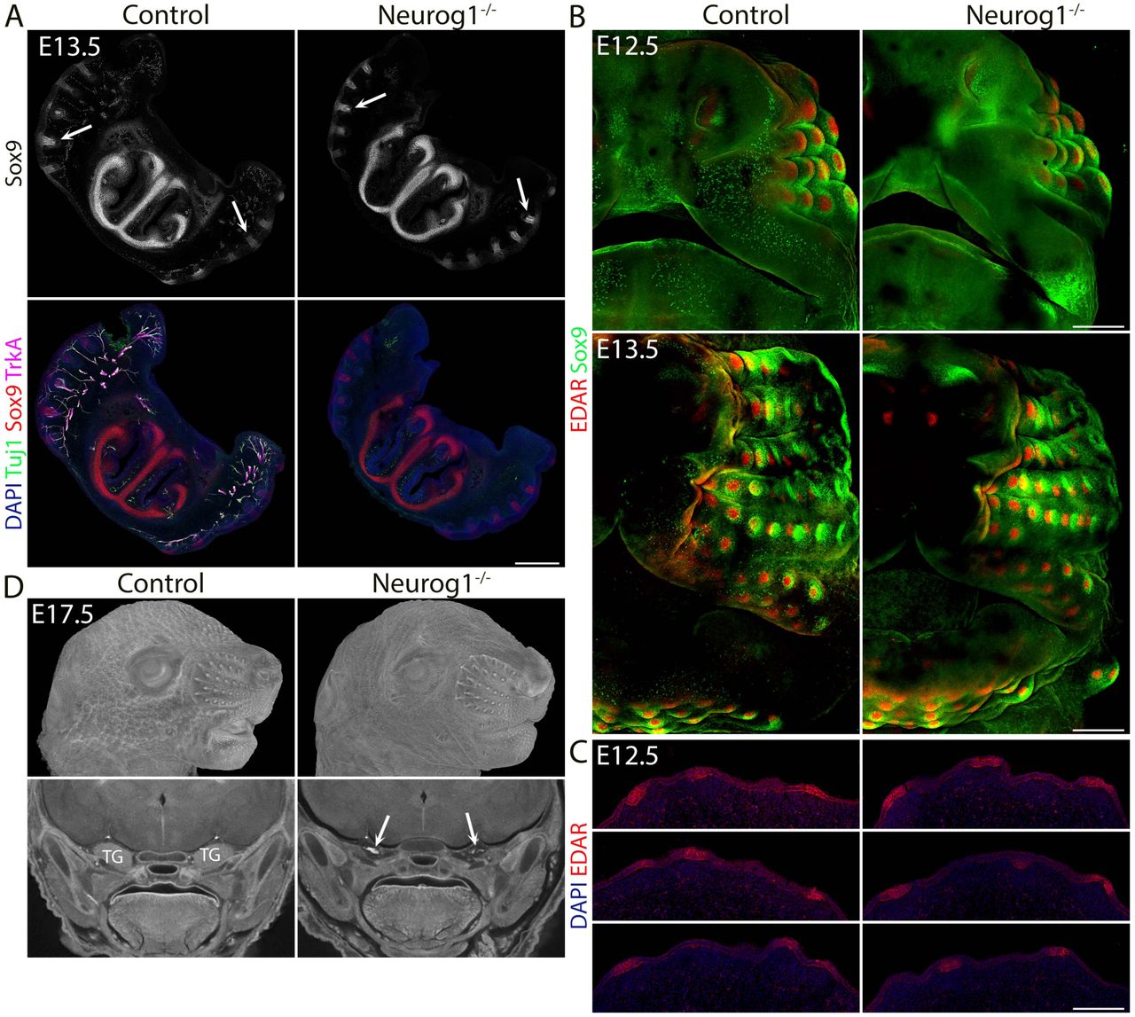
Mesenchymal Meis2 controls whisker development independently from trigeminal sensory innervation
Mehmet Mahsum Kaplan, Erika Hudacova, Miroslav Matejcek, Haneen Tuaima, Jan Krivanek, Ondrej Machon
Neuronal progenitors suffer genotoxic stress in the Drosophila clock mutant per0
Nunzia Colonna Romano, Marcella Marchetti, Anna Marangoni, Laura Leo, Diletta Retrosi, Ezio Rosato, Laura Fanti
Mutations in ErbB2 accumulating in the male germline measured by error-corrected sequencing
Atena Yasari, Monika Heinzl, Theresa Mair, Tina Karimian, Shehab Moukbel Ali Aldawla, Ingrid Hartl, Andrea J. Betancourt, Peter Lanzerstorfer, Irene Tiemann-Boege
Austin Rivera, Jou-Hsuan Roxie Lee, Shruti Gupta, Linda Yang, Raghuveera Kumar Goel, Joseph Zaia, Nelson C. Lau
Gal Finer, Mohammad D. Khan, Yalu Zhou, Gaurav Gadhvi, George S. Yacu, Joo-Seop Park, R. Ariel Gomez, Maria Luisa Sequeira-Lopez, Susan E. Quaggin, Deborah R. Winter
Naoki Hirono, Masakazu Hashimoto, Hinako Maeda, Hiromi Shimojo, Hiroshi Sasaki
Distinct checkpoint and homolog biorientation pathways regulate meiosis I in Drosophila oocytes
Joanatta G. Shapiro, Neha Changela, Janet K. Jang, Jay N. Joshi, Kim S. McKim
A whole-organism landscape of X-inactivation in humans
Björn Gylemo, Maike Bensberg, Colm E. Nestor
Epigenetic and transcriptional regulation of ovarian development altered in ErβKO ovaries
Ryan Mohamadi, Kevin Vo, Yashica Sharma, Amelia Mohamadi, Elizabeth S. Bahadursingh, Patrick E. Fields, M. A. Karim Rumi
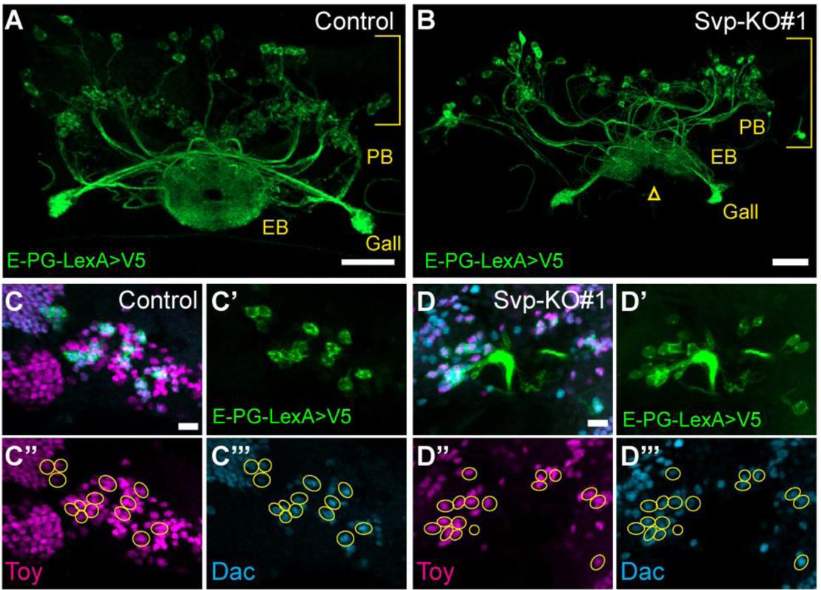
Castor is a temporal transcription factor that specifies early born central complex neuron identity
Noah R. Dillon, Chris Q. Doe
Dynamic convergence of autism disorder risk genes across neurodevelopment
Meilin Fernandez Garcia, Kayla Retallick-Townsley, April Pruitt, Elizabeth Davidson, Yi Dai, Sarah E. Fitzpatrick, Annabel Sen, Sophie Cohen, Olivia Livoti, Suha Khan, Grace Dossou, Jen Cheung, P.J. Michael Deans, Zuoheng Wang, Laura Huckins, Ellen Hoffman, Kristen Brennand
H3K4me2 distinguishes a distinct class of enhancers during the maternal-to-zygotic transition
Matthew D. Hurton, Jennifer M. Miller, Miler T. Lee
Early Hox Gene Expression in Echinoderms
Olga V. Ezhova, Natalya V. Ageenko, Konstantin V. Kiselev, Anastasiya I. Lukinykh, Vladimir V. Malakhov
Simeiyun Liu, Andrew D. Holmes, Sol Katzman, Upasna Sharma
Izumi Oda, Yutaka Satou
Gal Finer, Mohammad D. Khan, Yalu Zhou, Gaurav Gadhvi, George S. Yacu, Joo-Seop Park, R. Ariel Gomez, Maria Luisa Sequeira-Lopez, Susan E. Quaggin, Deborah R. Winter
Haley E. Brown, Brandon P. Weasner, Justin P. Kumar
A dual ribosomal system in the zebrafish soma and germline
Arish N Shah, Friederike Leesch, Laura Lorenzo-Orts, Lorenz Grundmann, Maria Novatchkova, David Haselbach, Eliezer Calo, Andrea Pauli
| Stem cells, regeneration & disease modelling
Emer Aisling King, Eleanor Jacobsen, Nicholas Woolner, Joaquín de Navascués, Owen J Marshall, Jerome Korzelius
PDK-1/S6K and mTORC1 bypass systemic growth restrictions to promote regeneration
Ananthakrishnan Vijayakumar Maya, Liyne Nogay, Lara Heckmann, Isabelle Grass, Katrin Kierdorf, Jörg Büscher, Anne-Kathrin Classen
Janine Hoffmann, Theresa M. Schütze, Annika Kolodziejczyk, Annekathrin Kränkel, Susanne Reinhardt, Razvan P. Derihaci, Cahit Birdir, Pauline Wimberger, Haruhiko Koseki, Mareike Albert
Serotonin neuromodulation directs optic nerve regeneration
Kristian Saied-Santiago, Melissa Baxter, Jaffna Mathiaparanam, Michael Granato
Yuki Ishii, Jessica C. Orr, Marie-Belle El Mdawar, Denise R. Bairros de Pilger, David R. Pearce, Kyren A. Lazarus, Rebecca E. Graham, Marko Z. Nikolic, Robin Ketteler, Neil O. Carragher, Sam M. Janes, Robert E. Hynds
A Sox2 Enhancer Cluster Regulates Region-Specific Neural Fates from Mouse Embryonic Stem Cells
Ian C Tobias, Sakthi D Moorthy, Virlana M Shchuka, Lida Langroudi, Mariia Cherednychenko, Zoe E Gillespie, Andrew G Duncan, Ruxiao Tian, Natalia A Gajewska, Raphaël B Di Roberto, Jennifer A Mitchell
The level of HAND1 controls the specification of multipotent cardiac and extraembryonic progenitors
Adam T Lynch, Naomi Phillips, Megan Douglas, Marta Dorgnach, I-Hsuan Lin, Antony D Adamson, Zoulfia Darieva, Jessica Whittle, Neil A Hanley, Nicoletta Bobola, Matthew J Birket
The microbiota affects stem cell decision making in Hydra
Jinru He, Alexander Klimovich, Sabine Kock, Linus Dahmke, Sören Franzenburg, Thomas C.G. Bosch
Shubham Haribhau Mehatre, Harsh Agrawal, Irene Mariam Roy, Sarah Schouteden, Satish Khurana
Morgan E. McCartney, Gavin M. Wheeler, Audrey G. O’Neill, Jeet H. Patel, Zoey R. Litt, S. John Calise, Justin M. Kollman, Andrea E. Wills
Chromatin activity of IκBα mediates the exit from naïve pluripotency
Luis G. Palma, Daniel Álvarez-Villanueva, María Maqueda, Mercedes Barrero, Arnau Iglesias, Joan Bertran, Damiana Álvarez-Errico, Carlos A. García-Prieto, Cecilia Ballaré, Virginia Rodriguez-Cortez, Clara Bueno, August Vidal, Alberto Villanueva, Pablo Menéndez, Gregoire Stik, Luciano Di Croce, Bernhard Payer, Manel Esteller, Lluís Espinosa, Anna Bigas
Heather K. Le Bleu, Rea G. Kioussi, Astra L. Henner, Victor M. Lewis, Scott Stewart, Kryn Stankunas
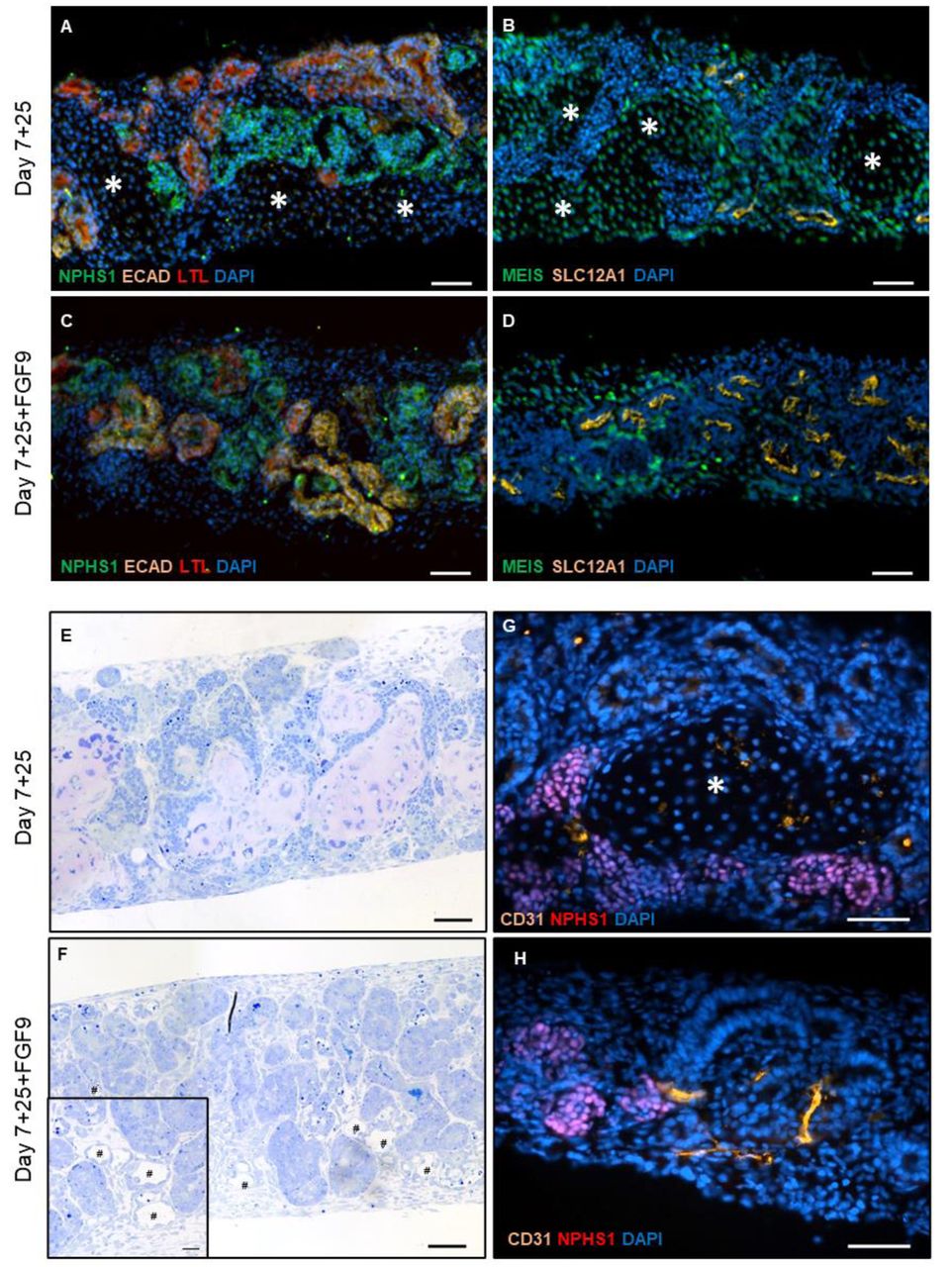
FGF9 treatment reduces off-target chondrocytes from iPSC-derived kidney organoids
Virginie Joris, Anika Schumacher, Paula Marks, Maria Eischen-Loges, Martijn van Griensven, Vanessa L.S. LaPointe
Vincent Truong, Jackson Brougher, Tim Strassmaier, Irene Lu, Dale George, Theodore J. Price, Alison Obergrussberger, Aaron Randolph, Rodolfo J. Haedo, Niels Fertig, Patrick Walsh
Aishwarya Prakash, Maneesha S. Inamdar
Inês Caramelo, Catarina Domingues, Vera M. Mendes, Sandra I. Anjo, Margarida Geraldo, Carla M. P. Cardoso, Mário Grãos, Bruno Manadas
Metadichol induces CD14 Glycoprotein Expression in Human Embryonic Stem Cells and Fibroblasts
P. R. Raghavan
Nuclear actin is a critical regulator of Drosophila female germline stem cell maintenance
Nicole M. Green, Danielle Talbot, Tina L. Tootle
Zhiyuan Cao, Lining Qin, Kaixuan Liu, Chen Yao, Enhong Li, Xiaoyu Hao, Molin Wang, Baichun Jiang, Yongxin Zou, Huili Hu, Qiao Liu, Changshun Shao, Yaoqin Gong, Gongping Sun
Posterior specification of multi-lineage axial assembloids from human pluripotent stem cells.
Nigel Kee, Mélanie Leboeuf, Silvia Gómez, Charles Petipré, Irene Mei, Salim Benlefki, Daniel W Hagey, José Dias, François Lallemend, Samir EL Andaloussi, Johan Ericson, Eva Hedlund
| Plant development
Yuki Hata, Nicola Hetherington, Kai Battenberg, Atsuko Hirota, Aki Minoda, Makoto Hayashi, Junko Kyozuka
Knockout of the tomato HAIRY MERISTEM 4 alters phloem-characteristics and impairs development
Jackson Khedia, Abhay Pratap Vishwakarma, Ortal Galsurker, Shira Corem, Suresh Kumar Gupta, Tzahi Arazi
Marlon Enrique López, Raphael Ricon de Oliveira, Lillian Magalhães Azevedo, Iasminy Silva Santos, Thales Henrique Cherubino Ribeiro, Dapeng Zhang, Antonio Chalfun-Junior
Physiological and Molecular Responses of Projected Future Temperatures on Potato Tuberization
Abigail M. Guillemette, Guillian Hernández Casanova, John P. Hamilton, Eva Pokorná, Petre I. Dobrev, Václav Motyka, Aaron M. Rashotte, Courtney P. Leisner
Jakob Maximilian Horz, Katharina Wolff, Ronja Friedhoff, Boas Pucker
Sebastián R. Moreno, Martin O. Lenz, Elliot M Meyerowitz, James CW Locke, Henrik Jönsson
Youngwoo Lee, Heena Rani, Eileen L. Mallery, Daniel B Szymanski
Viral delivery of an RNA-guided genome editor for transgene-free germline editing in Arabidopsis
Trevor Weiss, Maris Kamalu, Honglue Shi, Zheng Li, Jasmine Amerasekera, Zhenhui Zhong, Benjamin A Adler, Michelle Song, Kamakshi Vohra, Gabriel Wirnowski, Sidharth Chitkara, Charlie Ambrose, Noah Steinmetz, Ananya Sridharan, Diego Sahagun, Jill Banfield, Jennifer Doudna, Steven E. Jacobsen
Sana Dieudonne Dr., Ruth Kristianingsih Ms, Stephanie Laine Ms, Beline JESSON Ms, Veronique VIDAL Ms, Rachel Wells Dr., Richard Morris Pr., Fabrice Besnard Dr.
MAC3A and MAC3B modulate FLM splicing to repress photoperiod-dependent floral transition
Yu-Wen Huang, Chih-Yen Tseng, Yi-Tsung Tu, Hsin-Yu Hsieh, Yu-Sen Wang, Yun-Tung Ly, Yu-Zhen Chen, Shih-Long Tu, Chin-Mei Lee
Conserved role of the SERK–BIR module in development and immunity across land plants
Yijia Yan, Jaqueline Mellüh, Martin A. Mecchia, Hyung-Woo Jeon, Katharina Melkonian, Clemens Holzberger, Anne Harzen, Sara Christina Stolze, Rainer Franzen, Yuki Hirakawa, Ana I. Caño Delgado, Hirofumi Nakagami
Carlos Henrique Cardon, Victoria Lesy, Catherine Fust, Thales Henrique Cherubino Ribeiro, Owen Hebb, Raphael Ricon de Oliveira, Mark Minow, Antonio Chalfun Junior, Joseph Colasanti
Chengzhi Ren, Jule Bodendorf, Jurgen Kleine-Vehn
Natural alleles of LEAFY and WAPO1 interact to regulate spikelet number per spike in wheat
Junli Zhang, German F Burguener, Francine Paraiso, Jorge Dubcovsky
Developmental variability in cotton fiber cell wall properties linked to important agronomic traits
Michael C Wilson, Alexander H Howell, Anika Sood, Youngwoo Lee, Pengcheng Yang, Heena Rani, Elena Yu, Eileen L. Mallery, Sivakumar Swaminathan, Corrinne E. Grover, Jonathan F. Wendel, Olga A. Zabotina, Jun Xie, Chelsea S. Davis, Daniel Szymanski
Flower bud cooling protects pollen development and improves fertility during heatwaves
Martijn J. Jansen, Stuart Y. Jansma, Klaske M. Kuipers, Wim H. Vriezen, Frank F. Millenaar, Teresa Montoro, Carolien G.F. de Kovel, Fred A. van Eeuwijk, Eric J.W. Visser, Ivo Rieu
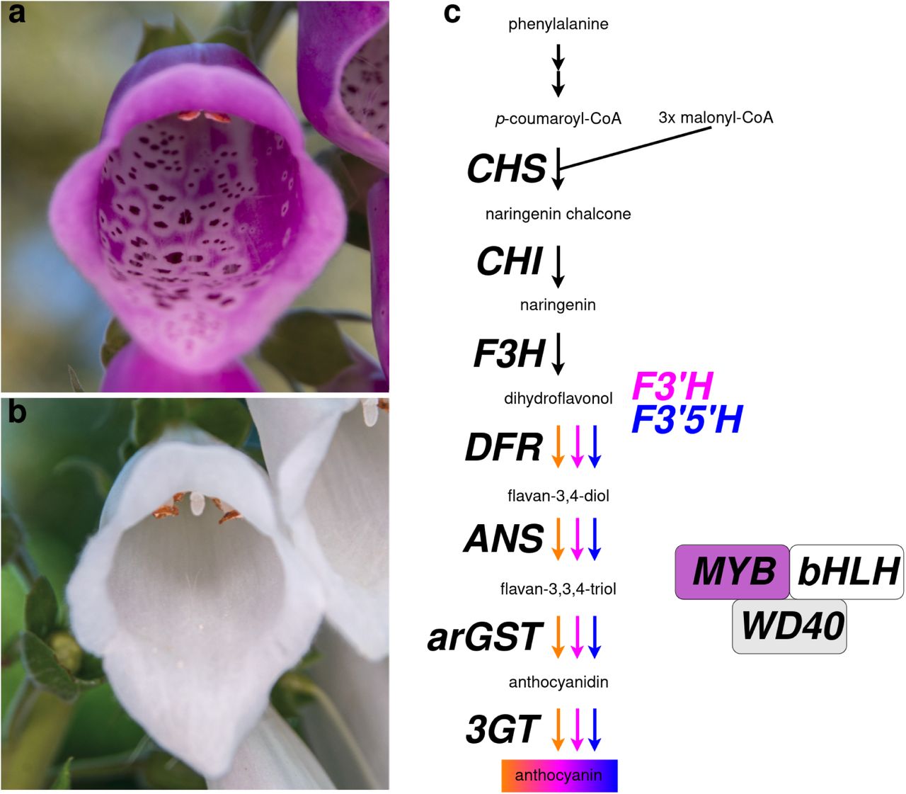
Jurriaan M. de Vos, Yannick Woudstra, Ilia J. Leitch, Oriane Hidalgo
Victoria Spencer, Eva-Sophie Wallner, Katharina Jandrasits, Natalie Edelbacher, Magdalena Mosiolek, Liam Dolan
Xiangzi Zheng, Qingzheng Lu, Yuling Luo, Jiaxuan Xu, Weiqi Wang, Min Tan, Dongmei Liao, Wuqiang Hong, Sirong Chen, Chuheng Lin, Xiaoli Wang, Chunlan Fan, Habiba, Xiaowei Wang, Yanyun Li, Yu Zhang, Wenfang Lin, Ying Miao
Soya Nakagawa, Atsushi Hoshino, Kazuyo Ito, Hiroyo Nishide, Katsuhiro Shiratake, Atsushi J Nagano, Yasubumi Sakakibara
Guard-cell phytosterol homeostasis is critical for proper stomatal development
Chih-Chung Yen, Ya-Wen Hsu, Kuan-Chieh Leu, Sheau-Shyang Chen, Tzu-Yun Chen, Chien-Ta Juan, Chi Kuan, Jei-Fu Shaw, Chin-Min Kimmy Ho, Guang-Yuh Jauh
The receptor-like kinase ALE2 promotes giant cell formation in the sepal epidermis
Frances K Clark, Jessica McGory, Nicholas Russell, Pau Formosa-Jordan, Adrienne H. K. Roeder
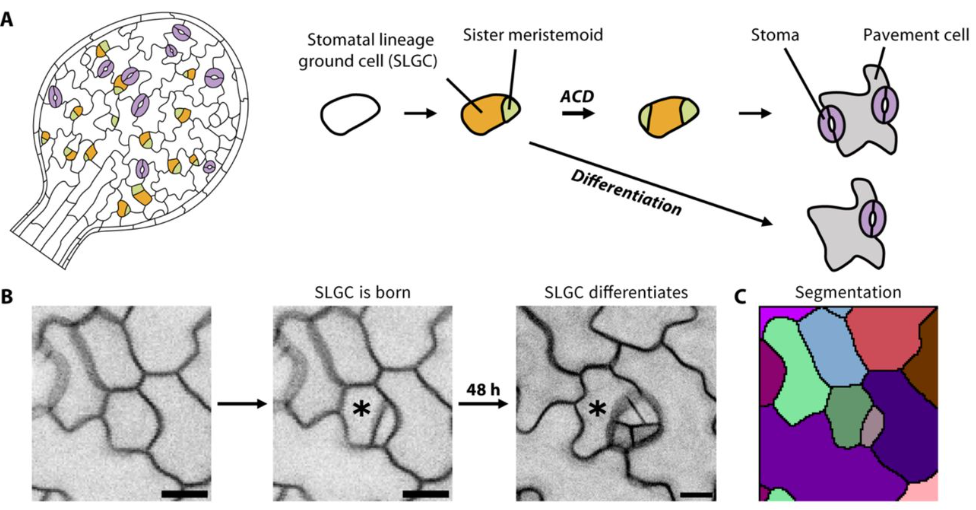
Hannah F. Fung, Gabriel O. Amador, Renee Dale, Yan Gong, Macy Vollbrecht, Joel M. Erberich, Andrea Mair, Dominique C. Bergmann
| Evo-devo
Reverse development in the ctenophore Mnemiopsis leidyi
Joan J. Soto-Angel, Pawel Burkhardt
Protein degradation shapes developmental tempo in mouse and human neural progenitors
Shota Nakanoh, Despina Stamataki, Lorena Garcia-Perez, Chiara Azzi, Hayley L Carr, Alexandra Pokhilko, Lu Yu, Steven Howell, Mark Skehel, David Oxley, Simon Andrews, James Briscoe, Teresa Rayon
A punctuated burst of massive genomic rearrangements and the origin of non-marine annelids
Carlos Vargas-Chávez, Lisandra Benítez-Álvarez, Gemma I. Martínez-Redondo, Lucía Álvarez-González, Judit Salces-Ortiz, Klara Eleftheriadi, Nuria Escudero, Nadège Guiglielmoni, Jean-François Flot, Marta Novo, Aurora Ruiz-Herrera, Aoife McLysaght, Rosa Fernández
Jade Whitlam, Pascal Flohr, Amy Bogaard, Mike Charles, Bill Finlayson, Cheryl A. Makarewicz
Nadja Milivojev, Camila L. Velastegui Gamboa, Gabriele Andreatta, Florian Raible, Kristin Tessmar-Raible
Emily E. K. Kopania, Nathan L. Clark
Microglia cannibalism and efferocytosis leads to shorter lifespans of developmental microglia
Hannah Gordon, Zachary Schafer, Cody J. Smith
Sara Wighard, Hanh Witte, Ralf J. Sommer
Gaspar Sánchez-Serna, Jordi Badia-Ramentol, Paula Bujosa, Alfonso Ferrández-Roldán, Nuria P. Torres-Águila, Marc Fabregà-Torrus, Johannes N. Wibisana, Michael J. Mansfield, Charles Plessy, Nicholas M. Luscombe, Ricard Albalat, Cristian Cañestro
Cell Biology
Juhi G. Narula, Sarah M. Wignall
Huanhuan Liu, Anupama Binoy, Siqi Ren, Thomas C. Martino, Anna E. Miller, Craig R. G. Willis, Shivakumar R. Veerabhadraiah, Abhijit Sukul, Joanna Bons, Jacob P. Rose, Birgit Schilling, Michael J. Jurynec, Shouan Zhu
Actin dynamics switches two distinct modes of endosomal fusion in yolk sac visceral endoderm cells
Seiichi Koike, Masashi Tachikawa, Motosuke Tsutsumi, Takuya Okada, Tomomi Nemoto, Kazuko Keino-Masu, Masayuki Masu
Cara Moravec, Francisco Pelegri
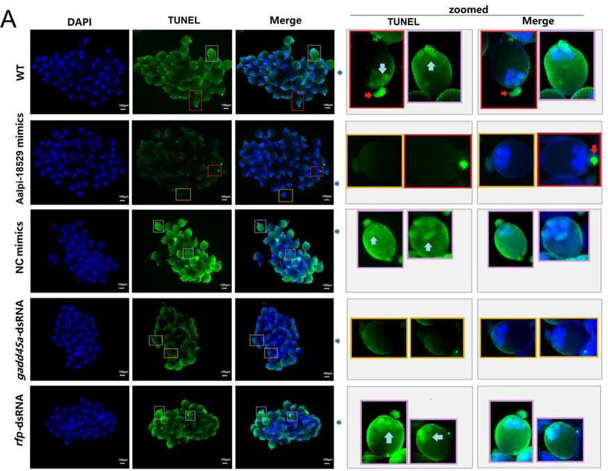
Lu Yang, Yonghui Gao, Yulan Chen, Shuyi Ren, Yifan Guo, Peiwen Liu, Khadija Batool, Jianxia Tang, Jinbao Gu
Elham Alzyoud, Dóra Németh, Viktor Vedelek, Titanilla Szögi, Viktória Petra Tóth, Mónika Krecsmarik, Edit Ábrahám, Zoltán Lipinszki, Rita Sinka
Proteome asymmetry in mouse and human embryos before fate specification
Lisa K. Iwamoto-Stohl, Aleksandra A. Petelski, Maciej Meglicki, Audrey Fu, Saad Khan, Harrison Specht, Gray Huffman, Jason Derks, Victoria Jorgensen, Bailey A.T. Weatherbee, Antonia Weberling, Carlos W. Gantner, Rachel S. Mandelbaum, Richard J. Paulson, Lisa Lam, Ali Ahmady, Estefania Sanchez Vasquez, Nikolai Slavov, Magdalena Zernicka-Goetz
The PIDDosome controls cardiomyocyte polyploidization during postnatal heart development
M Leone, N Kinz, F Eichin, D Obwegs, VC Sladky, D Rizzotto, C Manzl, K Moos, ED Jacotot, C Savko, MA Sussman, M Boerries, A Villunger
Natascha Schippel, Jing Wei, Xiaokuang Ma, Mrinalini Kala, Shenfeng Qiu, Peter Stoilov, Shalini Sharma
Sustained fertility from first-wave follicle oocytes that pause their growth
Bikem Soygur, Eliza A. Gaylord, Mariko H. Foecke, Steven A. Cincotta, Tegan S. Horan, Anna Wood, Paula E. Cohen, Diana J. Laird
Modelling
Physical modeling of embryonic transcriptomes identifies collective modes of gene expression
Dominic J. Skinner, Patrick Lemaire, Madhav Mani
Yuchen Wen, Hang He, Yunxi Ma, Lorie Chen Cai, Huaquan Wang, Yanmei Li, Baobing Zhao, Zhigang Cai
Somya Mani, Tsvi Tlusty
Role of Data-driven Regional Growth Model in Shaping Brain Folding Patterns
Jixin Hou, Zhengwang Wu, Xianyan Chen, Dajiang Zhu, Tianming Liu, Gang Li, Xianqiao Wang
A Computational Framework for Modeling Emergence of Color Vision in the Human Brain
Atsunobu Kotani, Ren Ng
Streamline tractography of the fetal brain in utero with machine learning
Weide Liu, Camilo Calixto, Simon K. Warfield, Davood Karimi
Multiple Notch ligands in the synchronization of the segmentation clock
Marcos Wappner, Koichiro Uriu, Andrew C. Oates, Luis G. Morelli
Tools & Resources
Silk-Ovarioids: Establishment and characterization of human ovarian primary cells 3D-model system
Valentina Di Nisio, Tianyi Li, Zhijie Xiao, Kiriaki Papaikonomou, Anastasios Damdimopoulos, Ákos Végvári, Filipa Lebre, Ernesto Alfaro-Moreno, Mikael Pedersen, Terje Svingen, Roman Zubarev, Ganesh Acharya, Pauliina Damdimopoulou, Andres Salumets
Phylogeny, morphology, and behavior of the new ciliate species Stentor stipatus
D. Rajan, B. Lee, A. Albright, E. Tang, A. Maravillas, C. Vargas, W. F. Marshall, D. Cortes
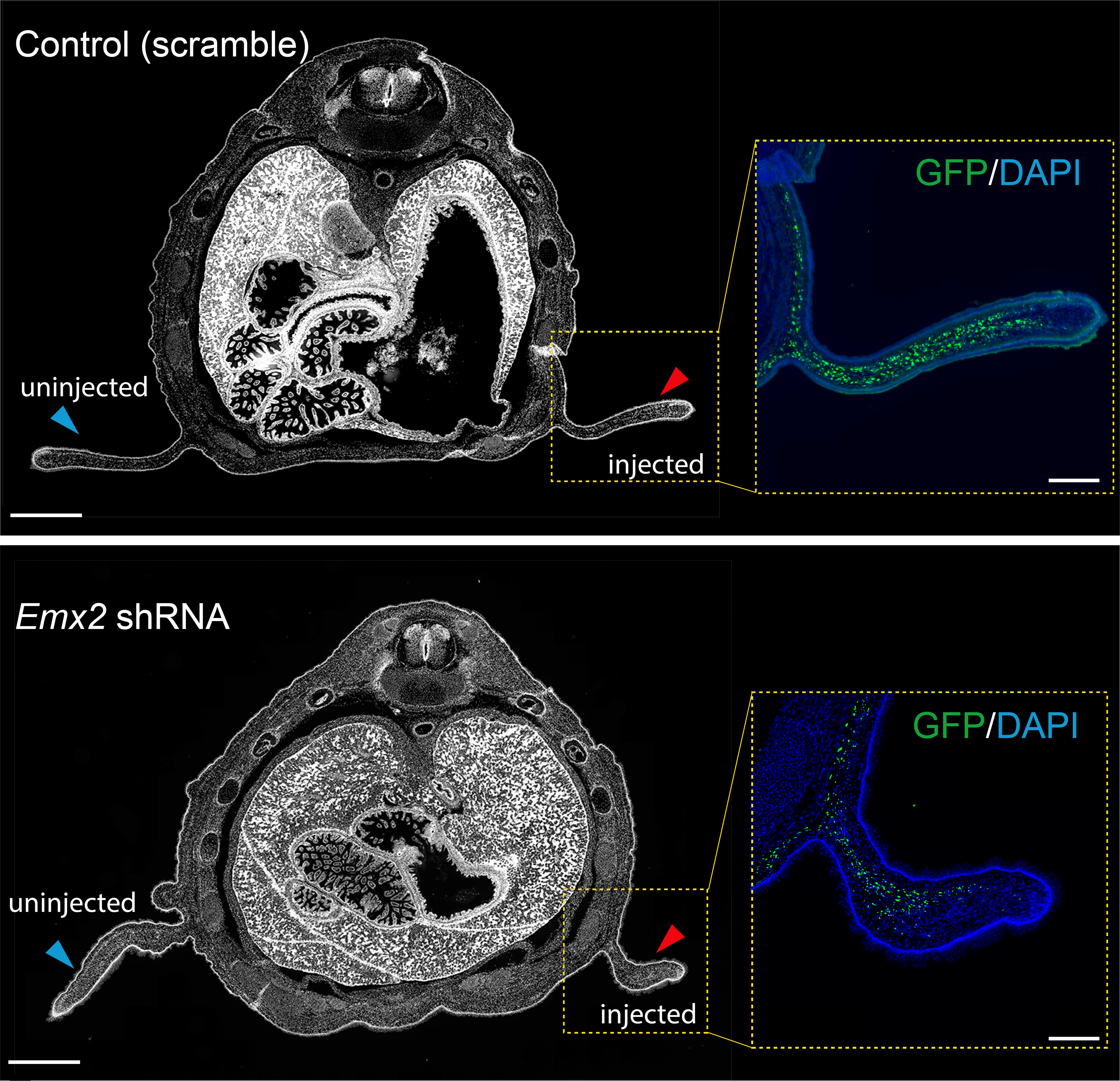
CRISPR-Cas13d as a molecular tool to achieve targeted gene expression knockdown in chick embryos
Minyoung Kim, Erica J. Hutchins
Compatibility of time-lapse dry incubator on in vitro production of bovine embryos
Haruhisa Tsuji, Hiroki Nagai, Sayaka Kobinata, Hinata Koyama, Atchalalt Khurchabilig, Noritaka Fukunaga, Yoshimasa Asada, Satoshi Sugimura
Shiyu Sun, Yi Zheng, Yung Su Kim, Zheng Zhong, Norio Kobayashi, Xufeng Xue, Yue Liu, Zhuowei Zhou, Yanhong Xu, Jinglei Zhai, Hongmei Wang, Jianping Fu
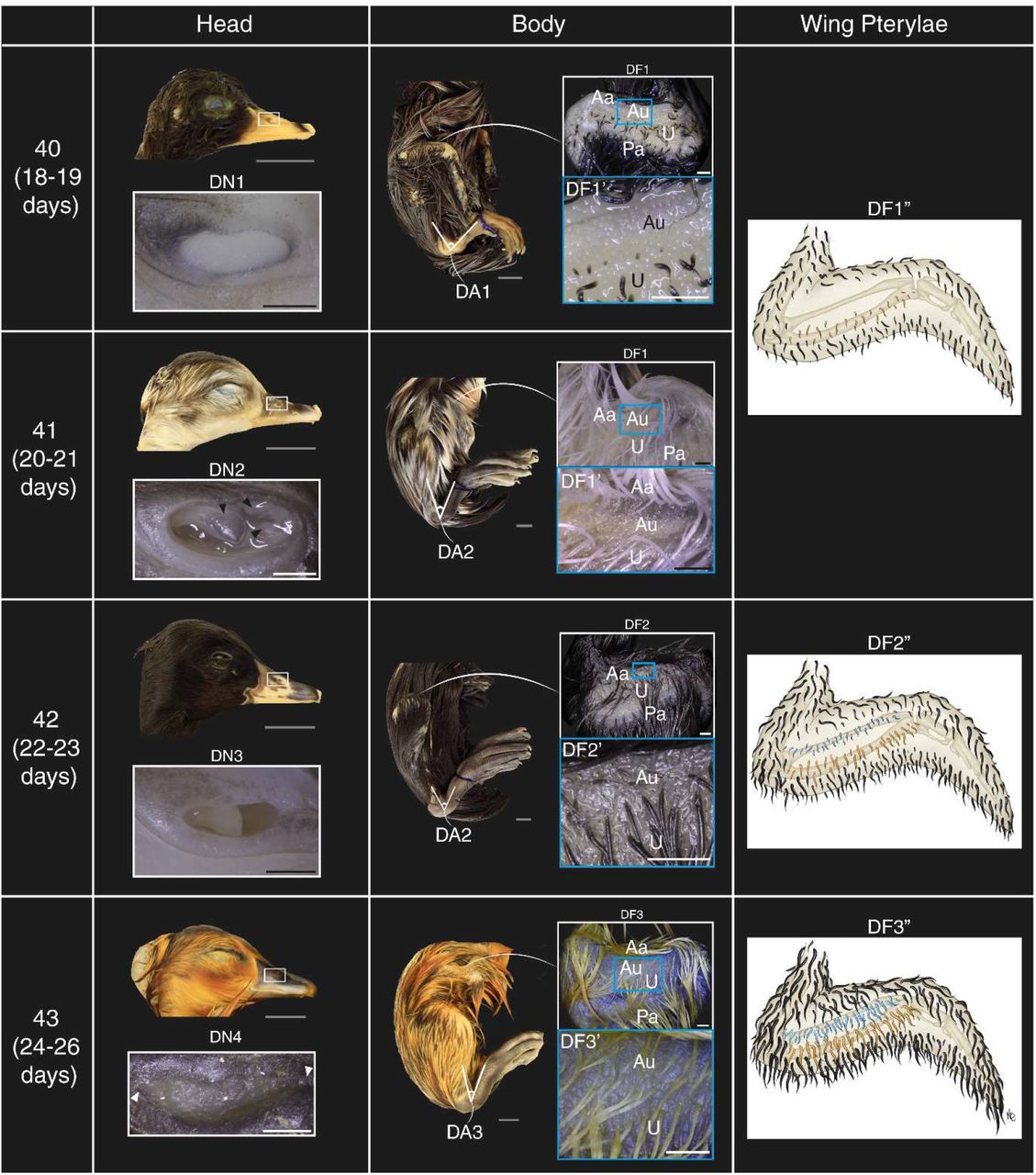
Bassel Arnaout, Kaylen Brzezinski, Benjamin Steventon, Daniel J. Field
Juliana I. Candelaria, Ramon C. Botigelli, Carly Guiltinan, Ariella Shikanov, Anna C. Denicol
Zhen Qi, Zhanguang Zuo, Yi Dong, Jingyu Shao, Chong Wang, Rosanna Zhang
Caroline Hookway, Antoine Borensztejn, Leigh K. Harris, Sara Carlson, Gokhan Dalgin, Suraj Mishra, Nivedita Nivedita, Ellen M. Adams, Tiffany Barszczewski, Julie C. Dixon, Jacqueline H. Edmonds, Erik A. Ehlers, Alexandra J. Ferrante, Margaret A. Fuqua, Philip Garrison, Janani Gopalan, Benjamin W. Gregor, Maxwell J. Hedayati, Kyle N. Klein, Chantelle L. Leveille, Sean L. Meharry, Haley S. Morris, Gouthamrajan Nadarajan, Sandra A. Oluoch, Serge E. Parent, Amber Phan, Brock Roberts, Emmanuel E. Sanchez, M. Filip Sluzewski, Lev S. Snyder, Derek J. Thirstrup, John Paul Thottam, Julia R. Torvi, Gaea Turman, Matheus P. Viana, Lyndsay Wilhelm, Chamari S. Wijesooriya, Jie Yao, Julie A. Theriot, Susanne M. Rafelski, Ruwanthi N. Gunawardane
Birthe Thuesen Mathiesen, Mayu Ohta, Boris Pinto De Magalhaes, Chiara Castelletti, Vincenzo Perria, Lionel Christiaen, Naoyuki Ohta
Three-dimension transcriptomics maps of whole mouse embryo during organogenesis
Mengnan Cheng, Huiwen Zheng, Qi Fang, Yinqi Bai, Chao Liu, Hailin Pan, Zhewei Zhang, Qin Lu, Chang Shi, Tianyi Xia, Zehua Jing, Huanlin Liu, Ning Feng, Guojun Fu, Yumei Li, Jing Feng, Zepeng Li, Jingjing Wang, Yuanyuan Chen, Lianying Wang, Zhonghan Deng, Mei Li, Longqi Liu, Ao Chen, Xun Xu
A Human Biomimetic Intestinal Mucosa Model to Study Gastrointestinal Development and Disease
Alessandro Dei, Carlemi Calitz, Joep Korsten, Nina Johannesson, Eline Freeze, Allen Eaves, John Stingl, Ryan K Condor, Wing Chang, Dasja Pajkrt, Katja C. Wolthers, Adithya Sridhar, Salvatore Simmini
Moult cycle and setal development of the Atlantic ditch shrimp Palaemon varians Leach, 1814
Kenneth Kim, Jonathan Antcliffe, Allison C. Daley, Marc Robinson-Rechavi
Whole-embryo Spatial Transcriptomics at Subcellular Resolution from Gastrulation to Organogenesis
Yinan Wan, Jakob El Kholtei, Ignatius Jenie, Mariona Colomer-Rosell, Jialin Liu, Joaquin Navajas Acedo, Lucia Y. Du, Mireia Codina-Tobias, Mengfan Wang, Ahilya Sawh, Edward Lin, Tzy-Harn Chuang, Susan E. Mango, Guoqiang Yu, Bogdan Bintu, Alexander F. Schier
Dissecting the regulatory logic of specification and differentiation during vertebrate embryogenesis
Jialin Liu, Sebastian M. Castillo-Hair, Lucia Y. Du, Yiqun Wang, Adam N. Carte, Mariona Colomer-Rosell, Christopher Yin, Georg Seelig, Alexander F. Schier
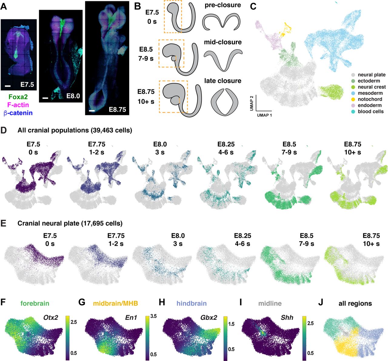
A single-cell atlas of spatial and temporal gene expression in the mouse cranial neural plate
Eric R. Brooks, Andrew R. Moorman, Bhaswati Bhattacharya, Ian Prudhomme, Max Land, Heather L. Alcorn, Roshan Sharma, Dana Pe’er, Jennifer A. Zallen
 (No Ratings Yet)
 (3 votes)