This is the latest dispatch from a recipient of a Development Travelling Fellowship, funded by our publisher The Company of Biologists.
Learn more about the scheme, including how to apply, here, and read more stories from the Fellows here.
Barbara Swierczek
I am a PhD student at the University of Warsaw in Poland. In my home lab, headed by Prof. Maria Anna Ciemerych-Litwinienko, I study the signalling pathways affecting pluripotency and differentiation in mouse embryonic stem cells, focusing on Wnt proteins. Wnts were first described more than 30 years ago, and since then became an intensively studied yet challenging field of cell biology. Why? First of all, there are 19 Wnt proteins found in vertebrates that activate a wide range of signalling pathways. Additionally, they often act antagonistically, creating a complicated and complex network of interactions. Next, Wnts play a key role during development, when their activity is orchestrated in a very precise manner: a defined Wnt has to be expressed at a right moment in a defined place.
It has long been appreciated that Wnts, particularly those dependent on β-catenin transduction (so called ‘canonical’ Wnts), are crucial for gastrulation, the process by which the three germ layers are formed in the embryo. However, despite well-documented function of Wnts in this process, it is not fully understood exactly how these proteins are regulated during gastrulation. Expression of Wnts can be regulated by inhibitors, other Wnts acting antagonistically or miRNAs. The latter are short (approximately 20 nucleotide long) non-coding RNAs, which block the expression of target genes. While studying Wnts and their regulation during mouse embryonic stem cell differentiation I came across some evidence that Wnt signalling can be regulated by miRNAs targeting either Wnts themselves or Wnt receptors or transducers. I found it very interesting and decided to pursue this link further. Also, I wanted to learn how to study this link in a developing embryo.
Around this time, I heard that the The Company of Biologists funds Travelling Fellowships for a short research visit. I decided to give it a go, and applied for three-month long fellowship in the Professor Andrea Münsterberg’s lab at the University of East Anglia in Norwich, Great Britain. Professor Münsterberg is a leading specialist in miRNA functions during development, especially their functions in modulating signalling pathways in skeletal muscle and the heart. During my stay in Norwich I had an opportunity to work on a model organism which was different for me: a chick embryo. Chick is a potent model: in contrast to mammalian development which occurs mostly within the female’s body, chick development can be studied and manipulated easily in the egg, even during later stages of development. Working with a new model was quite a challenge at first, but also an exciting perspective in the same time.
Me isolating embryos
UEA in Spring
UE covered in snow
Goodbye party
My project concerned the role of miRNAs in regulating Wnt pathway in the gastrulating chick embryo. I focused on Wnt5a, which is a double-faced Wnt: it can bind to different receptors and activate both canonical and noncanonical signalling pathways. I studied the relationship between this Wnt and an miRNA which I selected as a putative regulator as a result of sequence analyses. During my fellowship I learned how to isolate chick embryos and how to manipulate them. Since the group of Professor Münsterberg studies different aspects of chick development, I became familiar with various stages of this process, from gastrulation to more advanced stages. I was able to learn a lot from people in the group: real specialists in working on chick development. Also, I learned a lot about miRNAs and the methods of miRNA analysis which will be beneficial for my future research.
Thanks to my short-term fellowship at UEA, I had a great opportunity to experience an international scientific environment. I enjoyed my stay in Norwich which is not a big city – there are about 100,000 people living there – and for this reason a peaceful and quiet one. Thanks to the Development Travelling Fellowship I was able to learn a lot about the techniques I had not used before, something that encouraged me to seek new challenges for my future research. But what I think is the most important aspect of my visit was making new connections and meeting people working in the field. I wanted to thank once again everyone in the Münsterberg group for helping me – I hope to work with you again!
From the MRC Weatherall Institute of Molecular Medicine blog.
Stem cell turnover and tissue maintenance is a stochastic process. This means that a randomly occurring mutation has an unknown chance of becoming fixed and spreading within a tissue. Clonal mutations have been observed in apparently healthy tissue, increase in frequency with age and – in some cases – have been described as a pre-malignant state (e.g. clonal haematopoiesis). In certain tissues, such as the colonic epithelium, the contribution of mutations in stem cells to neoplastic transformation remains unclear.
This process is a major interest of Ed Morrissey, who joined the MRC WIMM Centre for Computational Biology in late 2016. His group has recently published a mathematical model that aims to address how functional mutations can contribute to altered stem cell dynamics, with the hope of understanding precisely how these rare mutations accumulate in the lead up to cancer.
The Bloomekatz laboratory in the Department of Biology at the University of Mississippi in Oxford, MS is seeking a research associateto assist in our investigations of the fundamental mechanisms underlying cardiac morphogenesis and disease using zebrafish. Please see our website thebloomekatzlaboratory.org for further details on our research. The successful candidate will have an opportunity to be involved in all aspects of our dynamic innovative research program; from experimental design to data analysis and publication. Duties may involve – conducting developmental/cell biological experiments, analyzing imaging and next-generation sequencing data, zebrafish husbandry, and mentoring undergraduate laboratory members.The candidate will work closely with and be trained by Dr. Bloomekatz. Interested in joining our group, apply online: https://careers.olemiss.edu, keyword cardiac.Salary dependent on experience. This position is eligible for benefits. The University of Mississippi is an EOE/AA/Minorities/Females/Vet/Disability/Sexual Orientation/Gender Identity/Title VI/Title VII/Title IX/ADA/ADEA employer.
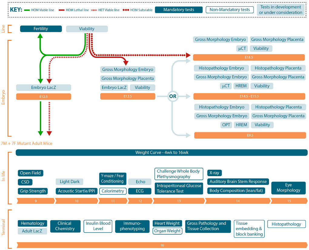
The entire genome of many species has now been sequenced, but the function of the majority of genes still remains unknown. This is where the International Mouse Phenotyping Consortium (IMPC) comes in, with the goal of characterising all 20,000 or so protein-coding mouse genes. To achieve this, genes are systematically inactivated then mice are put through a standardised phenotyping platform, with tests undertaken across a broad range of biological systems.
The consortium is comprised of 19 research institutions, 5 national funders and 11 countries. Each centre focuses on particular genes, applies standardised tests and then records the resulting data. After this, phenotype analysis is conducted and the resulting data and statistics made freely available to the research community. As well as completing large scale comparative studies, the overall aim of the project is to create a platform for this data where researchers and clinicians can search for genes, phenotypes or diseases of interest to help them understand human biology, health and disease.
Professor Steve Brown, the IMPC chair says “The IMPC is rising to the challenge of generating a complete functional catalogue of the mouse genome. Since its inception in 2011, it has made great strides with a third of the genome already analysed. Moreover, many startling and hitherto undiscovered features of the mammalian genome landscape have been revealed.”
There are now over 6,000 genes with mouse mutant data on an isogenic genetic background (C57BL/6N) on the IMPC website, all of which can be viewed and downloaded for free. In its initial stages the knock out lines used for IMPC were all made in ES cells by homologous recombination, all containing a lacZ reporter and many of them generating conditional mouse lines. However, as for many areas of developmental biology, new gene editing technologies, in particular CRISPR/Cas9, have condensed the process of generating knockout mouse lines. Advancements such as this have improved production and will allow all 20,000 genes to be characterised in the next few years.
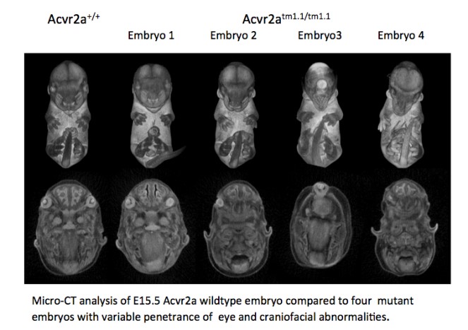
Around a third of knockout genes are embryonic lethal and consequently developmental biology is an integral part of the IMPC project. In particular, there is an extensive embryo phenotyping pipeline that includes systematic harvest of embryos at set stages, capture of morphology by 3D imaging (OPT or microCT, depending on embryonic stages) and evaluations of morphological abnormalities in mutant embryos. These procedures can allow direct insight into the window of lethality for each mouse line, but they also provide valuable information on gene function. For example, accurate measurements of organ size and shape can be collected using microCT scan data, or macroscopic observations undertaken by a trained researcher. Importantly, all 3D data sets are available to download from the website for further in-depth analysis by specialist researchers.
In the last few years the IMPC has made major discoveries about parts of the genome that were up to now unexplored, with novel genes discovered relating to areas such as embryonic development, deafness, diabetes, and rare diseases. Recent high profile publications have included research focusing on inferring mammalian gene function, studies on specific human conditions, sexual dimorphism in mouse research, and even using IMPC data to help in wildlife conservation. New methods and analysis tools have also been developed under the umbrella of the IMPC, such as PhenStat, an extensive library of functions that analyse the phenotypical data. Another example is illustrated by a recent article that highlights a new bioimage informatics platform for high-throughput embryo phenotyping. Although this platform was built for the IMPC, the software tools that facilitate the analysis and dissemination of 3D images can be used by other researchers, and is available under an open-source licence. Indeed, sharing resources across the research community is a crucial aspect of the IMPC, and mutant mouse lines can be obtained from the website.
The IMPC is continuing to deliver data and mouse models for the developmental biology field and ultimately will be part of the effort to understanding and treating genetic conditions in humans. More information on the latest research of the IMPC can be found on our blog, and you can search the IMPC database for free at https://www.mousephenotype.org/.
A Technician position in Molecular Biology is available starting November 2018 in the group of Thomas Lecuit at the Developmental Biology Institute of Marseille (IBDM, CNRS UMR7288), Marseille, France. Funding is provided by an ERC grant. The initial appointment will be made for 1 year, with a possible extension to up to 5 years.
We are seeking a highly-motivated candidate with some practical experience in molecular biology with a specialized training as a laboratory technician recognized by a diploma (Bachelor +2 : BTS, DUT,…). Since the working language in the laboratory is English, a rather good understanding in English would be appreciated. The Technician will perform Genetic Engineering in Drosophila using recent molecular biology cloning and CRISPR mediated editing techniques. This position would suit a recent graduate with some practical experience in molecular biology and seeking training in recent Genetic Engineering techniques.
Are you interested in metabolism/nutrition/organ size control/developmental neurobiology? A postdoctoral researcher position is available in the Cheng Lab at the Peter MacCallum Cancer Institute from the beginning of 2019 in sunny Melbourne, to work on organ cross talk, metabolism, or developmental neurobiology related projects in Drosophila. We are looking for postdoc candidates with expertise in metabolism/imaging or other areas of developmental/cell biology, experience with flies preferred but not essential. You need to be finishing or have recently finished a PhD in related subjects, with an excellent track record. The lab is located within the Parkville precinct, home to the University of Melbourne and other top research institutes. The position is for 3 year initially (with possibility of extension), internationally competitive salary (A$78k-A$94k) + benefits. This position is suitable for creative, ambitious, independent, highly organised, self-motivated and hard working individuals. Please contact: Louise.cheng[at]petermac.org for informal discussions and application.
Most solid tumours with metastatic potential show a high degree of chromosomal instability.
A study published in the journal Developmental Cell demonstrates that chromosomal instability itself promotes invasive behaviour.
The researchers identify the oncogene Fos and the tumour suppressor Capicua as necessary molecular elements mediating this invasive behaviour.
Barcelona, 26 September 2018.- Metastatic tumours are characterised by a high degree of chromosomal instability—that is to say a constant change in the number and structure of chromosomes they hold. In spite of this observation, it is unknown whether chromosomal instability contributes to the metastatic process.
A study at the Institute for Research in Biomedicine (IRB Barcelona) using Drosophila melanogaster has demonstrated that chromosomal instability itself can induce invasive behaviour in epithelial cells and has identified the underlying molecular mechanisms involved.
To perform this study, the team directed by ICREA researcher Marco Milán generated a fruit fly model with chromosomal instability. “The cells started to actively invade adjacent tissues,” explains Marco Milán, group leader of the Growth Control and Development lab at IRB Barcelona and head of the study.
The aim was to study whether chromosomal instability itself has the capacity to stimulate invasive behaviour in epithelial cells. The results published in Developmental Cell indicate that indeed this is the case and they describe a series of molecular and cell mechanisms that favour cell migration and invasion of other tissues.
In particular, the researchers have demonstrated that invasive cells use the actin cytoskeleton and activate the ERK and JNK signalling pathways to trigger a pro-invasive transcriptional programme executed by the oncogene Fos and repressed by the tumour suppressor Capicua.
“These findings are relevant in the context of cancer because they highlight the causal relationship between aneuploid genomes and cell invasion and they identify the molecular elements involved in the process,” say Najate Benhra and Lara Barrio, first authors of the study and postdoctoral researchers at IRB Barcelona.
Milán adds, “the results increase our understanding of the effects of chromosomal instability on the metastatic potential of solid human tumours and identify molecular targets for the development of future treatments”.
This research has been funded by the Ministry of Science, Innovation and Universities.
Reference article:
Najate Benhra, Lara Barrio, Mariana Muzzopappa, and Marco Milán
Chromosomal instability induces cellular invasion in epithelial tissues
Stockholm University offers a multicultural environment in one of the world’s most dynamic capital cities. With more than 60,000 students and 5,000 staff, the University facilitates individual and societal development by providing top quality education that is tightly linked to its internationally recognized research programs.
The Department of Molecular Biosciences, The Wenner-Gren Institute (MBW) unites 30 independent research groups pursuing fundamental questions in molecular cell biology, infection and immunobiology, and integrative biology. The department carries out experimental research primarily investigating the function of genes and cells in tissues and organisms.
Research project
We are looking for a highly motivated and focused young scientist to carry out postdoctoral research studies in Assistant Prof. Qi Dai’s research group. The research project aims at understanding how key transcription factors determine cell fates using Drosophila as a model system.
Qualification requirements
The successful candidate must have received a doctoral degree from an accredited college/university outside Sweden latest in 2016. The degree must be in the fields of developmental biology, genetics or equivalent. The ideal candidate should have documented experience in fly genetics, tissue manipulation and molecular biology skills. Hands-on experience in work with bioinformatics and in high-throughput assays will be an asset. Excellent English language skills, both written and spoken, are a requisite.
Terms of the fellowship
The terms of this fellowship are regulated by the rules for postdoctoral fellowships at the Stockholm University (http://www.su.se/mbw/mbw-internal/scholarship/guidelines-for-scholarship). The fellowship is for full-time postdoctoral studies during one year, with possibility for extension up to a maximum of two years. The starting date is negotiable and the fellowship is available to start immediately.
Additional information
Further information about the research project and about the conditions of this fellowship can be obtained from Assistant Prof. Qi Dai, qi.dai@su.se, tel. +46 8 16 4149.
Application
Applications should be sent electronically as a single PDF file to the following addresses:
qi.dai@su.se
The applications should comprise the following parts:
Complete CV, including full contact information, date of birth, and copy of the PhD title
List of publications
Personal statement describing research interests (1-2 paragraphs), research
experience (1–2 paragraphs) and career goals (1-2 paragraphs)
Please provide also the names, e-mail addresses and telephone numbers of 2-3 references
Here, Mylène Lancino and myself will introduce our motivation to investigate and delve deeper into one essential and very peculiar process of Stem Cell Biology: the de novo genesis of hematopoietic stem cells, according to a process referred to as the Endothelial-to-Hematopoietic Transition (the EHT, named initially by Kissa & Herbomel 2010). The work led to a paper published in eLife at the end of last month (https://doi.org/10.7554/eLife.37355). We will explain the technical difficulties inherent to the live imaging and image analysis approaches that have been undertaken. We will also summarize some of our main findings and underline their impact on fundamental and conceptual challenges for the hematopoiesis topic.
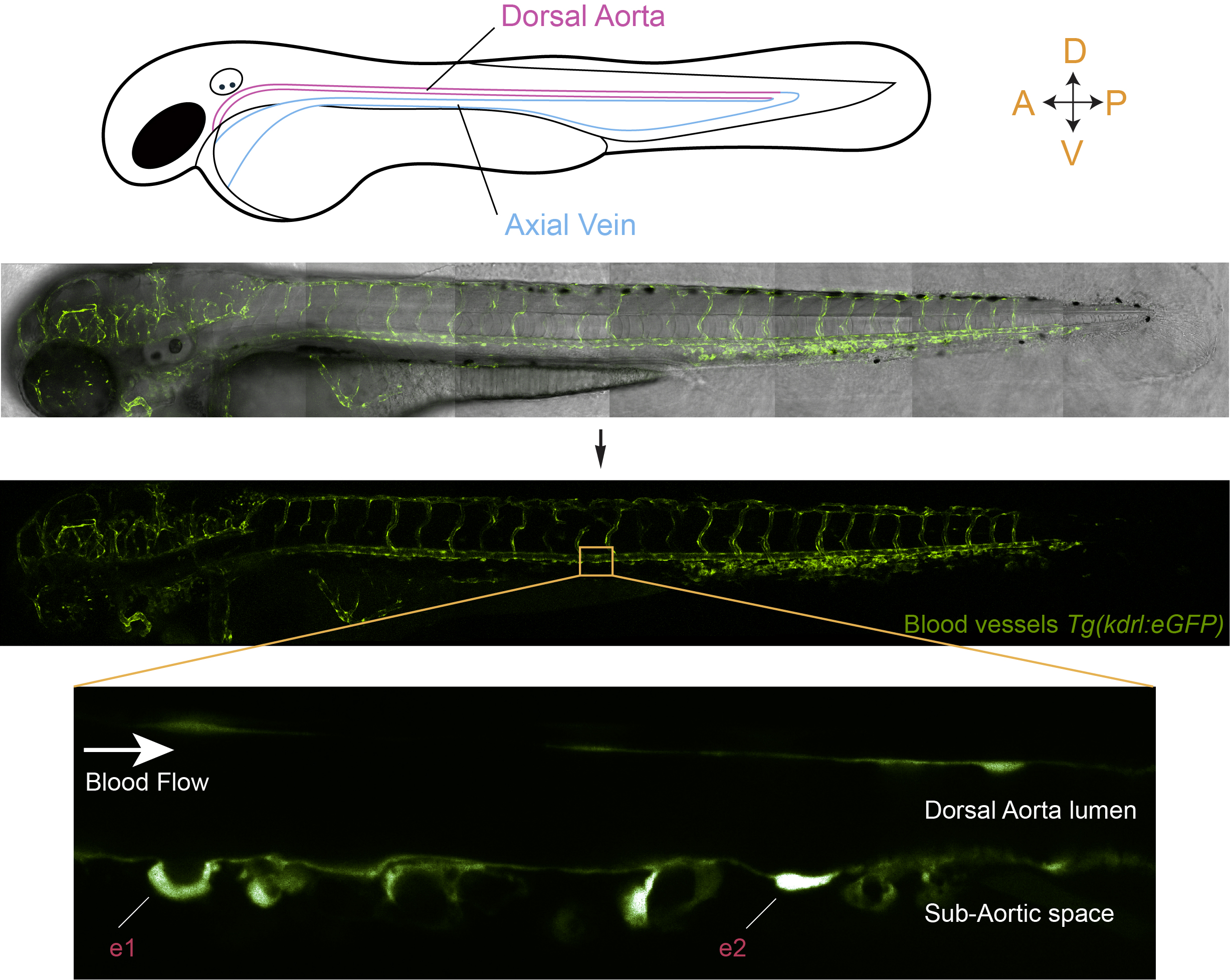
In the year 2010, several important papers that unambiguously demonstrated the vascular origin of hematopoietic stem cells were concomitantly published, which shed light on a long standing debate initiated at the beginning of the 20thcentury (Jordan 1917, Sabin 1917; and for a recent review on hematopoiesis reporting on the findings of 2010 by Bertrand et al; Boisset et al; Kissa & Herbomel; Lam et al, see Klaus & Robin 2017). The breakthrough was possible owing to the power of technical imaging approaches and in particular confocal time-lapse fluorescent microscopy on the living animal, more specifically the zebrafish embryo. This model organism offers unique advantages owing to its small size, its fast development and, last but not least, its transparency. This latter advantage offers unique opportunities for in-depth live imaging and was essential to achieve the goal that we had in the mind when we started our recently published work; i.e imaging the emergence of hematopoietic stem cells from the aortic wall at sufficient spatio-temporal resolution to capture potentially short-lived events and critical intermediate stages so as to be able to propose a comprehensive model of the EHT. In addition, we intended to perform an in–depth descriptive study of the sequential steps of the EHT without disconnecting it from its natural environment. Hence, we aimed at visualizing the morphodynamic changes of endothelial cells, meaning the whole aortic landscape surrounding hemogenic regions, concomitantly to the EHT. The rationale behind this is that some of the EHT unique features when the process is compared with any other cellular dynamic event leading to the extrusion of a cell from an organized epithelium (among which the very peculiar bending of the emerging cell, primarily visualized in Kissa & Herbomel 2010, see also figure 1), suggested that it is adapted to the biomechanical properties of the aortic wall, made of very flat endothelial cells subjected to high mechanical load and exposed to the multidirectional forces exerted by the blood flow. Beyond this and on more conceptual grounds stands also the fundamental issue of the influence of the blood flow on the fate of hemogenic and hematopoietic stem cells (see also later).
Figure 1: In vivo visualization of hematopoietic stem cell emergence via the EHT, from the aortic floor, in a live zebrafish embryo. The middle panels of the figure show the organization of the vascular system in a transgenic line that expresses eGFP under the control of a vascular promoter. The bottom panel shows a magnification of a region of the dorsal aorta in the trunk of the embryo and highlights the emergence of hematopoietic cells via the EHT (e1 and e2; e1 is a more advanced stage than e2).
Let’s come back to the work now and share with the reader our motivation to invest time and energy on the project as well as telling about its experimental challenges.
When Mylène started her PhD, she did not have a strong technical expertise on imaging approaches but was already well acquainted with the zebrafish model. She undertook however the challenge of imaging the EHT more in depth “I was fascinated by the way hematopoietic stem cells are born. I was also really determined to find out the most appropriate set up to visualize as clearly as possible the dynamics of intracellular components during the EHT within the live embryo. Here began a very long journey during which I learned from many imaging specialists and started to realize how tricky in vivo imaging is“, as she said. From my side, when I started with this topic, about a year after Mylène did, with a strong expertise in cell biology and biological membrane dynamics as well as a strong motivation to modelling the EHT, we joined forces and started to analyze the series of time-lapse sequences that she had accumulated in the meanwhile and that were aimed at focusing on two aspects of cell biological features of EHT undergoing cells: the dynamics of their luminal and basal membranes as well as intracellular actin organization (by using double transgenic fish line expressing the membrane marker ras-mCherry and the actin reported Lifeact-eGFP, respectively). But looking into the movies made us realize that few of them would actually allow visualizing the entire EHT process with enough resolution (in particular for the sealing of the aortic floor and the release steps) and be able to reproduce several times each significant observation. This is because we had to face a series of major issues: (i) the stochastic initiation of the EHT (EHT cells are born from a sub-population of endothelial cells belonging to the so-called hemogenic endothelium but you never know if and when a cell will initiate the EHT; to circumvent the problem, we often started imaging when the cell had already begun its typical bending (which we refer in our work as to the cup-shaped stage)), (ii) the stochastic length of the process (that varies very much, ranging from approximately 4 to more than 15 hours, hence raising the issue of phototoxicity after long periods of laser exposure, see our paper for more details), (iii) the growth of the embryos that triggers drifts as well as the movements associated with the beating of the heart. We also had to fight against mosaicism (not all hemogenic regions of the aortic floor were expressing the fluorescent marker of interest, thus decreasing the chance to image the relevant events successfully). Mosaicism hampered quite significantly the work when using transient transgenesis or another fish line that we had wished to use to follow intercellular contacts between EHT undergoing cells and their endothelial neighbours (and that finally brought us most valuable information; a transgenic line that expresses the junctional protein eGFP-ZO1 that Mylène had managed to obtain, after several rounds of selection (meaning 2 years of efforts to select the best expressing fishes)). So, on several occasions when the project was developing we have been thinking that the outcome of the fastidious work should better be worth the effort !!! At the end, when we made the final selection of time-lapses that would constitute the body of the publishable work, we used less then a 100 of them and had collected more than 500 over 4 years (which makes …. 5 TB to scrutinize !).
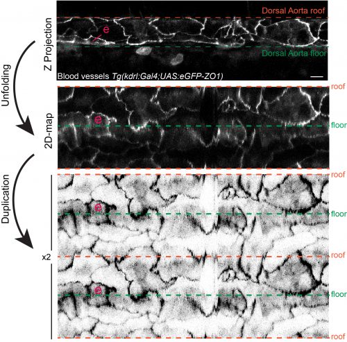
Apart from the visualization of the EHT sequential steps and key features, as mentioned above, our aim was also to picture the EHT in the developmental vascular landscape. This is not a simple issue if one wants to get a clear picture because during the EHT time-window (that lasts from approximately 30 hrs post-fertilization (hpf) to 60 hpf), the aorta shrinks in diameter, must adapt to the loss of cells constituting the hemogenic endothelium and aortic cells undergo cell-shape changes as they elongate to adapt to mechanical tension (Lagendijk et al 2017). To standardize as much as possible the analysis, we started imaging at 48 hpf (which is also the timing at which the EHT is culminating). When one looks through the Z-stacks of an acquisition, even with a 3D-rendering view, one realizes that our brain experiences difficulties embodying the information and morphometrics becomes quite tricky. Things become even more difficult if one wants to explore the dynamics and inter-relation of objects through time, which is what we wanted to do. To fulfil our aim, we initiated an essential collaboration with experts in image analysis and physics from the Pasteur campus, namely Jean-Yves Tinevez and Fabrice de Chaumont. This interdisciplinary collaboration was most fruitful. The team developed an algorithm capable of deploying the aortic wall to project the fluorescent signals onto a 2D-plane (see figure 2) and of accurately re-iterating the aortic projections through time (taking into account distortions of the aorta and its morphological changes). With this algorithm, we have been able to exploit several of our time-lapse sequences and in particular the ones performed with eGFP-ZO1 expressing fishes that allowed following the cell boundaries and inter-cellular contacts. With this, we managed to visualize unambiguously the symmetric division of EHT undergoing cells (an amazing event when you think about the tension that must be exerted on the emerging cells !) as well as the dynamic interplay between EHT cells and their endothelial neighbours. We observed that the number of endothelial cells contacting EHT ones decrease with time, most probably to minimize the risk of leakage upon sealing of the endothelium. Importantly, the clear dynamic views provided by the 2D-projections revealed that the cells from the hemogenic endothelium, around 48 hpf, are rather elongated (in comparison to the surrounding endothelial cells) and that progression of the emergence is characterized by the contraction of the interface with neighbours that proceeds along the antero-posterior axis (the interface delimitating the EHT cell apex facing the aortic lumen). This axis parallels the blood flow, raising in our mind the idea that the direction of the contraction, anisotropic since oriented preferentially along a specific axis, may be minimizing the exposure to hemodynamic forces. Finally, the 2D-mapping of our time-lapse sequences performed on embryos expressing the actin reporter Lifeact also revealed anisotropy in the organization of sub-cortical actin, with its densification at sub-plasmalemmal regions of hemogenic and EHT cells enriched in junctional proteins. This suggested that these regions (whose orientation is perpendicular to the blood flow) are the most exposed to mechanical tension. These interpretations are now awaiting being tackled by further modelling of the forces at play.
Figure 2: 2D-map representation of the dorsal aorta after its deployment using the TubeSkinner plugin of the ICY software. “e” highlights a cell undergoing the EHT. The transgenic fish expresses eGFP-ZO1, ZO1 being part of the complex building the tight junctions. For more details on the 2D-algorithm see our paper (https://doi.org/10.7554/eLife.37355).
Overall, our results strongly suggest that anisotropy, because it organizes according to the blood flow axis, may be dictated by the mechanical constraints imposed by the aortic environment and in particular the blood flow. Thus, what would happen if blood flow is inhibited from the very beginning ? To answer to this question, we prevented blood flow by blocking heart beating (amazingly, the zebrafish embryo can survive several days without heart beat and circulation, by passive diffusion of gas through the skin). In this situation, we observed the impairment of actin cytoskeleton anisotropic organization and destabilization of junctional complexes. Quite surprisingly, hemogenic cells were still capable of escaping from the aortic wall (with, surprisingly also, the apparent maintenance of their localization on the ventral side of the aorta). However, they managed to do so both toward the sub-aortic space – as they do under normal physiological conditions – and the aortic lumen. The intra-aortic emergence most probably took place because the force that normally applies to the aortic wall and that is perpendicular to the arterial axis (the so-called mechanical strain) does not act anymore to push the emergence toward the sub-aortic space. Hence, even in the absence of blood flow, a population of aortic cells retains the ability to escape from the endothelium. This was unexpected because the emergence of hematopoietic stem cells was shown to depend on the activation of a transcription factor essential for hematopoiesis and whose expression is induced by the blood flow (Runx1, see Adamo et al 2009; see also for a seminal paper on the influence of blood flow on hematopoiesis North et al 2009). However, we do not know if those cells, released in the absence of blood flow, do retain the bona fide properties of hematopoietic stem cells, meaning the capacity to differentiate and replenish the entire repertoire of immune cells of the adult body. It is probable that they don’t and that they will end up dying, which is what we observed for some of them, few hours after the release. What these results suggest in addition is that, even if essential pathways are impaired, cells programmed to become hematopoietic may retain some of their abilities, such as escaping from the endothelial environment. This is possibly the reason why in vitro settings aimed at producing hematopoietic stem cells for regenerative purposes and using pluripotent stem cells produce hematopoietic-like cells that ultimately fail to express full hematopoietic potential. Indeed, hematopoietic stem cells are among the rare ones that cannot be produced yet and our work reinforces the idea that the mechanical constraints of the aortic environment are required to produce bona fide hematopoietic stem cells. It would be very interesting in the future to address the question as of the influence, on the fate of hematopoietic stem cells, of mechanical forces taking place contemporarily to the emergence.
So, at the end, our investment was really worth the effort and we gleaned interesting results and ideas from the hundreds of time-lapse sequences that were accumulated by Mylène during her PhD. Currently, we hope that our work, beside its contribution to the understanding of the Cell Biology and Biomechanics of cell extrusion processes, will bring valuable knowledge for reproducing at will the genesis of hematopoietic stem cells.
Anne Schmidt & Mylène Lancino
Developmental and Stem Cell Biology Department
CNRS UMR3738
Macrophages and Development of Immunity
INSTITUT PASTEUR
25 rue du Dr. Roux
75724 Paris Cedex 15 France
anne.schmidt@pasteur.fr
mylène.lancino@pasteur.fr
_______________
References
Adamo, L., O. Naveiras, P. L. Wenzel, S. McKinney-Freeman, P. J. Mack, J. Gracia-Sancho, A. Suchy-Dicey, M. Yoshimoto, M. W. Lensch, M. C. Yoder, G. Garcia-Cardena, and G. Q. Daley.2009. Biomechanical forces promote embryonic haematopoiesis. Nature 459:1131-5.
Bertrand, J. Y., N. C. Chi, B. Santoso, S. Teng, D. Y. Stainier, and D. Traver.2010. Haematopoietic stem cells derive directly from aortic endothelium during development. Nature 464:108-11.
Boisset, J.C., van Cappellen, W., Andrieu-Soler, C., Galjart, N., Dzierzak, E., and Robin, C.2010. In vivo imaging of haematopoietic cells emerging from the mouse aortic endothelium. Nature464, 116-120.
Jordan, H. E.1917. Aortic Cell Clusters in Vertebrate Embryos. Proc Natl Acad Sci U S A 3:149-56.
Kissa, K., and P. Herbomel.2010. Blood stem cells emerge from aortic endothelium by a novel type of cell transition. Nature 464:112-5.
Klaus, A., and C. Robin.2017. Embryonic hematopoiesis under microscopic observation. Dev Biol 428:318-327.
Lagendijk, A. K., G. A. Gomez, S. Baek, D. Hesselson, W. E. Hughes, S. Paterson, D. E. Conway, H. G. Belting, M. Affolter, K. A. Smith, M. A. Schwartz, A. S. Yap, and B. M. Hogan.2017. Live imaging molecular changes in junctional tension upon VE-cadherin in zebrafish. Nat Commun 8:1402.
Lam, E. Y., C. J. Hall, P. S. Crosier, K. E. Crosier, and M. V. Flores.2010. Live imaging of Runx1 expression in the dorsal aorta tracks the emergence of blood progenitors from endothelial cells. Blood 116:909-14.
North, T. E., W. Goessling, M. Peeters, P. Li, C. Ceol, A. M. Lord, G. J. Weber, J. Harris, C. C. Cutting, P. Huang, E. Dzierzak, and L. I. Zon.2009. Hematopoietic stem cell development is dependent on blood flow. Cell 137:736-48.
Sabin FR.1917. Preliminary note on the differentiation of angioblasts and the method by which they produce blood-vessels, blood-plasma and red blood-cells as seen in the living chick. Anat. Rec. 13:199-204.
During early mouse development, a series of signalling interactions breaks the symmetry of the egg cylinder, spatially organising the embryo into territories that define the future axes of the body. Symmetry breaking can also be observed in embryonic stem cell (ESC) colonies cultured on micropatterned substrates, which thus provide a powerful system to test the role of signalling as well as other features of the cell population in early patterning. A recent paper in Development uses micropatterned colonies to test the role of geometry in symmetry breaking. We caught up with three of the paper’s authors – Guillaume Blin, Manuel Thery and Sally Lowell – to hear all about the history.
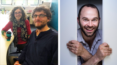
Sally, Guillaume and Manuel
Guillaume, can you give us your scientific biography and the questions that drive your research?
GB I did my undergrad in the south of France where I was trained as a biochemist and biophysicist. I spent one of my first lab experience with Prof. Catherine Picart (Material biophysicist) and Dr. Christian Roy (Cell Biochemist) in the DIMNP lab in Montpellier. I worked on a biomimetic system to model and quantify the interaction between the protein ezrin (known for its role in cell polarity) and PIP2 (a constituent of the plasma membrane). This work was truly interdisciplinary and as I built skills in good old biochemistry, I also had the chance to learn about techniques more alien to the biologist world I came from – zeta potential, DLS, FCS, QCM, this was fascinating.
Catherine, Christian, as well as Prof. Andrea Parmeggiani (theortical physicist and teacher who changed my understanding of bio-molecular interactions), succeeded in creating a synergistic environment spiced with intrinsic ‘cultural differences’ between actors which meant that the coffee table was often the scene of some of the most hilarious banter between physicists and biologists. I am very grateful to them as this experience gave me the will and confidence to keep on building my own interdisciplinary skill set later on.
A question that came out of this work is how macroscopic phenomenon emerge as a result of microscopic events and for this reason I next changed system scale and went on to study cell biology in the lab of Prof. R. Foisner as an Erasmus student in Vienna. Again I am grateful to the people who have trained me Dr. Andy Brachner and Dr Josef Gotzmann. I don’t think I would have adapted well to what would come next if I had not spent such a great time with them in the Max Perutz lab. My Viennese experience coincided with the publication of seminal papers from the D. Discher and C. Chen lab showing that the fate of stem cells could be largely influenced by the physical properties of their environment.
And so, I then started a PhD with Catherine who had elaborated a collaboration with stem cell scientist Dr. Michel Puceat, in order to test the influence of stiffness on the cell fate of embryonic stem cells. During my PhD, I was also involved in another project aiming at modeling cardiac development with ESC to isolate a cardiac progenitor population to use as a source for cell therapy. This jump from purely basic science to a more translational approach has also brought its load of teaching and I am grateful to Michel for introducing me to this important dimension of research.
While working with ESC I noticed that even a clonal population of cells would create a wide diversity of behaviors in the dish and at that time the literature was flourishing with examples of heterogeneous expression of genes associated with cell fate biases. As a result, the next question I became interested in is how can we reconcile the propensity of stem cells to diversify in a seemingly erratic fashion in the dish with the remarkable reproducibility and robustness of developmental patterning in vivo.
Inspiring work came out from the lab of P.W Zandstra who was using micropatterns to dissect endogenous signaling in ESC and the role of the micro-environment. This gave me the idea that we may be able to guide patterning with geometrical confinement. I was also very impressed with Manuel’s work and so I contacted Manuel to learn how to make micropatterns. Manuel was even more enthusiastic and supportive than what I could have hoped for. I learned a lot with him and I must say, his good mood and energy are very contagious, beware! The first preliminary results were obtained as early as 2010. However, this work also coincided with the end of my PhD and the work was held back by the writing of my thesis and also by some technical challenges I was facing (notably in imaging).
Motivated by the will to better understand the origin of the heterogeneous response of stem cells to differentiation signals, I then joined the lab of Dr. Sally Lowell in the MRC-Center for Regenerative Medicine in Edinburgh. I was awarded a 4-year Wellcome Trust post-doctoral fellowship in order to determine how changes in tissue geometry may feedback onto early cell fate decisions. As part of my post-doc project, I developed new image analysis tools to quantify patterning in complex 3D cell populations at single cell resolution. With this new computational tool in hand, we decided to resume the story initiated about 6 years earlier. Sally has been incredible at providing support to ‘resurrect’ this project. Like the dream PI for a post-doc, she provided the pitch-perfect environment to reveal the hidden potential of this work, a bit like geometry in our study!
So there I stand now, looking to establish my own group in order to continue identifying general principles about how the cells self-organise using synthetic and quantitative methods.
Sally, what are the main aims of the Lowell Lab, and what led you to investigate the role of geometry in patterning?
SL I’m fascinated by how cells talk to each other to generate patterns. We think that when these conversations are disrupted, for example through changes in adhesion and tissue morphology, this can help to reinforce differentiation decisions to make development more robust. Paradoxically these same mechanisms may well be confounding our ability to direct differentiation in cells in culture. My wonderful and creative lab members are working hard to understand how all this is happening. When Guillaume joined the lab he set to work revolutionising the way we were able to approach these questions. For example, thanks to a set of image analysis tools that Guillaume has developed we can now measure cell-cell interactions and changes in morphology at single cell resolution even within enormous (to us) post-gastrulation mouse embryos. Watch this space for a couple more of Guillaume’s papers which will report these tools to the community. The idea to investigate the role of geometry came entirely from Guillaume, who is the brains and the driving force behind this project. Indeed he very presciently began this project eight years ago during his PhD, taking inspiration from the work of Manuel Thery. Manuel will be able to tell you more about how it all began…
And how did you become involved with the project Manuel?
MT I was working on cell architecture and developing micropatterning methods to play with cell shape and adhesion. Meanwhile I was reading the papers by Christopher Chen who has always been a great source of inspiration for me. He was constraining cell groups in crazy shapes where mesenchymal stem cells were differentiating toward adipocytes or osteoblast depending on their position in the group. Osteoblasts appeared at the periphery while adipocytes sat at the center of the colony. In a very thoughtful experiment he plated cells on sinusoidal avenues. All cells were close to the periphery, but osteoblast appeared along convex edges while adipocytes appeared in concave edges. The proof was shown that stem cells sense geometrical cues and not only the local cell density. I was super excited by this results and used it a lot as an example of the importance of geometry to cell physiology. But I could not work on it. In 2010 I was starting my group with two students and we were focusing on intra-cellular architecture. It was very frustrating. Then Guillaume run into my office and proposed to study the role of geometry in ES cell colony! I jumped on my chair like the wolf facing Little Red Ridding Hood in the nightclub in Tex Avery’s cartoons. We immediately started to draw shapes that could contradict the density dogma. The issue was that Guillaume was not equipped to perform microfabrication experiments in his lab. We had to set up a new protocol. Guillaume worked a lot and got very enthusiastic with what I would consider pretty unsatisfying results. So he insisted more and more, tried new chemicals and got it to work. From there I started reading a lot on ES cells but it was too complex to me. Guillaume was visiting me and teaching me mouse embryonic development. I learned so much! It was really great. I became fascinated by the gastrulation and started working on epithelial-to-mesenchymal transition.
The experimental rationale of the paper
What was known about the role of geometry in early mammalian patterning prior to your work?
GBGeometry is known to have a strong influence during the 1st cell fate decision, when the blastomeres become segregated either to the ICM or to the TE. Geometry dictates cell polarity which in turns define the activity of key transcription factors. This is a phenomenon that is now well documented.
In the first article Mesnard and colleagues have shown that the axis defined by the anterior and posterior domains aligns with the longest axis of the embryo. They also show that this is a dynamic process as the AP axis initially aligns with the shorter axis early on.
In the second article, the authors grow pre-gastrulating mouse embryos ex-vivo within agarose microwells to mimic constraints from the endometrium. They show that the soft physical constraints provided by microwells facilitate the elongation of the embryo resulting in the most distal region of the embryo ‘moving’ far from the source of BMP signalling (the extra-embryonic ectoderm). According to the authors, the resulting decrease in BMP signalling in the distal region participates in the specification of the DVE.
Overall, I believe that everyone has this intuition that geometry participate in defining the shape of signalling gradients, but there is still work to be done to understand how the cells sense the physics of their environment locally and how changes in geometry may feedback onto morphogenetic events.
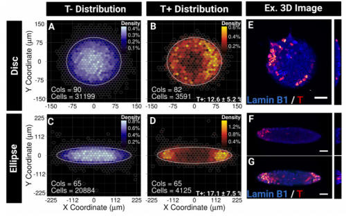
T+ expression in circle versus ellipses
Can you give us the key results of the paper in a paragraph?
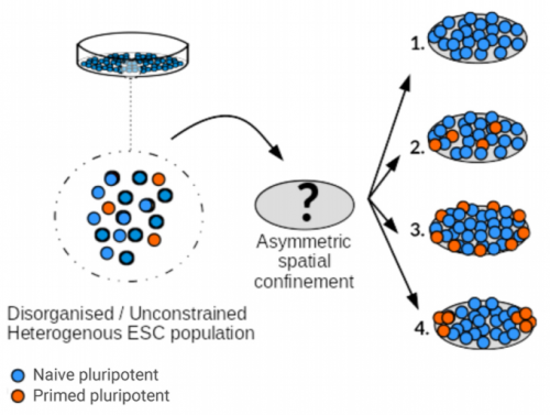
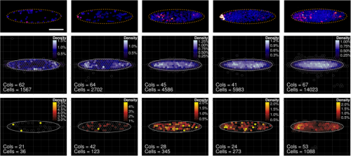
GB, MT, SL We show that imposing physical boundaries to ESC colonies in culture drives the patterning of a T+ primitive streak-like population of cells which is initially present but spatially disorganised. Importantly, if we introduce asymmetry in the shape of colonies, the resulting positioning of the cells follows the asymmetry which raises the possibility that geometry plays an important role in guiding precise positioning of the cells at the streak.
We also show that patterning is robust to changes in the relative number of T+ cells present initially. This is an important result because it indicates that the mechanisms which regulate the number of cells which will ingress into the streak may be distinct from the mechanism which regulates the precise positioning of the cells. To me, this is an indication that it is important to reflect on the notion of control during development and how processes are articulated with one another. Having a global signal triggering local events leading to patterning rather than tightly controlled signalling gradients micromanaging both patterning and specification is an elegant solution. The need for control is minimised and a certain level of robustness is achieved by the system. I can only recommend one of my favourite reviews on the topic from Arthur Lander ‘Pattern Growth and Control’
These ideas can also have great implications for our ability to engineer synthetic systems and actually one of our plans is to see if we can induce patterning in cells which do not normally sort.
Have you got any ideas for how your T+ cells end up at the periphery of the colonies?
GB/SL/MT There are quite a few hypothesis that we can formulate. A possibility is that the cells change fate if they end up in the ‘wrong place’. There may be some selective apoptosis going on as well. If the cells are surrounded by cells with a different level of T, maybe the cells are eliminated. If the cells reorganise, then again this can happen in various ways. As you suggest, cell sorting via differential adhesion may be involved or alternatively, T+ cells may become more motile and migrate faster and with more directional persistence than T- cells. This differential motility may be sufficient to drive sorting even in the absence of chemotaxic cues. It may be a combination of all of the above, we know that the embryo can use a diversity of cell behaviours in order to achieve patterning in the late blastocyst (Plusa et al, Development 2008). Our plan is to use mathematical modelling in combination with live imaging in order to further address this question.
Emergence of pattern in elliptical colonies
Can you translate your findings to the early embryo – where might the guiding hand of geometry be acting?
GB, MT, SL This is a great and difficult question because it is still unclear how the shape of the streak is maintained as the embryo grows. In the chick, a community effect mediated by Nodal signalling and cell-cell interactions as well as Wnt-PCP, have been identified as important elements regulating this process (Voiculescu et al., eLife 2014). The apical polarity regulator crumbs2 has also been involved (Ramkumar et al, NCB 2016). These are examples of molecular determinants which may be part of the machinery which enable the cells to sense the folding of the streak. This geometry sensing may be key to maintain the stable geometry of the streak as the embryo grows. However much work remains to be done to fully understand this process and to assess inter-species differences as well.
When doing the research, did you have any particular result or eureka moment that has stuck with you?
GB The first one is when I saw T+ cells localising at the tips of the ellipses. Despite variability between colonies, the patterning was very clear under the microscope! I remember thinking, I need to quantify this, maybe I am just imagining it, so when we found a way to create the density maps and saw the figure showing a clear result, I was both a little a bit euphoric and very intrigued.
And what about the flipside: any moments of frustration or despair?
GB I had moments when I felt the results were not coming as fast as I wanted, this was mainly due to the fact that imaging was a bottleneck in our initial setup. As a result I was spending very long hours at the confocal in the (cold and dark) room of the imaging facility, skipping activities with family or friends. This was particularly difficult in the autumn when the colours outside were so vibrant. Not being able to see the season pass by was frustrating. But in the end we managed to streamline the whole process and this solved the issue. This was not so bad but I am lucky to have a very understanding and supportive family.
Guillaume’s hollow ellipses
What next for you after this paper?
GB I am currently working on publishing other outputs from my post-doc which include methodological papers describing computational tools for quantitative image analysis, I hope you will be able to hear soon about it and that my tools will be useful to the community. I am also trying to establish my own group in order to continue working on synthetic approaches to identify general principles about how the cells self-organise.
Any plans for future Franco-Scottish collaboration?
MT, SL Yes definitely! We will continue to work together, the combination of microfabrication and stem cells offers a fertile ground with almost limitless possibilities. We will look into remodellable materials and 3D solutions to further guide the development of the cells in vitro. Maybe this is a good place to advertise our recent review on the topic also (Laurent et al. NBE 2018).
Finally, let’s move outside the lab – what do you like to do in your spare time in Edinburgh and Paris?
GB I like to spend time with my family, whatever we do does not matter as long as my 2 daughters keep being cheeky with their sly faces hinting at their next mischiefs. When I find time I also enjoy cycling in the beautiful moors and hills around Edinburgh.
MT I like to get high, and I know many ways for it. Apart from the classical ones, I enjoyed art exhibitions very much. Artists observe the world, analyse it interpret it, and then try to fing original means to convey their thoughts into something that will question people imagination and help them to consider reality differently. Isn’t it exactly what we do every day in our labs? Except that we have much more tools to play with … ;-) Artists also want to understand how things work, how light makes things visible, how images print in our mind, how interactions change intrinsic properties, how context change identity. So I am pretty sure most of them would enjoy Guillaume’s paper very much. Too bad they don’t dare reading Development, while we all go to the Tate.
SL There is no such thing as spare time, but during my not-doing-science time my kids have been teaching me how to mix music on a computer. I’ve only had two lessons so far but probably I will soon become an internationally famous and unfeasibly wealthy DJ. My other hobby is choosing theme tunes for papers that I like and then posting them on twitter, as my service to the developmental biology community*.
*The theme song for Guillaume’s Development paper, “Down on the Corner” by Creedence Clearwater Revival, was suggested by new Development Editor in Chief and incurable hippy James Briscoe, for which we thank him.


 from a recipient of a Development Travelling Fellowship, funded by our publisher The Company of Biologists.


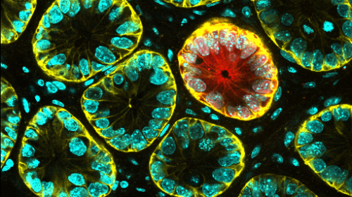


 Barcelona, 26 September 2018.- Metastatic tumours are characterised by a high degree of chromosomal instability—that is to say a constant change in the number and structure of chromosomes they hold. In spite of this observation, it is unknown whether chromosomal instability contributes to the metastatic process.