If you look at, especially say, identical twins, if one person does testing, you are automatically finding out for your twin what their genetic status is. And so that’s tough, right? Because you’re deciding for them.
Kira Dineen, host of DNA Today
In the latest episode of the Genetics Unzipped podcast, we’re discussing Chris Hemsworth’s recent Alzheimer’s risk findings and the pros and cons of direct-to-consumer DNA testing with genetic counsellor and host of the podcast DNA Today, Kira Dineen.
If you enjoy the show, please do rate and review on Apple podcasts and help to spread the word on social media. And you can always send feedback and suggestions for future episodes and guests to podcast@geneticsunzipped.com Follow us on Twitter – @geneticsunzip
We recently announced that we will be working with three newly appointed the Node correspondents, who will be helping us to develop and write content for the Node in 2023. We caught up with each of them to chat about their research backgrounds and the topics that they’re excited to write about over the course of the coming year.
First up is Brent Foster, a technician at the Whitney Laboratory for Marine Bioscience, University of Florida, where he is studying the evolutionary origin of nervous systems using a range of marine invertebrates. As you will see, Brent has a longstanding interest in creative writing alongside his scientific career and began to blend the two by writing for his local newspaper. We talked about how his experience of using unconventional experimental organisms influences his writing, and the importance of being part of a science writing community.
Congratulations on being selected as a the Node correspondent. Why did you decide to apply for the role?
I decided to apply for it mostly because I had such a good experience with The Company of Biologists’ Creative Science Writing Workshop that I went to last year. I respect the mission of The Company of Biologists and their whole philosophy, and I thought this would be a really cool opportunity to expand beyond the readership I’ve had so far with my science writing.
What led you to first become interested in science writing? And what sort of science writing have you been doing so far?
I’ve always liked science. My dad was a high school biology teacher, and we lived in a rural area. I got to know the local wildlife, local trees and all of that fairly well. I really enjoyed the outdoors and I’ve always loved learning how nature works. And then on the flip side, once I started my undergraduate degree, I discovered that I enjoyed writing as well. And I had never really thought of pairing the two – I just considered writing as a hobby and assumed science would be my career, and that was my understanding going forward.
When I started working in the lab at the UF Whitney Laboratory, I met up with a local news editor. We struck up a conversation and I found out he was interested in science, but didn’t have a science background, and I was interested in writing, but hadn’t really done any formal writing, other than a few personal essays in college. And he basically issued a challenge to write something for the Palm Coast Observer, which is the local newspaper here. So, I approached my supervisor, and I asked him if it would be ok to write up some articles about what the lab is doing for the newspaper. From there, I discovered that I really enjoyed writing about the sciences, maybe even a little bit more than I enjoyed doing the experiments in the laboratory, and so I’ve expanded from there. The past two years I’ve presented some work at the Society for Integrative and Comparative Biology (SICB) meeting, and there I met some people who write for the SICB blog, and so I’ve contributed a few posts there. Last year I applied to the Creative Science Writing Workshop, hosted by The Company Biologists, loved it, and I’ve kind of snowballed into larger and larger projects. In October, I went to a science writing conference in Memphis, where I met a few folks and learned a little bit about the life of a science writer.
Thinking about what you were saying there about going to workshops and conferences focused on science writing, have you found that meeting likeminded scientists who also have an interest in creative writing has influenced you?
Yeah, absolutely. That was one of the big things from the Workshop last year: I was kind of shocked at how all of these brilliant scientists really just seemed to want to have some sort of creative outlet. And not always just about the work that they’re doing themselves, which showed me how creative science and scientists can be. So that was really eye opening. And then in January, when I was at SICB, I announced that I was a correspondent for the Node. I had a couple of people come up to me afterwards and we were throwing ideas back and forth about some different writing projects. That was a lot of fun. So, it has opened up some doors as well as friendships that I don’t think I would have found otherwise.
In terms of your own research, what has your career path been so far? And what’s your current research focus?
This seems to be the story for a lot of people now, but my career path is very, very windy. When I got started as an undergraduate student, I wanted to be an epidemiologist. After my first semester, I deferred my education and lived in Brazil for two years. I learned the language and the richness of that culture, and after just two months I noticed that I started dreaming in Portuguese. When I returned to my University, I attended a club meeting for deaf and hearing-impaired students. I only know rudimentary American Sign Language (ASL), so I’m sure my fingers were just fumbling with an accent. To offset the clumsiness of my hands, I would mouth the words I was trying to spell. It was only after the meeting when I realised that the words I mouthed were in Portuguese, not English. Somehow ASL and Portuguese had gotten tangled up in my brain, and both of them seemed sectioned off from English. I thought that was pretty fascinating. So, in the end, my undergraduate degree was in neuroscience, with minors in linguistics and creative writing.
I was especially interested in neurolinguistics, so how language is processed in the brain. That was my attempt to try to marry my interest in writing and science. I did some eye tracking studies, MRI studies, and EEG. And as I was getting all these great research experiences, I was a little disappointed in how little we could actually tell from those particular methods. I mean, you can tell a lot by each of them, but there were no clear causal effects, right? It was all “well, this is happening, and we are interpreting it to mean maybe this.” It wasn’t very definite, and that was a little disappointing for me when trying to understand some basic biology.
Then my wife was accepted to a PhD programme at the University of Florida, and so we moved down here. Originally, we were on the main campus, and I was in an MRI lab where we did a lot of cognitive neuroscience research related to attention and anxiety. They were really, really cool projects, and I’m grateful for the experience that I had there, but again, I had the same feeling of, “man, I’m just not sure how much we can actually tell from what we’re doing.” My wife and I stayed on main campus for the first year of her PhD. And then she moved out here to the Whitney Lab, and I didn’t want to have to commute back and forth. So, I actually reached out to the director of the lab, Mark Martindale (who’s now my supervisor), and I told him about my research experience. I had no wet lab experience – all of the studies that I’d done were human studies and nothing at all relevant to what they do here at the Whitney Laboratory. But I was interested in learning and asked if I could come down even just on the weekends to get some wet lab experience. And he turned around and offered me a job, and that’s how I started here.
So, I’ve switched from neurolinguistics to basic marine biology and developmental biology. And I love it. I feel like I can understand the outcomes of the experiments that I do a little bit clearer than I could within the broader cognitive sciences. I don’t mean to disparage other sciences as I think there are a lot of valuable things that we can learn from them – it just wasn’t for me.
Do you find that the science you’re doing now is more inspiring for your writing than what you were studying before? Because I imagine the sort of organisms you’re working on are a lot more interesting from that creative science writing perspective.
Yeah, that’s a great question. I think people could relate more to what I was doing before, when I was asking questions like “what’s the brain doing?” and “how do people process language?” In the eye-tracking lab, I came to understand a little bit about how people read, which has been helpful in the technical part of crafting my writing in a way that’s easy to read. But thinking back to what I found cool about science when I was a kid, I was always drawn to weird, quirky animals that do weird, quirky things – nature’s a real creative playground. Now I get to tell people that I work with sea anemones, and they’ll say, “I don’t know if I know anyone who works with a sea anemone,” or I mention that I work with comb jellies, and they say, “oh, what’s a comb jelly?” That’s a really fun type of interaction, to hear people saying, “that’s so cool. I never knew that.” And I try to capture that fun as much as I can in my writing.
Thinking about the sort of things that you’re going to be writing for the Node, what topics are you excited to write about? And do they relate to your own work or are you going to be branching out a bit into other areas?
Well, like I said, the quirky biology is what gets me excited, so I’m hoping to focus on non-model organisms, organisms that not everyone knows or hears about. This will probably involve a little bit of the work in our lab, because there are a lot of us here at the Whitney Laboratory who work with non-model organisms that are great for answering very specific questions but often get glossed over just because they’re not so well established.
My secondary focus is related to the idea of working with non-model organisms. Because these organisms are not as well established, I want to highlight labs that have to develop their own technologies or develop their own techniques or adapt existing techniques for their animals. This is hard and I think a lot of times underappreciated, both by the public and even by other scientists. With Drosophila, for example, you can do all sorts of things which you might take for granted. By contrast, I just talked to a scientist a few weeks ago who is trying to adapt transgenic approaches for cuttlefish, and they’ve spent almost a year trying to get something to work. So, I want to show those two aspects of something that I don’t think gets shown a lot, or at least doesn’t get into the major headlines. I want to give non-model organisms their due, I guess!
What are you hoping to gain from the experience of being a the Node correspondent?
Most of my science writing so far has been either hyperlocal or very specific to one group of biologists from a single conference, so one thing I’m hoping to gain as a correspondent for the Node is reaching a broader audience. I know the Node is pretty popular internationally, and I haven’t written for an international audience before. So I hope to stretch myself a bit in that area. I am looking forward to meeting other scientists in my field, hearing their stories and helping to share those stories.
I am also excited to interact with the Node team and other correspondents to learn about the craft of science writing a bit more. I don’t really have any formal training in science communication so I’m looking forward to participating in workshops. And of course, I’ve mentioned this several times now, but just having a community is huge. I didn’t realise how big the science writing community is until last year, and it’s gratifying to feel welcomed to it. Before I discovered it, I almost felt like an outsider. I was always asking myself, “is there a place for these two different aspects of my identity that everyone else seems to think of as a contradiction?” It’s fun to see that there are indeed other people who are combining their interests in science and writing.
Apart from writing, what do you enjoy doing in your spare time?
Lately, I have been doing a lot of writing! But I do enjoy the outdoors, particularly kayaking or canoeing – there are some lovely springs around here. In fact, I have a kayak trip this weekend and I’m hoping to see some manatees. I enjoy playing the piano, too. Primarily classical piano, though I don’t get a lot of opportunities to do that these days. And I enjoy reading. As a student, you kind of feel as if you either don’t have the time to read or that sometimes you’re reading about things that you’re not too excited about. In the gaps between studies, it’s been fun to be able to read some things just for the pleasure of reading.
Do you find that this sampling of other people’s writing has an influence on your own work?
Yeah, absolutely. Because I’ve gotten so interested in science writing, a lot of what I’ve been reading lately is by other science writers. That’s been informative in learning how other people approach science writing, but I also enjoy regular literature. I’ll pick up a piece of fiction or even poetry sometimes. And to be honest the poetry reading that I do, and the creative writing classes I took as an undergraduate that taught me a little bit about poetry, have actually informed my ability to write concisely in a way that I’m not sure I would have picked up from other genres. So, I think that by dabbling a little bit in a novel here, a short story there, some poetry, all of those things, you can take something from each one of those genres that together can make a science writing piece pretty powerful.
At the Gurdon Institute, which is part of the University of Cambridge. We are located in the city centre of Cambridge, UK.
Research summary:
EmmaRawlins: We ask, how are our lungs built and maintained? We have a particular focus on stem cell biology of the developing and adult lungs and asking how cell-cell interactions regulate growth, patterning and repair. We have also recently started collaborating with human geneticists to explore the genetic contribution to chronic lung diseases in humans, and whether these have their origins in infancy, or the adult, or both.
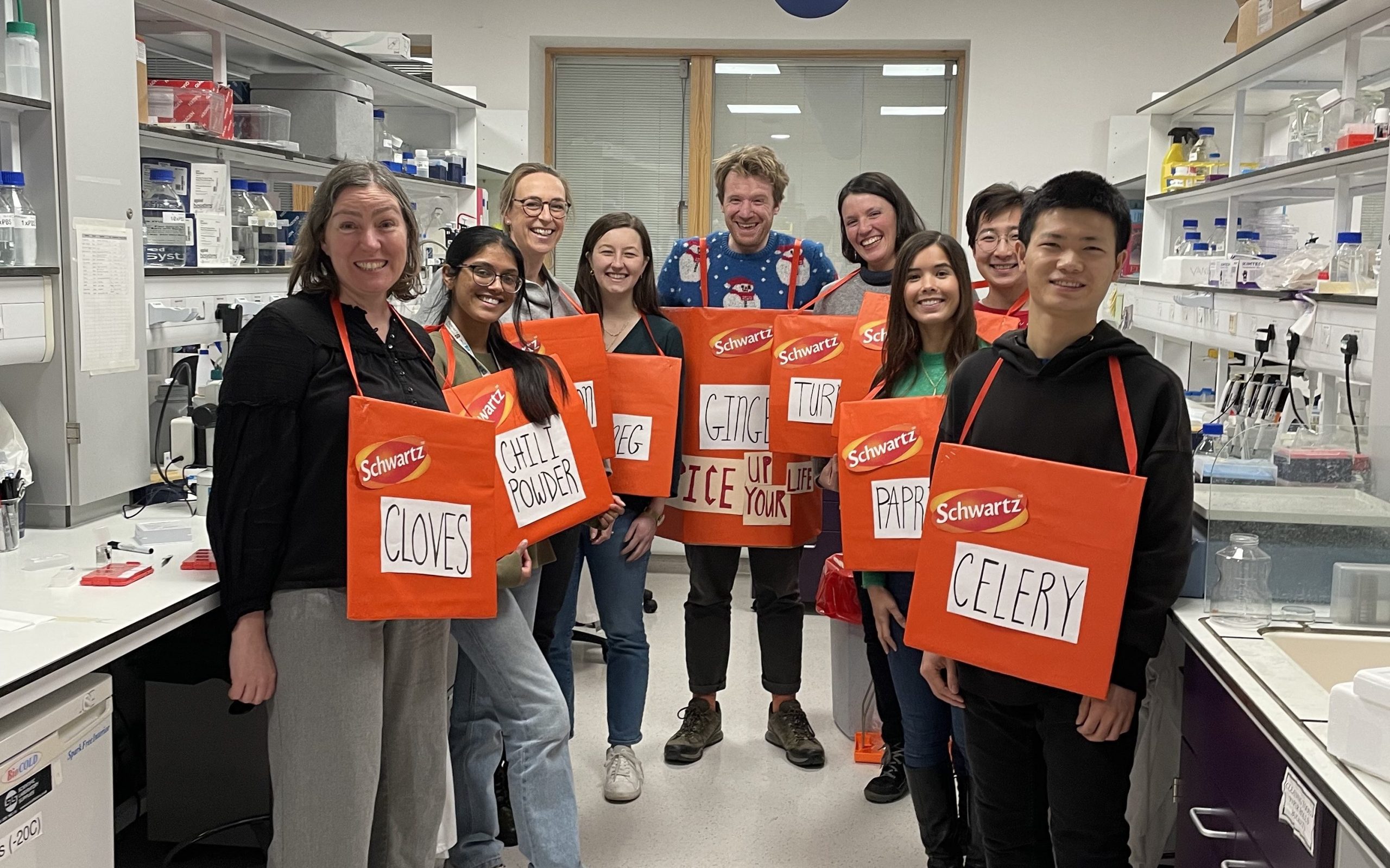
Rawlins lab, Christmas party 2022
Lab role call:
Claire: 1st year PhD student investigating airway homeostasis in humans.
Emma: PI – does all the paperwork.
Hannah: Clinical lecturer – has completed an MD and a PhD and is now balancing postdoctoral work with clinical duties, working on lineage decisions in the developing airway.
John: Postdoc interested in morphogenesis of the developing human lung.
Kyungtae: Postdoc interested in cell-cell signalling during lung alveolar development.
Odara: 1st year PhD student jointly supervised by Vito Menella who is interested in analysis of volumetric electron microscopy and using human organoids to study toxicology.
Quitz: Lab manager who keeps us all organised and enjoys thinking about computational biology methods.
Tessa: PhD student working on epithelial cell fate decisions in the developing mouse lung, and optimal imaging techniques for visualizing lung development.
Vanesa: PhD student (just submitted her thesis!) jointly supervised by Kristian Franze and works on the role of stiffness in cell fate decision making during human lung development.
Yihong: Visiting final year PhD student from Zhejiang University who is interested in tuft cells.
Ziqi: PhD student working on the role of hypoxia in cell fate decision making during normal human lung development.
Favourite technique, and why?
Emma: Clonal lineage-tracing combined with genetic manipulation and microscopy, and now often coupled to single cell transcriptomics. In my opinion, this is the most elegant method to study normal and aberrant cell fate decisions due to the presence of mutant and control cells in the same tissue. One current lab challenge is how to apply this technique to human samples.
Apart from your own research, what are you most excited about in developmental and stem cell biology
Emma: The advances being made in understanding of regeneration from organisms that regenerate on an impressive scale like Axolotls.
How do you approach managing your group and all the different tasks required in your job?
Emma: Most of the time it feels like neither managing my group, or the other admin and teaching tasks, are being done properly. It’s important to recognise that good-enough is sufficient for many tasks we must do, particularly the administrative ones. I try and prioritise my lab as it’s the science that excites me. I block time out in my diary to ensure that I have a detailed science conversation with every lab member every 2 weeks, and other tasks are fitted in around this schedule. I also have annual career planning meetings with everyone in the lab (the University’s appraisal scheme) and try and make sure that I keep up to date with any changing aspirations in addition. That way I/we can be looking out for the best career development opportunities for each person.
What is the best thing about where you work?
Claire: Working in Cambridge offers so much rich scientific history alongside cutting-edge techniques. It’s incredible to train in a such a collaborative space, and the Rawlins lab has been such a supportive and kind group with lots of advice and guidance as I begin my PhD.
Emma: Cambridge has a really rich scientific environment, it’s very easy to network to find collaborators in pretty much any research area that you are interested in.
Kyungtae: The best place in the world to do science – especially developmental biology. Also, it’s easy to get connected between fields to fields and person to person, leading to fantastic interdisciplinary studies.
Tessa: The lab environment is very supportive and collaborative, it’s an exciting place to do science and work with like-minded colleagues who are happy to help you troubleshoot or discuss new ideas.
What’s there to do outside of the lab?
Claire: As a PhD student, I’m part of a college here at Cambridge, and you have the opportunity to meet peers across different subject areas over weekly dinners and different social events. My favourite thing to do is explore Cambridge by going on runs around town and trying all the different restaurants!
Kyungtae: There are great walks and places around the Cambridge with beautiful nature which I can feel the universe – that is why it is the University of Cambridge, in Cambridge. Work and walk and feel the Cambridge.
Tessa: Cambridge has a lot of green space, and (weather permitting!) you can spend lots of time outdoors on the meadows or walking through the backs of the Colleges and admiring the architecture.
Browse through other ‘Lab meeting’ posts featuring developmental and stem cell biology labs around the world.
We are delighted to announce a new series on the Node called ‘Lab meetings’! In these posts, we will be highlighting developmental and stem cell biology labs across the globe. We aim to build up a directory of labs, which will not only showcase the exciting research and researchers in the community, but will also provide a useful resource for anyone looking for their next job. We ask the group leaders about their research and mentoring style, while the group members share what they like about the lab and what they like to do away from work.
Our first ‘Lab meeting’ is with the Rawlins lab, at the Gurdon Institute in Cambridge, which you can read here. You’ll be able to see all our full directory on our ‘Lab meetings‘ page.
Drop us an email if you would like to nominate a lab (including your own), especially if you have (or will soon have) open positions. We look forward to working with you all to build up a useful resource!
It’s not ridiculous to suggest that the Y chromosome might eventually become so mutation-addled that it disappears entirely. In fact, it’s already happened…in the Amami spiny rat.
Please join us for the International Symposium on Women in Tunicata Biology. The Tunicata are invertebrate chordates, several of which are model organisms in developmental biology (e.g. Ciona intestinalis, Botryllus schlosseri). The symposium will be Tuesday, March 28 and Wednesday, March 29, starting at 6 am Pacific Time/9 am Eastern Time. Speakers will be honoring the work of retired researchers and presenting their own research. All researchers are welcome to attend. Please email Marie Nydam (mnydam@soka.edu) or Anna Di Gregorio (adg13@nyu.edu) for a schedule and Zoom link.
Public Engagement with Research (PER) encompasses ways of engaging the public with the design, conduct and dissemination of research. Engagement is, by definition, a two-way process to generate mutual benefit for society and the research community by enhancing research quality and socio-economic impact (See the National Co-ordinating Centre for Public Engagement webpage for more information).
Engaging with the public can take many forms. The format is chosen according to the audience, the aim, the nature of the research and the capacity of the PER team. Examples of public engagement activities are:
citizen science experiments
panel discussions
interactive exhibitions
festival participation
school collaborations
co-creation with artists
contributions to mainstream and social media
There is no hierarchy of engagement approaches. If meaningful and two-way, they are all valid in their own format, and very often, an event or an activity will contain a blend of these approaches and purposes.
The mutual benefit for society and the research community is essential to PER. Benefits might include learning, acquiring new skills, gaining new insights or ideas, developing better research, generating a new network, raising aspirations, or being inspired.
The ways scientists interact with the public across the world differ. Some countries continue with the ‘outreach’ approach, which is mainly a one-way communication to generate attention. In contrast, other countries develop citizen science programmes with multiple educational, social, and economic impacts.
So, we’ve established that public engagement in the UK is more than just educating the public about science – crucially, it is two-way, and involves finding out what the public think about our science too. But how did public engagement come to have this emphasis on two-way communication in the UK?
The story of genetically modified food
A big part (but not the only part) of this story started in the late 1990’s, when there was widespread scepticism and anxiety about genetically modified (GM) food. (Rowe et al., 2005). This public concern around GM food was not just about the science, either: press coverage at the time reveals it was also a political, environment and consumer issue (Durant & Lindsey, 2000).
In 1998, to ease public scepticism, the UK government decided that GM crops wouldn’t be grown commercially in the UK until a set of experiments on farms had been completed. However, this didn’t dampen public opposition: local communities were angry about not being consulted about these farm experiments in the first place (Mayer, 2003). Then, in 1999, major UK food producers and retailers responded to consumer pressure and removed GM ingredients from products on shelves (Mayer, 2003).
In the midst of the GM food controversy, Parliament also issued a report encouraging scientists to engage in dialogue with the public. They recommended openness and transparency in order to help regain public trust, placed greater emphasis on public attitudes, opinions and values and recognised that without public buy-in, science would struggle to move forward. (Science and Technology Select Committee, 2000).
In 2003, the government ran a series of public debates, in parallel with scientific and economic reviews of the issue of GM food. However, despite asking the public what they wanted, there was still widespread polarisation, opposition and scepticism (Mayer, 2003). And today, more than 20 years later, the legacy of anti-GM sentiment in the UK still lives on.
Lessons Learnt
So why didn’t this dialogue with the public work? One of the big reasons scholars give is that the public were involved much too late and it wasn’t clear how their input would be used in the decision-making process. As a result, engagement professionals today are keenly aware today that public engagement needs to be more than just window dressing: when science will impact people’s lives, scientists can’t just use ‘dialogue’ to try to legitimise a decision that has already been made. You have to ask people what their concerns are early enough so that you can respond to what they tell you in a meaningful way, ensuring that your research. and its potential outcomes. are aligned with societal priorities and expectations. (Mayer, 2003; Rowe et al., 2005; Singh, 2008; Marris, 2015; Morrison & de Saille, 2019; de Saille & Martin, 2018; Gjerris, 2008).
Another big take away from the GM story is that increasing public knowledge about biotechnology won’t necessarily mean that people agree with you about how and whether that biotechnology should be used. Disagreements can be fuelled by different interpretations of the facts, often reflecting individual and cultural values. While science can help us answer ‘can we’ questions, science can’t always directly answer ‘should we’ questions – this is why public input is so vital.
This history informs today’s aim to have two-way conversations with the public, regardless of how fundamental the research is, in order to ensure the ultimate outcomes of science are considered mutually beneficial.
Challenges of engaging with fundamental research
While mutual benefit sounds great, there are certainly some challenges to public engagement with fundamental research, when the research outcomes are often unknown. For example, engaging with the public about implications that are remote and uncertain can encourage ideology-based reactions, leading to polarisation and conflict (Tait, 2009; Tait, 2017).
And while today’s public engagement encourages dialogue over merely informing the public, when speaking with non-scientists about fundamental research, there is often a substantial need to build shared language and understanding of the topic before informed conversations can take place. Meaningful PE with fundamental research can take a lot of time and resources (Clements-Brod et al., 2022). However, it is not impossible!
Case studies
We’ve put together some examples of how we’re doing public engagement with fundamental research in hope to inspire others to do the same.
References
Clements-Brod, N., Holmes, L., and Rawlins, E. (2022). Exploring the challenges and opportunities of public engagement with fundamental biology. Development. 149 (18) https://doi.org/10.1242/dev.201170
de Saille, S. and Martin, P. (2018). Monstrous regiment versus Monsters Inc.: Competing imaginaries of science and social order in responsible (research and) innovation. In Science and the Politics of Openness: Here be Monsters (ed. B. Nerlich, S. Harley, S. Raman and A. Smith), pp. 148-166. Manchester: Manchester University Press. https://library.oapen.org/handle/20.500.12657/30733 [accessed 11 August 2021].
Gjerris, M (2008). The three teachings of biotechnology. In David, K.H. & Thompson, P.B. (eds.), What Can Nanotechnology Learn From Biotechnology?: Social and Ethical Lessons for Nanoscience From the Debate Over Agrifood Biotechnology and Gmos. Elsevier/Academic Press., pp. 91-105
Morrison, M. and de Saille, S. (2019). CRISPR in context: towards a socially responsible debate on embryo editing. Palgrave Communications 5, 110. https://doi.org/10.1057/s41599-019-0319-5
Rowe, G., Horlick-Jones, T., Walls, J. and Pidgeon, N. (2005). Difficulties in evaluating public engagement initiatives: reflections on an evaluation of the UK GM Nation? Public debate about transgenic crops. Public Underst. Sci. 14, 331-352. https://doi.org/10.1177/0963662505056611
Postdoc positions are immediately available in Guillermo Oliver’s lab in downtown Chicago to work in different aspects related to functional roles of the mammalian lymphatic vasculature in organogenesis.
Information about some of our recent work can be found in these links:
Highly motivated individuals who recently obtained a PhD. or MD degree and have a strong background in the use of mouse models, and molecular and developmental biology are encouraged to apply.
She might not see it the next day. At first glance, the slag in the saltwater aquarium is just a rock covered with purple jasmine polyps and green star polyps. Orange tendrils of finger corals wave with a silent current, an undulating calm that almost seems to slow the heart rate. Suddenly the rock flutters. A part of it shifts, seems to detach and hover in the tank, and the shape of a dwarf cuttlefish emerges as bands of light and dark ripple across its skin.
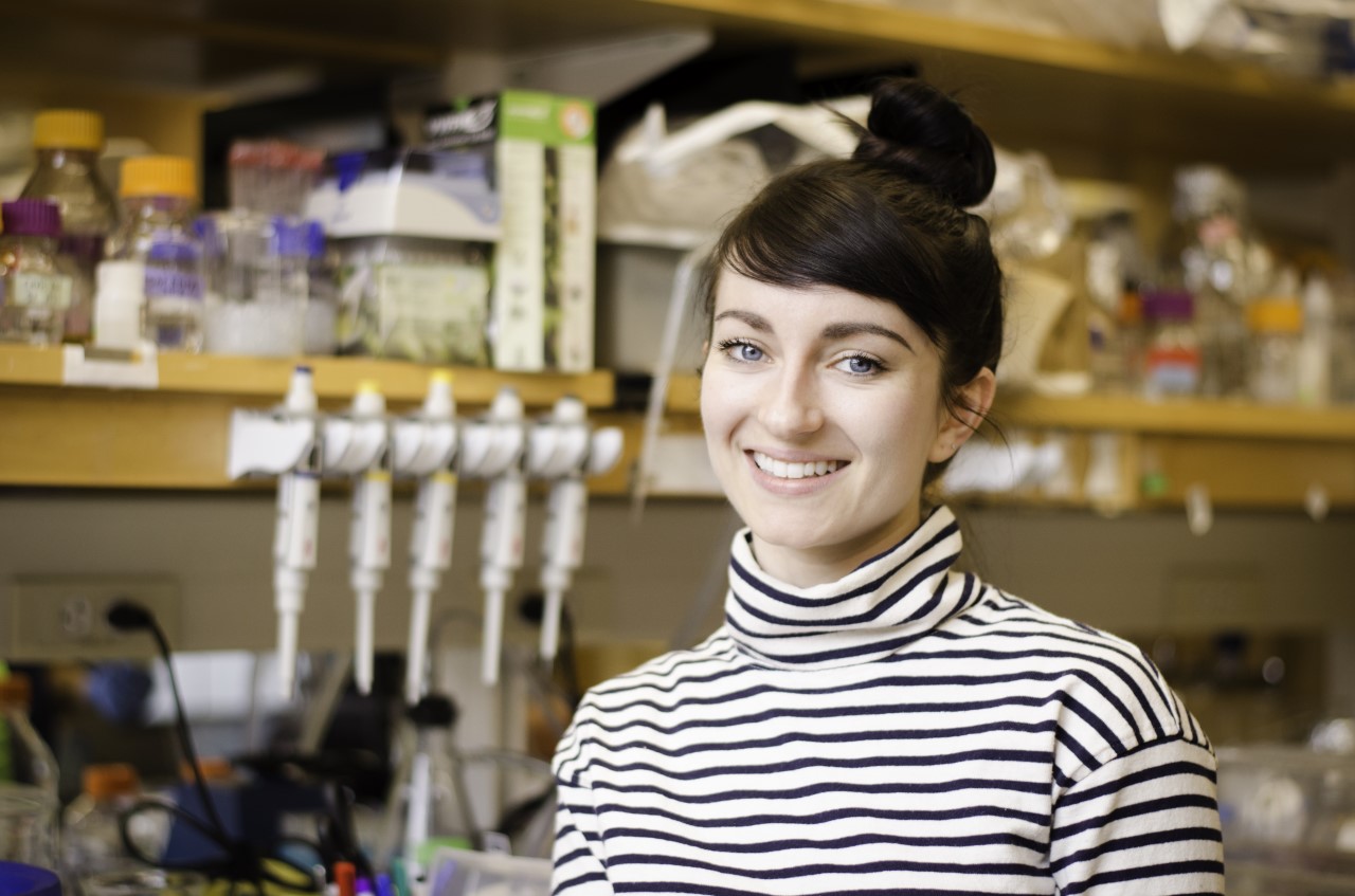
“They’re like little artists creating impressionist paintings on their skin,” said Tessa Montague, a post-doc in the Axel lab at Columbia University. Her latest scientific mission is to understand how the cuttlefish brain controls dynamic camouflage behavior.
From the moment a cuttlefish is born, its skin already contains thousands of pigment-filled sacs called chromatophores that expand and contract under direct control of the brain. And, compared to more established model organisms like the mouse, the dwarf cuttlefish brain is huge. Nearly 75% of its brain is optic lobe, indicating just how important vision is for cuttlefish to sense the world around them.
“When a cuttlefish sees an object or visual scene,” Tessa explained, “the brain must create an abstract representation of the scene using patterns of neural activity.” But through the secret language of neurons the cuttlefish undergoes a remarkable transformation and recreates an approximation of what it has seen on its skin through the fine control of its chromatophores. By studying this transformation, Tessa and colleagues have an opportunity to learn fundamental principles about how brains process the visual world.
Studying a brain as alien as the cuttlefish requires tools to manipulate the genome and record neuronal activity. The way to to do that is to utilize the cuttlefish’s development to create a transgenic animal. But it’s challenging to design experiments without knowing what’s possible or most likely to succeed. At the start of Tessa’s project, no one had sequenced the dwarf cuttlefish genome, and no one had adapted any of the myriad transgenic tools for cephalopods. How hard could it be?
Columbia postdoc Tessa Montague in the lab. (Photo credit: Fred Rubino)
Tessa and colleagues from UCSD and the Chan-Zuckerberg Initiative sequenced the dwarf cuttlefish genome — a 5.5 GB string of DNA, almost twice the size of the human genome. Then, with colleagues at the Marine Biological Laboratory in Woods Hole, Massachusetts, they sequenced RNA transcripts and assembled the cuttlefish transcriptome. Unfortunately, these were not sufficient to identify candidate transgenic drivers. Unfazed, they teamed up with a group at Columbia to perform ATAC-seq — a method that identifies regions of open chromatin in the genome, revealing hidden stretches of non-coding DNA near a gene that determines when and where that gene will be expressed. Scientists can hijack these promoter regions and repurpose them to drive a transgene of their choosing, usually something that glows. Find the right promoter to drive the right gene, and voila! You have a transgenic construct that will be expressed in a specific population of cells. Tessa’s goal is to drive GCaMP, a fluorescent indicator of neural activity, in the cuttlefish brain.
“After many trials and tribulations, we found a really good promoter within the actin gene,” Tessa told scientists at the Society for Integrative and Comparative Biology (SICB) 2023 Conference in January. “We also identified some promising neuronal promoters, which is very exciting.” But getting a promoter to transiently drive expression of a transgene isn’t good enough. To establish a stable transgenic line, the transgene needs to be integrated directly into the genome so it can be passed on to the offspring. And that requires the right tools.
There’s no shortage of possibilities. For decades, scientists have been fiddling with different transgenic tools, from transposases to Zinc-fingers to restriction enzymes to CRISPR-Cas9.
Tessa’s research team has tried several of these tools, with limited success. Most recently, while teaching the embryology course at the Marine Biological Laboratory, a student told her about a special protocol using the meganuclease I-SceI that creates transgenic frogs and fish very efficiently. Tessa’s testing it now in her dwarf cuttlefish.
“Every time I describe the method I’m using, someone says, ‘Have you thought about this?’ and they offer me a different approach,” said Tessa. “The community support has been amazing, but I try to avoid the temptation to constantly jump between methods because each one probably requires months of adaptations for the cuttlefish. And I won’t know for sure if any method has worked until months later.” It can be difficult balancing the decision of when to spend time and resources on giving something a fair chance versus when to move on.
Injecting cuttlefish embryos offers its own host of challenges, not least of which is getting the cuttlefish parents to cooperate in the first place. During mating, the male cuttlefish embraces the female with his eight arms and deposits his spermatophores into her cheeks. The female can then store the sperm for weeks, and she decides when to fertilize and lay her eggs at her own leisure.
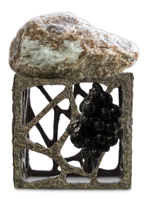
“They tend to want to do this on weekends and in the middle of the night, which is not very good for us,” Tessa told scientists at SICB. She came up with a simple trick of putting a box with large openings inside the tank. The cuttlefish generally don’t like the box because it’s too exposed. But place a rock on top and it suddenly becomes a safe haven. “Almost like clockwork, within three hours the cuttlefish swim inside and lay a bunch of eggs,” said Tessa.
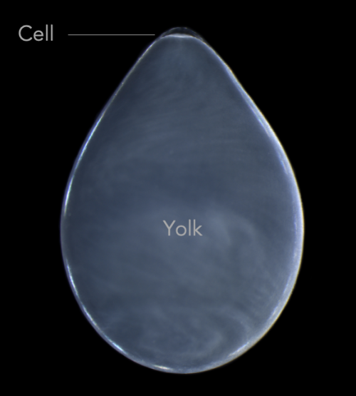
The eggs themselves are covered in dozens of layers of inky jelly that need to be removed before injecting. If you just tear away the jelly and try to grow the embryos in a dish, they die. The solution? Dunk the embryos in bleach and then gently rinse them off. After this bleach treatment, the ink jelly sloughs off easily and researchers can plop the embryo into a dish with antibiotics and squash it between two plastic tubes. This creates a positive pressure that, when combined with a quartz needle beveled at 15º to a very sharp 2.5-micron tip, allow researchers to punch through a tough chorion to reach the teensy cell at the top of a glob of yolk.
Left: The so-called “Holy Temple”: an open box with holes and a rock on top with an inky clutch of cuttlefish eggs. Right: Closeup image of a de-jellied cuttlefish egg. (Photo credits: Tessa Montague)
“No matter how delicately we do this, the vast majority of injected embryos die,” said Tessa, “but after years of practice, enough survive that we can do some experiments.” It takes four months, filled with lots of food and care, before the cuttlefish become sexually mature and Tessa can see if her transgenes make it to the germ line and are passed on to another generation of little cuttlefish.
Even without the worry of death by injection, it takes quite a lot of effort to keep cuttlefish alive. Two full-time animal specialists count grass and mysid shrimp and offer them one by one to the 200 dwarf cuttlefish housed in the Axel lab at Columbia University. Because of their fast metabolism, cuttlefish must be fed three times a day, resulting in lots and lots of poop. And they’re extremely sensitive to changes in water quality, meaning the animal tanks must feature myriad filtration systems and be cleaned every day.
“I’m very fortunate to have a team of six or seven people that work with me,” said Tessa. “Creating something out of nothing is very difficult. But every day that goes by, things get a little easier.”
Tessa wants to share her research to inspire other scientists whose burning scientific questions in new model organisms also require technological developments to answer them. To that end, she created Cuttlebase, a website that chronicles the scientific tools her team has developed, including a 3D brain atlas and a staging series of dwarf cuttlefish embryonic development. Her outreach extends beyond scientists to the general public — her personal website hosts a CuttleCam livestream where anyone in the world can watch these alien creatures from the comfort of their own home.
So, if you give a scientist a cuttlefish, know that she may embark on a journey to understand its secret language of colors rippling across its skin. It may be bumpy, but that journey will have at least a few moments that will take her breath away. And if you know where to look — if you can find the fluttering fins of a cuttlefish hidden in the rock — you might find yourself holding your own breath on your own journey. Maybe that just confirms there’s a scientist in all of us.
A dwarf cuttlefish moseys along and gives the viewer a side-ways glance. (Video credit: Tessa Montague) (3 votes) Loading...
In the February issue of Development, Niveda and her colleagues report on the identification and function of a previously unreported downstream effector of retinoic acid (RA) signalling in the chick forebrain. Niveda shares some insights into the story behind the paper and the science communication outreach initiated by her department at the Indian Institute of Technology Kanpur.
How did you get started on this project?
The genesis of this project was based on the findings of a study carried out in Prof. Amitabha Bandyopadhyay’s laboratory at the Department of Biological Sciences and Bioengineering (BSBE) at IIT Kanpur, wherein they performed a genome-wide expression screen of Metabolism-Related Genes (MRGs) in the chicken embryo. The result of this experiment was very interesting as the expression of MRGs peaked at the time of differentiation of the various tissues in the embryo. Intrigued by this result, we started to explore the role of Metabolism-Related Genes during the development of the chick brain.
We started the study by first examining the spatiotemporal expression profiling of the MRGs that were reported by the initial genome-wide screen. Our laboratory is interested in understanding the process of midline invagination of the forebrain roof plate, which leads to the formation of the cerebral hemispheres from the single forebrain vesicle. Thus, we were very intrigued when we discovered that one MRG known as Connector Enhancer Kinase Suppressor of Ras 2 (CNKSR2) was expressed very precisely in the middle of the invaginating roof plate of the chick forebrain. We then decided to examine its role in the process of midline invagination.
What was already known about the topic?
Before we started our research, the only information available about the process of separation of the cerebral hemispheres was that certain genes linked to holoprosencephaly, a devastating developmental disorder in humans, may be regulating this process. However, nothing was known about the molecular mechanism involved. In a previous study from our lab, we observed that during the process of midline invagination, the roof plate forms a characteristic W-shaped fold1. Also, it is through this process that the two hemispheres and the medially derived structures such as the hippocampus and choroid plexus are formed. This W-shaped invagination functions as a secondary signalling centre for pathways such as BMP2 and WNT3. In the paper published in 2015, we reported that Retinoic Acid signalling is detected in the middle loop of the W-shaped invagination of the roof plate and its inhibition leads to a flattened forebrain roof plate, a phenotype that resembles the human disorder holoprosencephaly1, where the hemispheres are improperly separated4. In this study, we found that the expression of CNKSR2 exactly coincided with the RA signalling domain in the chick forebrain roof plate, prompting us to investigate the role of this gene in this context.
Can you summarize your findings?
We can summarize our findings as follows: we found the expression of an MRG, CNKSR2, in the middle loop of the invaginating dorsal forebrain roof plate and overlapped with the active RA signalling domain. We manipulated RA signalling in the roof plate and found that the expression of the CNKSR2 transcript changed. This led us to infer that CNKSR2 is a downstream effector of RA signalling in this context.
Further, when we knocked down CNKSR2 from the invaginating roof plate using RNA-interference (RNAi) and obtained roof plate invagination defects which phenocopied loss of RA signalling. We found that the invagination defects upon knockdown of CNKSR2 were related to changes in cell proliferation and patterning of this region. Further, misexpression of mouse CNKSR2 was sufficient to ectopically induce the expression of roof plate midline markers in the lateral forebrain. This led us to conclude that CNKSR2 is necessary and sufficient for roof plate patterning. The final experiment that we performed revealed that CNKSR2 modulates Ras/Raf/MEK signalling to lower levels in the roof plate midline for proper patterning and subsequent chick forebrain morphogenesis.
When doing the research, did you have any particular result or eureka moment that has stuck with you?
We had three eureka moments during our study. The first was when we found that the knockdown of CNKSR2 in the chick embryo forebrain led to invagination defects with a holoprosencephaly-like phenotype. The second, was when the misexpression of the mouse CNKSR2 in the lateral forebrain was sufficient to induce the roof plate marker genes. And the third, and most unexpected result, was when ectopic downregulation of Ras/Raf/MEK in the lateral forebrain was sufficient to induce the expression of the patterning marker, Bmp7. I believe this third result was the final bit of evidence that helped us piece the story together.
Where will this story take the lab?
This story has helped to identify one important molecule-CNKSR2 in the bigger picture of understanding the process of midline invagination in the chick forebrain. Also, this gene may be used as both a proxy for RA signalling in the chick forebrain, as well as a roof plate midline marker. The process of midline invagination is complex with many aspects such as cell adhesion and cytoskeleton rearrangements also likely to be involved. We are currently investigating the possible role of CNKSR2 in each of these functions to understand the forebrain midline invagination process and the resulting separation of the cerebral hemispheres.
Science outreach and its importance
India is a country with enormous linguistic diversity. Our research group comprises members from across the country who are fluent in many languages. As our research is funded by taxpayers, we as a group believe that the general public in the country should be aware of the kind of research taking place in the lab and the resulting publications.
To fulfil this, the Department of Biological Sciences and Bioengineering (BSBE) at the Indian Institute of Technology Kanpur (IITK) decided to start a new initiative wherein the authors of a publication convey their research in their native language and English, all in layman’s terms. The authors of our study are fluent in Bengali, English, Hindi, Nepali and Tamil. Simple animated videos with narration in each of these languages were made and shared across social media for public awareness. We hope to continue this initiative with future publications, and we anticipate that it will be well-received by the viewers. In the end, we aim to inspire young students to actively consider becoming scientists and join us on this exciting journey!
1) Gupta S, Sen J. Retinoic acid signalling regulates development of the chick’s dorsal forebrain midline and the choroid plexus. Development. 2015 Apr 1;142(7):1293-8. doi: 10.1242/dev.122390. Epub 2015 Mar 10. PMID: 25758461.
2) Furuta Y, Piston DW, Hogan BL. Bone morphogenetic proteins (BMPs) as regulators of dorsal forebrain development. Development. 1997 Jun;124(11):2203-12. doi: 10.1242/dev.124.11.2203. PMID: 9187146.
3) Lee SM, Tole S, Grove E, McMahon AP. A local Wnt-3a signal is required for the development of the mammalian hippocampus. Development. 2000 Feb;127(3):457-67. doi: 10.1242/dev.127.3.457. PMID: 10631167.
4) Roessler E, Muenke M. The molecular genetics of holoprosencephaly. Am J Med Genet C Semin Med Genet. 2010 Feb 15;154C(1):52-61. doi: 10.1002/ajmg.c.30236. PMID: 20104595; PMCID: PMC2815021.

 (No Ratings Yet)
 (3 votes)