We have an opening for a motivated postdoctoral fellow with strong Drosophila molecular genetics experience and interest in post-transcriptional regulatory biology to join our team.
Although miRNAs are often thought to mediate “fine-tuning”, we revealed many examples of profound defects in neural miRNA mutants. These include switches of cell fates in diverse types of sensory organs and in the CNS, and disruptions of neural function and behavior.
We are particularly interested now to exploit precise CRISPR engineering of 3’UTRs of critical targets, to dissect target regulation in specific celltypes and circuits that underlie phenotypes, and integrate this with genomic approaches, including single cell RNA sequencing. Moreover, our interests in miRNAs dovetail with our studies of other modes of post-transcriptional regulation that are critical in the nervous system, including alternative polyadenylation, splicing, and RNA methylation.
The ideal candidate will be comfortable with classical and modern fly genetics and molecular methods, but will be interested to integrate biochemical, genomic, and/or computational strategies utilized by other lab members in a multidisciplinary and interactive environment.
Generous compensation, benefits, and housing package is available immediately (https://www.mskcc.org/education-training/postdoctoral/current-incoming). Please provide CV, motivation letter and 3 references to Eric Lai, laie@mskcc.org.
Welcome to our monthly summary of developmental biology (and related) preLights
In our last post of the year, we again have plenty of exciting research to feature, and would like to thank all the preLighters for their incredible work selecting and highlighting interesting preprints in a broad range of topics for the community. Early next year, we have two new ‘experiments’ coming at preLights, so stay tuned!
Morphogenesis
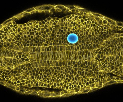
Sundar Naganathan wrote about endoderm morphogenesis in Drosophila and how local transcription activation of myosin and coupled mechanical changes regulate it. Staying with this model organism, Ivana Viktorinova’s preLight questioned whether the century-old Hertwig’s rule (stating that cells divide along their long axis) is universally valid. The authors of the study showed that in a population of the epidermis this is not the case, and that the tension exerted by actomyosin contractility – rather than the length of the axis – determines orientation of cell division.
The phenomenon of cell competition was initially described in Drosophila – in 1975 – and since then a number of its molecular details have been unravelled. This month, Rohan Khadilkar reviewed the inter-relationship between infection, the Toll-pathway induced innate response and cell competition. Sarah Bowling and Teresa Rayon explored a new role for this process during early mouse development, where unspecified cells are competitively eliminated during epiblast formation.
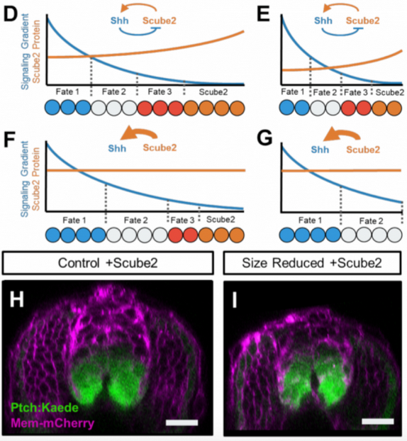
Moving to zebrafish, Teresa Rayon wrote about scaling of morphogen gradients and a preprint studying how in embryos of varying sizes the patterning of the neural tube remains proportional. The authors found that Shh and a protein required for Shh release – Scube2 – form a feedback loop that allows proportional patterning. From the world of plants, an intriguing kind of growth pattern, root circumnutation, and its regulation by a novel histidine kinase featured in the preLight by Martin Balcerowicz.
Figures taken from preprints by Hashimoto & Sasaki and Collins et al.,preLighted by Teresa Rayon and Sarah Bowling
From Development to Disease – scRNAs-seq studies in the spotlight
Sequencing the transcriptomes of individual cells in a tissue or organism has become a powerful way to gain new insights into development and disease and build valuable resources (watch out for the journal Development’s next special issue). A preLight by Leena Rasrado reported on mouse neural tube development at the single-cell level: with scRNA-seq, the authors unravelled the molecular mechanisms underlying spatial and temporal neuronal diversity.
Rob Hynds highlighted a study on the differentiation of the respiratory epithelium by scRNA-seq. In another preLight, he covered how the analysis of scRNA-seq datasets from cancer cells can identify specific targets of T cells for immunotherapy. Hannah Brunsdon often highlights cancer studies in model organisms, this month she featured work from bioRxiv’s ‘confirmatory results’ category where two groups validated and compared zebrafish models of B cell acute lymphoblastic leukemia.
The preprint Shikha Nayar wrote about combined single-cell transcriptomics with other ‘omics approaches to show that old fibroblast cultures are heterogeneous, and contain a larger proportion of ‘activated fibroblasts’. Remarkably, these cells secrete cytokines, which result in higher reprogramming efficiency.
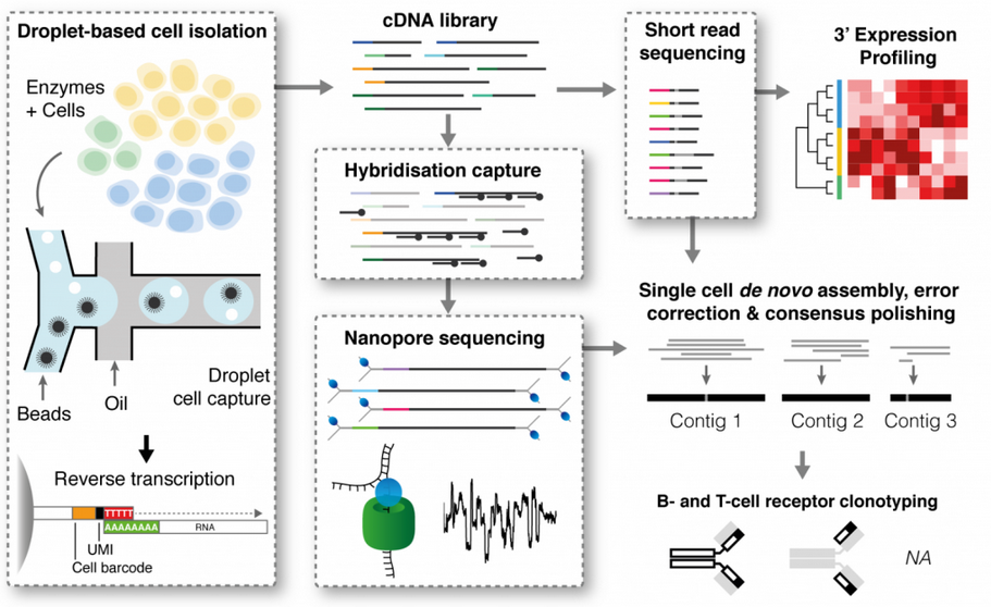
Finally, Samantha Seah covered two preprints that made important technological advances in droplet-based single-cell sequencing. The two new methods, RAGE-seq and DART-seq allowed researchers to capture sequence diversity at the 5’ end of a transcript and thus study the diversity of B-cell and T-cell receptors.
Figure taken from preprint by Singh et al., preLighted by Samantha Seah
Gene regulation & Chromatin
Genome-wide association studies have been useful in linking genetic variants (mostly SNPs) to diseases, however the majority of these SNP-s fall into non-coding regions with potential regulatory functions. Jesus Victorino preLighted work from the van Steensel lab assessing on a genome-wide scale which SNPs can modulate expression of a reporter. Carmen Adriaens and Clarice Hong teamed up to highlight another preprint from this lab, which used massively parallel reporter assays to study features of gene repression in heterochromatin at the nuclear periphery. Heterochromatin was also at the centre of Gabriel Aughey’s post, which discussed how cell cycle speed during early embryo development in Drosophila sets the pace for its establishment.
Visit the preLights website for much more – such as a highlight from Theo Sanderson about an emerging field of optics-free microscopy, or Ashrifia’s post on how favourable environmental conditions lead to an increase in languages spoken in a specific geographic area.
Two postdoctoral positions are available in the Boavida laboratory in the Department of Botany and Plant Pathology at Purdue University. The lab investigates the molecular mechanisms controlling gamete identity and function and cell-cell signaling in plant fertilization.
We are seeking highly motivated individuals who value achievement, are proactive, creative, detail-oriented, flexible and constructive team players. Candidates should have completed their Ph.D. degree on a relevant area in Biological Sciences within the last 2-3 years. Proven ability to present and publish research data and excellent English communication skills both oral and written are required.
Postdoc A. The recruited researcher will work in two main axes of investigation based on (1) identification of interaction partners of gamete-expressed proteins primarily through a high-throughput proteomic screening (NGS-Y2H); and (2) integration of PPI networks with derived NGS-transcriptomic approaches to establish high-confidence interaction maps for prediction and functional characterization of signaling networks operating in plant gametes and fertilization. The ideal candidate should have experience in molecular biology and protein biochemistry (membrane protein expression and purification from various cellular systems, Y2H, BIFC, co-immunoprecipitation, etc.). Knowledge of integrative computational analysis, structural bioinformatics, protein docking and characterization of regulatory networks is an asset.
Postdoc B. The postdoctoral researcher will use an exciting combination of techniques including flow cytometry (Microfluidics and FACs), genomics, transcriptomics, epigenomics, and molecular biology to study mechanisms underlying cell identity and differentiation of plant gametophytes in a spatiotemporal context. He/she will also be involved in the characterization of a cell-based functional assay to study the cellular functions of candidate genes in plant fertilization. Prior experience in flow cytometry, confocal microscopy, molecular genetics, functional genomics, and bioinformatics is highly desired. Candidates with interest in sexual reproduction and/or documented experience in Arabidopsis, tomato and maize are especially encouraged to apply.
Application
Applications will be accepted until the position is filled and should include CV, contact details of at least 2 references and a cover letter outlining how your previous research experience/contribution integrate with the laboratory research projects. Incomplete applications will not be considered for evaluation. Relevant publications and information can be found here.
Please send your application as a single PDF file to lboavida@purdue.edu. Include in the subject header PlantRepro2018_A (or B) (according to your interest in Postdoc position A or B). The appointment is initially for 12 months with the possibility of extension depending on research performance and available funds. Starting date is flexible and can be as early as February 1st, 2019.
Purdue University is an Equal Opportunity Employer, has a strong commitment to diversity and actively encourages applications from candidates from groups underrepresented in higher education.
A PhD position is available in the laboratory of Néva Meyer at Clark University in Worcester, MA USA. Research in Dr. Meyer’s lab is currently focused on understanding how the central nervous system develops in annelids with the goal of gaining a better understanding of how nervous systems evolved.
The successful applicant will develop a project focused on molecular control of neural fate specification in the annelid Capitella teleta, but this can be expanded to include other spiralians and different avenues of research depending on the applicant’s interests and goals. Additional information is available on the Meyer lab website.
The anticipated start date is late August 2019. Previous experience in molecular biology and working with marine larvae and/or bioinformatics is desirable. The successful applicant will be guaranteed funding for five years through a combination of research assistantships and teaching assistantships; two years of research assistantship for this position are currently available.
Please email a cover letter explaining your interest in the position and qualifications and a CV to: nmeyer[a]clarku.edu.
When we examined the kinetics of Rho GTPase activity in endothelial cells in response to receptor stimulation (Reinhard, 2017), we noticed considerable cell-to-cell heterogeneity. In the original work we published graphs with the average response, reflecting the response of the whole cell population. However, these graphs fail to show the cellular heterogeneity. What is the best way to visualize these data? I don’t know, but I will explore several options to visualize all individual traces from the experiment.
1. All together
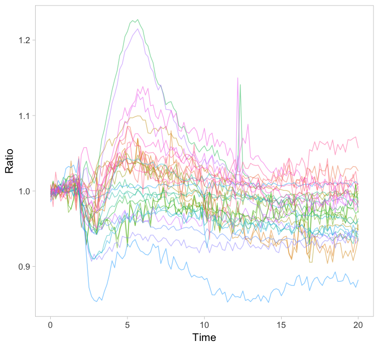
The original paper displayed the average responses of the Rho GTPases RhoA, Rac1 and Cdc42 and the graphs were remade with R/ggplot2 in this blog. Since the Rac1 data showed the largest differences between cells, I took this data to explore different ways of visualizing it. One can simply plot all the individual lines together with the average response in a single graph (figure 1). How this can be achieved with R/ggplot2 is explained in this blog. The graph may look cluttered when there are many lines and therefore it is advisable to use thin gray lines. To enable the identification of individual traces one may use a unique color for each trace. Putting all traces from a single experiment in the same graph is a simple but effective method to depict variability. However, putting all data in a single graph for multiple conditions is usually makes the graph difficult to understand.
Figure 1: Ordinary graphs showing the data from individual cells as thin lines on top of each other. In the left panel the average is shown as a thicker black line. In the right panel the data from each line has a unique color to improve identification of cells.
2. Ridges
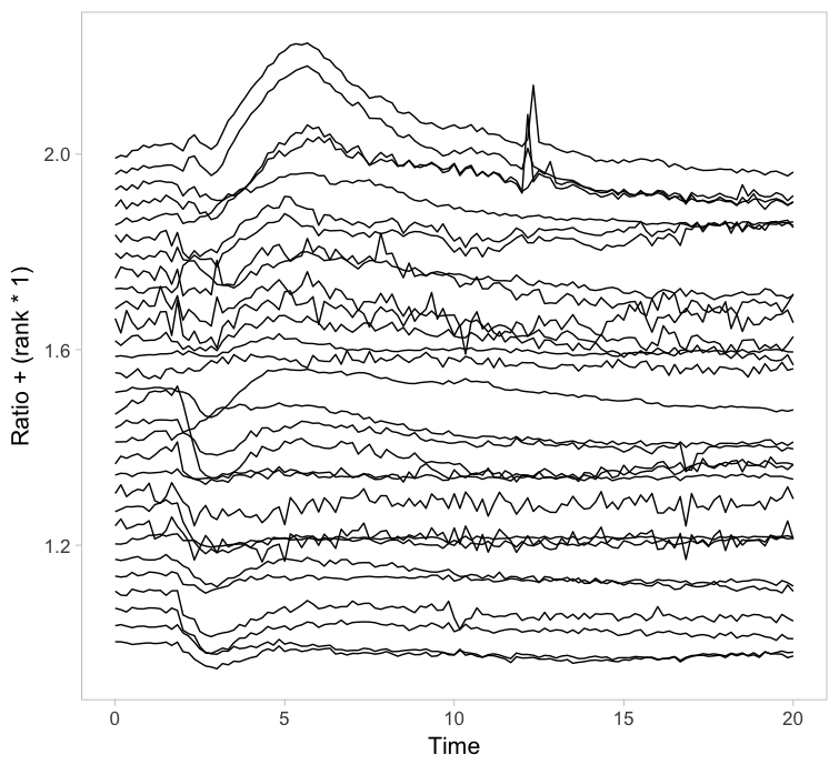
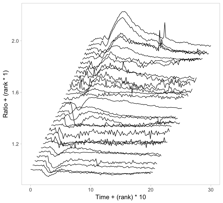
Instead of putting all graphs on top of each other, an offset along the y-axis is introduced, which reduces overlap. This type of plot is called a ridgeline plot (formerly known as a joyplot). By adding an increasing offset for the x-axis, the curves are further separated and a 3D effect is generated. This may result in a nicer plot to look at, but it comes at the cost of impeding quantitative interpretation. For example of the use of ridgeline plots fro kymographs see this blog.
Figure 2: Ridgeline plots showing the individual cellular responses with an offset in the vertical direction (left) or an offset in both vertical and horizontal direction (right).
3. Small multiples
Instead of plotting all lines in a single graph, it is also an option to show all lines in individual plots. This type of design is coined the “small multiple” and it was popularized by Edward Tufte. The presentation of small multiples is not limited to line graphs, but be used to depict other types of data, e.g. several still images from a movie.
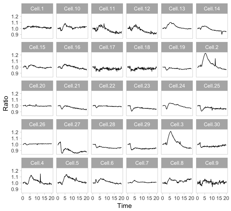
Small multiples can be reproduced in R/ggplot2 by using ‘faceting’ and this has been briefly highlighted in a previous blog (near the end). Figure 3 (left) shows the standard output of ggplot2, with labels for each plot. In my opinion, the small multiple is at its best when it is trimmed down to the bare minimum. As an example, I made a minimalistic variant with a high data-ink ratio (figure 3, right). Although numbers are absent, this visualization still allows for quantitative comparison and it shows the heterogeneity. In summary, the small multiple is a great way to depict the individual data, but it requires quite some space.
Figure 3: The small multiple presentation of the time-lapse data shown as the standard output of ggplot2 with facets (left) and a minimalistic version (right) with improved data-ink ratio.
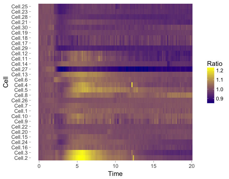
4. Heatmap
The ‘heatmap’ is well-known for the visualization of microarray data. It can also be used for different types of data including time-series data. To generate a heatmap, the actual numbers of each of the line graphs are color coded and these are presented as a line. Every line (row) of the heatmap represents a single trace from a time series experiment. The result is an image in that shows the responses encoded by colors (figure 4).
As the data is encoded by color, the quantitative comparison of the data is limited to comparing colors. This comparison is inherently non-quantitative. As such, the downside of a heatmap is that it only allows qualitative interpretation. Still, heatmaps enable the visualization of a lot of data on a relatively small space.
Figure 4: Heatmap style presentation in which each row represents the data from a cell. The response is color coded according to the legend. The data is presented in order of acquisition (left) or clustered based on similarity (right).
Sorting out the mess
The data from single cell experiments can be very heterogeneous (as in this example) and may look like a mess. To structure the data, it can be sorted or grouped. Sorting uses a feature of the data that can be used to rank the individual traces. For instance, for the ridgeline plot (figure 2) the maximum response of every trace was determined. Subsequently, the plots were ordered based on the maximum response (ratio) that was observed. Any other parameter can be used to order the data, e.g. integrated response, timing of the maximum response or noise of the baseline.
Another method to structure the data is grouping or clustering. For the data presented in the heatmap a hierarchical clustering was performed (figure 4, right), which determines the pairwise difference for each of the traces. The ouctome is an order of traces that groups similar responses together. There are many ways to cluster data and information on this topic can be found elsewhere (for instance here, here and here). In the end, the sorting or clustering of the time-lapse data may structure the data and aid interpretation. It can precede any of the visualizations shown above, but it makes most sense for ridgeline plots or heatmaps.
Howto
This blog explores several options to visualize data, but does not go into detail on how to achieve it. The code (and data) that was used to generate the graphs is available at Zenodo (http://dx.doi.org/10.5281/zenodo.2211123) and should give some idea of how to achieve the different visualizations in R/ggplot2. Alternatively, some of the plots shown in this blog (including small multiples and heatmaps) can be made with a web-based tool PlotTwist that uses R/ggplot2 but does not require coding skills. A nice feature of the web tool is that the presentation of the data (e.g. visibility of the lines and their color) can be adjusted in realtime.
Final words
Showing all of the data is important for full appreciation of experimental results.Since cell-to-cell variability is observed in every single imaging experiment, methods to visualize the heterogeneity are needed. Here, I have highlighted several methods to visualize data from time-lapse imaging. These methods may be combined with some way of sorting or clustering of the data. Which approach works best for your data needs to be examined on a case-by-case basis and will depend on what you want to communicate. The different visualizations shown here may serve as a starting point to explore how to best visualize your data from time-lapse experiments.
A position for a postdoctoral fellow is available in the lab of Dr. David Parichy at University of Virginia to study functions of thyroid hormone and other circulating factors during the development and homeostasis of adult neural crest stem cells and their derivatives in zebrafish.
Major interests lie in understanding how specific hormones drive disparate cellular behaviors across cell types, including Schwann cell precursors, glia, melanocytes, and novel populations recently identified. Additional questions relate to how these hormones contribute to stem cell maintenance and fate specification during ontogeny and regeneration, and consequences of hormonal signaling for cancer initiation and metastasis.
The group is equipped with lab-dedicated infrastructure for Next Generation Sequencing, single-cell RNA-seq, super-resolution time-lapse imaging, and high throughput genetic and transgenic screening. More information about the lab and the highly interactive Departments of Biology and Cell Biology can be at: http://dparichy.as.virginia.edu/.
Applicants must have or be pursuing a Ph.D. in biology, endocrinology, cell biology or related fields. Prior experience with zebrafish is not required.
Applicants should provide: CV’ contact information for three references; cover letter briefly describing interests, experience and career goals
We are seeking outstanding postdoctoral candidates to join the Campas lab at the University of California, Santa Barbara. Our group uses interdisciplinary and quantitative approaches to study the formation of tissues and organs during zebrafish embryonic development. We are interested in connecting the molecular and cellular process that orchestrate embryogenesis with the physical/mechanical processes that sculpt tissues and organs into their functional morphologies. To quantify and perturb local tissue mechanics we employ unique microdroplet techniques that we have recently developed (Campàs et al., Nature Methods, 2014; Serwane et al., Nature Methods, 2017). These techniques offer unprecedented opportunities to study tissue morphogenesis quantitatively (see e.g., Mongera et al., Nature, 2018).
We are specifically seeking independent, passionate, and motivated applicants for a postdoctoral position to study the interplay between the molecular and mechanical processes that shape embryonic tissues in zebrafish. The candidate will be able to work in a collaborative manner with a highly interdisciplinary group of researchers, including physicists, engineers and developmental biologists. A Ph.D. in the biological sciences (or related fields) with at least 3 years of laboratory research experience in zebrafish developmental biology is required. Experience in quantitative biology or biophysics, in addition to experience in zebrafish development, will be considered positively, but is not required.
This is a renewable, two-year position with full benefits, that will be extended as needed upon good performance of the candidate. Salary will be competitive and dependent on the level of experience of the candidate. Applicants should email a CV and a description of research interests to Prof. Campas (campas@ucsb.edu), and should also arrange for at least two references to submit letters of recommendation of their behalf. Applications submitted by February 15th, 2019 will receive priority consideration, but the position will remain open until filled. Start date is flexible.
The University of California, Santa Barbara provides an exceptional, interdisciplinary and collaborative environment for scientists interested in quantitative biology and systems biology (including exposure to the Santa Barbara/KITP Summer School on Quantitative Biology).
We are seeking independent, passionate and creative students to join the López-Schier group at the Helmholtz Zentrum in Neuherberg/Munich, Germany.
The position is to work, at either experimental or theoretical level, on the cellular, genetic and mechanical aspects of multicellular self-organisation and their relevance to sensory-organ morphogenesis and regeneration. This project is ideal for someone with a strong theoretical background in biology, physics or engineering. Knowledge of computer programming and optics would be ideal but not essential.
Qualifications & skills
– University studies in biology-related sciences, physics or engineering
– A good command of the English language is essential
Please, apply via electronic mail only, including a cover letter with a short statement of research interests and motivation, a Curriculum Vitae including a list of names and email-addresses for two/three academic references, to:
Dr. Hernán López-Schier
Unit Sensory Biology & Organogenesis, Helmholtz Zentrum München
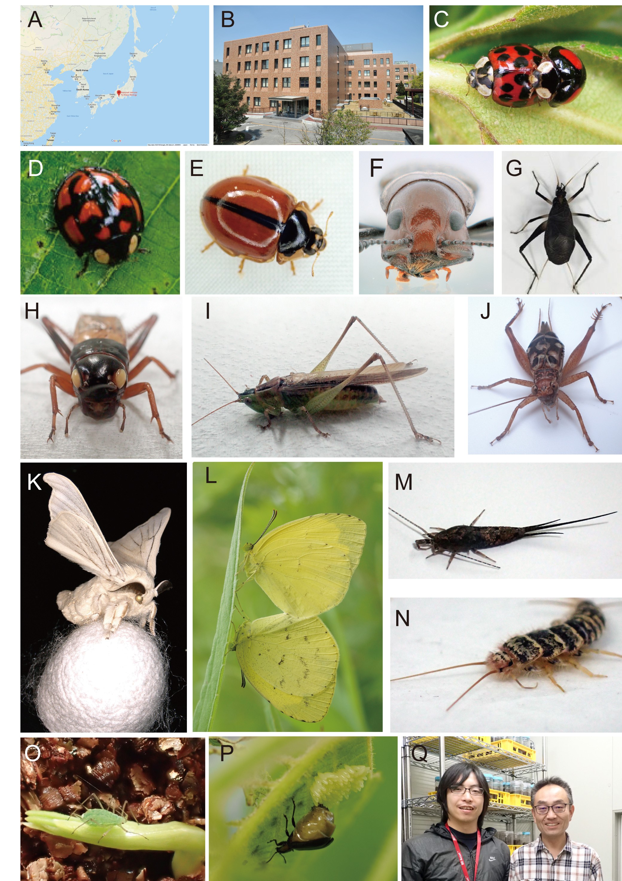
I am Shinichi Morita, a postdoctoral researcher in Teruyuki Niimi’s lab at the National Institute for Basic Biology, Japan (Fig. 1A, B). Our research interests focus on the evolutionary novelties that insects have acquired, and how various insect morphologies have arisen during evolution (Fig. 1C-P).
Fig. 1 Location of the Niimi ‘s laboratory and our experimental insects (A) Location of National Institute for Basic Biology, Okazaki, Japan (https://www.google.co.jp/maps). (B) Picture of the National Institute for Basic Biology building. (C-P) The insects we work on in the lab: (C) Harmonia axyridis, (D) Aiolocaria hexaspilota, (E) Propylea japonica, (F) Doubledaya bucculenta, (G) Homoeogryllus japonicus, (H) Gryllus bimaculatus, (I) Conocephalus maculatus, (J) Cardiodactylus guttulus, (K) Bombyx mori, (L) Eurema mandarina, (M) Pedetontus nipponicus, (N) Thermobia domestica, (O) Acyrthosiphon pisum, (P) Gastrolina depressa. (Q) The horn evo-devo team: Teruyuki Niimi (right) and myself.
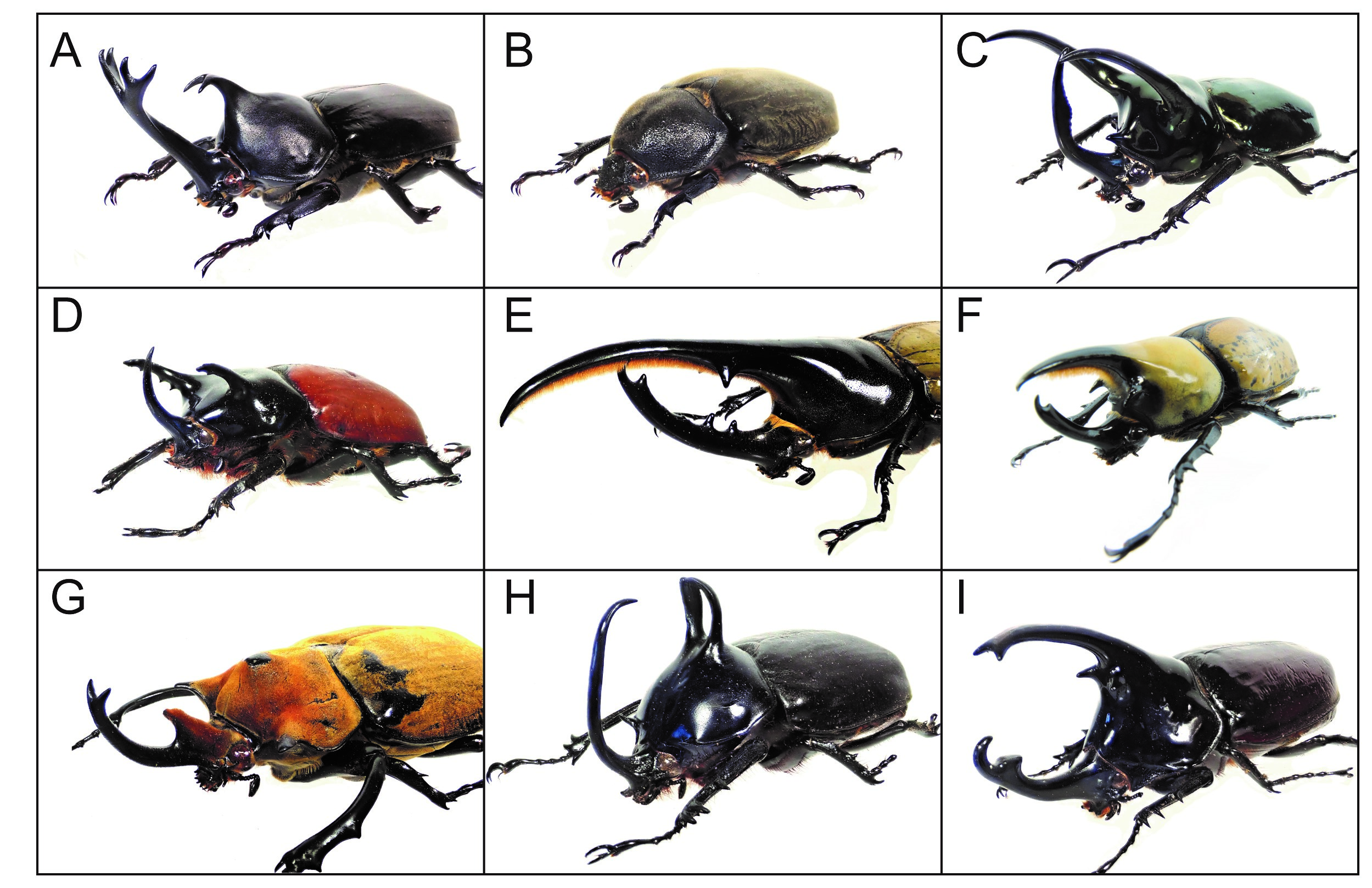
Beetle horns are thought to be an evolutionary novelty and are used as weapons for intraspecific combats between males. Beetle horns display sexual dimorphism in many Scarab beetles, and their shapes, numbers, sizes and forming regions are highly diverged even among closely related species (Fig. 2). Elucidating how these novel traits were acquired in Scarab species will lead to better understanding of the mechanisms of morphological diversification during evolution. My postdoc project is to understand how exaggerated horns are acquired and formed in Japanese rhinoceros beetles (Trypoxylus dichotomus) (Fig. 2A, B).
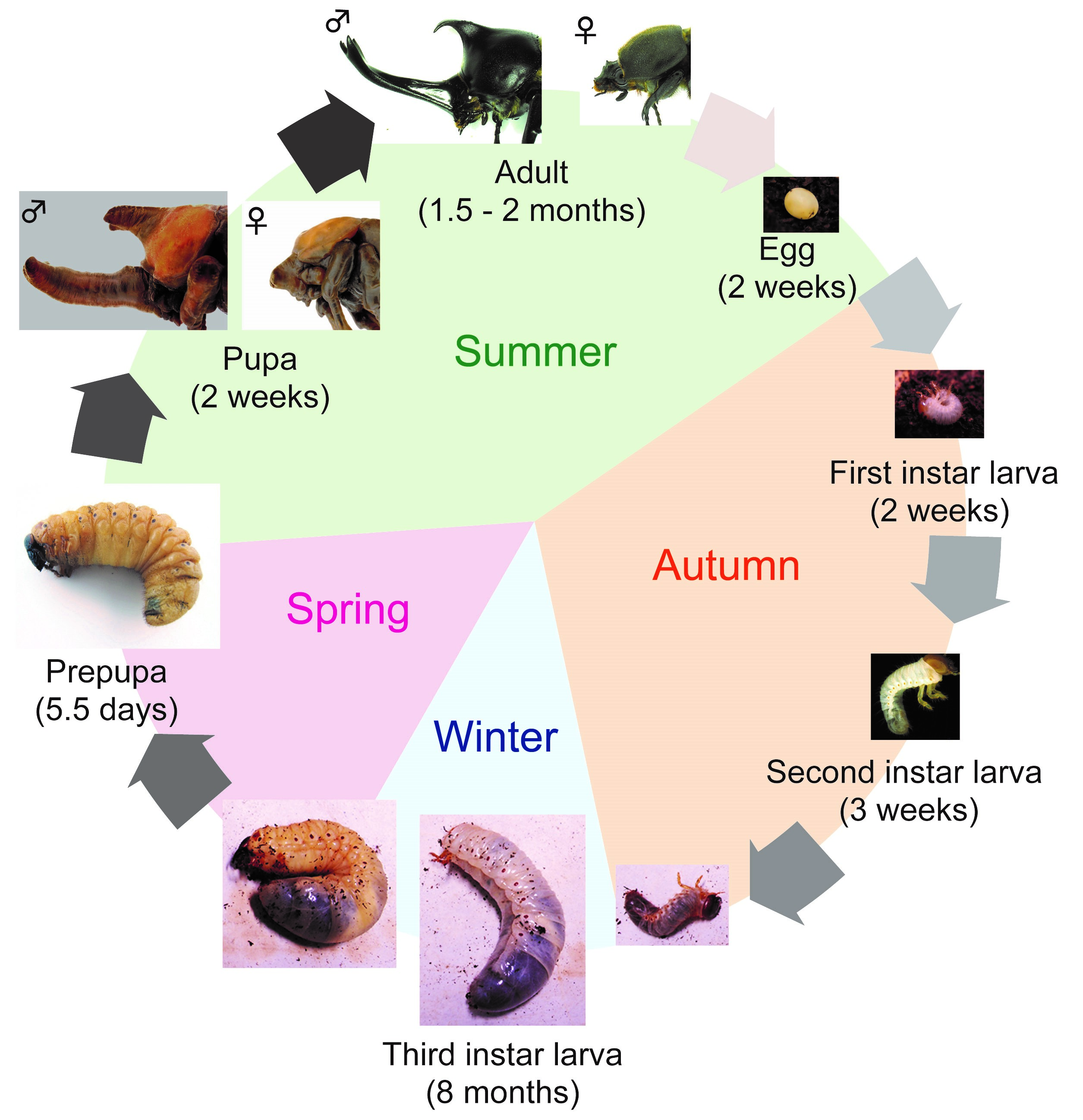
First, I will introduce T. dichotomus. In Japan, T. dichotomus is a popular and familiar insect to children and adults, and is sold as a pet at department stores and DIY shops during the summer. The Japanese name for T. dichotomus is Kabuto-mushi, and it is also used as one of the season words of “Haiku” (Haiku is a traditional short Japanese poem with seventeen syllables in the pattern of 5-7-5, including a season word in it). T. dichotomus is a holometabolous insect that has egg, larval, pupa and adult stages. Its life cycle is about 12 months (Fig. 3). Adults emerge in early summer and lay eggs in the soil at the end of summer. Larvae hatch from the eggs in about 2 weeks (Fig. 3, Egg) and feed on humus. The larval period is about 8 months and they pupate in the next spring (Fig. 3, First – Third instar larva). At the end of third (last) instar, they make pupal chamber (Fig. 3, Prepupa). During the prepupal period, horn primordia are formed in the head and the thorax. After about 2 weeks of pupal period (Fig. 3, Pupa), they become adults in the early summer (Fig. 3, Adult). T. dichotomus male adults have exaggerated horns on the head and prothorax (Fig. 2A), whereas females do not have these structures (Fig. 2B). The head horn is shaped like a plow with a long stalk, and bifurcated twice at the distal tip, while the prothoracic horn is shorter than the head horn, and bifurcated once at the distal tip.
Fig. 3 The life cycle of T. dichotomus
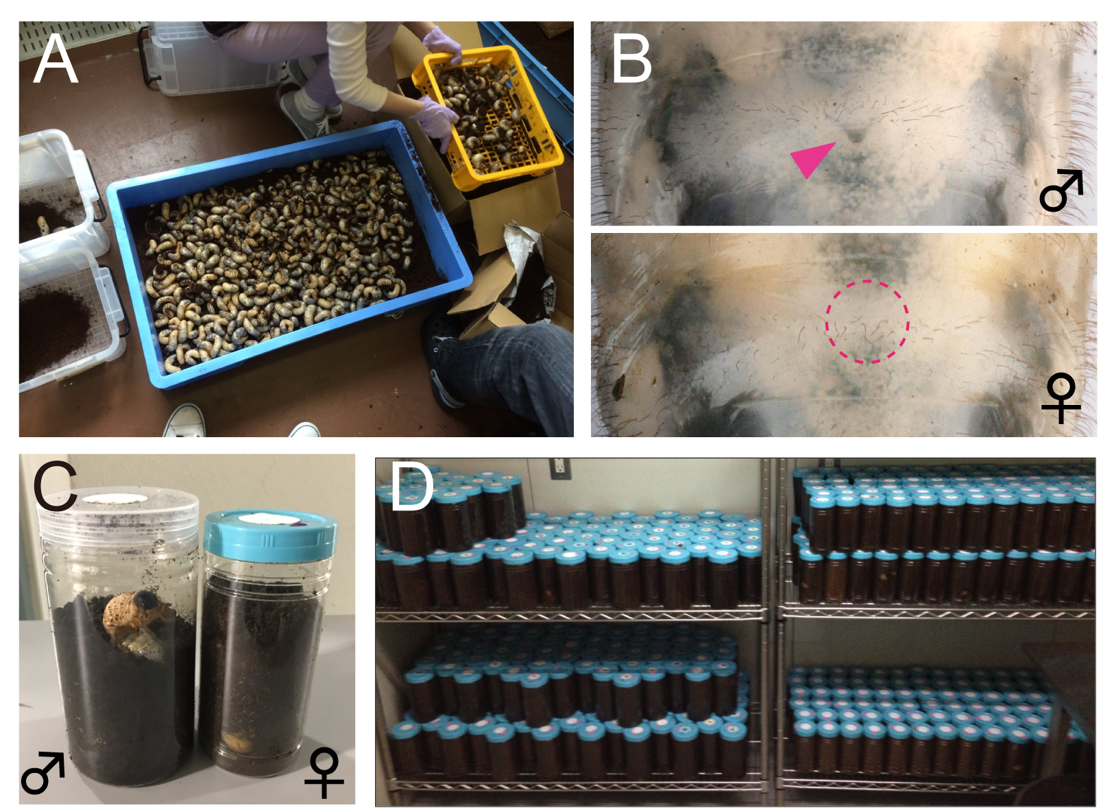
Here, I share my daily activities in the laboratory. As mentioned above, since the life cycle of T. dichotomus is one year, our daily life differs depending on season. In April, we purchase about 3,000 – 4000 last instar larvae of T. dichotomus from insect supplier (Fig. 4A). With help of our lab members, the sex of these larvae are determined (Fig. 4B) and they are packed individually in a bottle filled with breeding mats (Fig. 4C). They are stored at 10 ° C until use (Fig. 4D). Therefore, we can use T. dichotomus as an experimental material in any season.
Fig. 4 Handling of T. dichotomus larvae (A) Purchesed last instar larvae of T. dichotomus. (B) The male and female larvae are discriminated by the presence (male) or absence (female) of the Herold’s organ. (C) The larvae are reared individually in a container (140 mm in height and 95-mm in diameter for males, 130 mm in height and 75 mm in diameter for females) filled with breeding mats. (D) Larvae are stored in a low-temperature room.
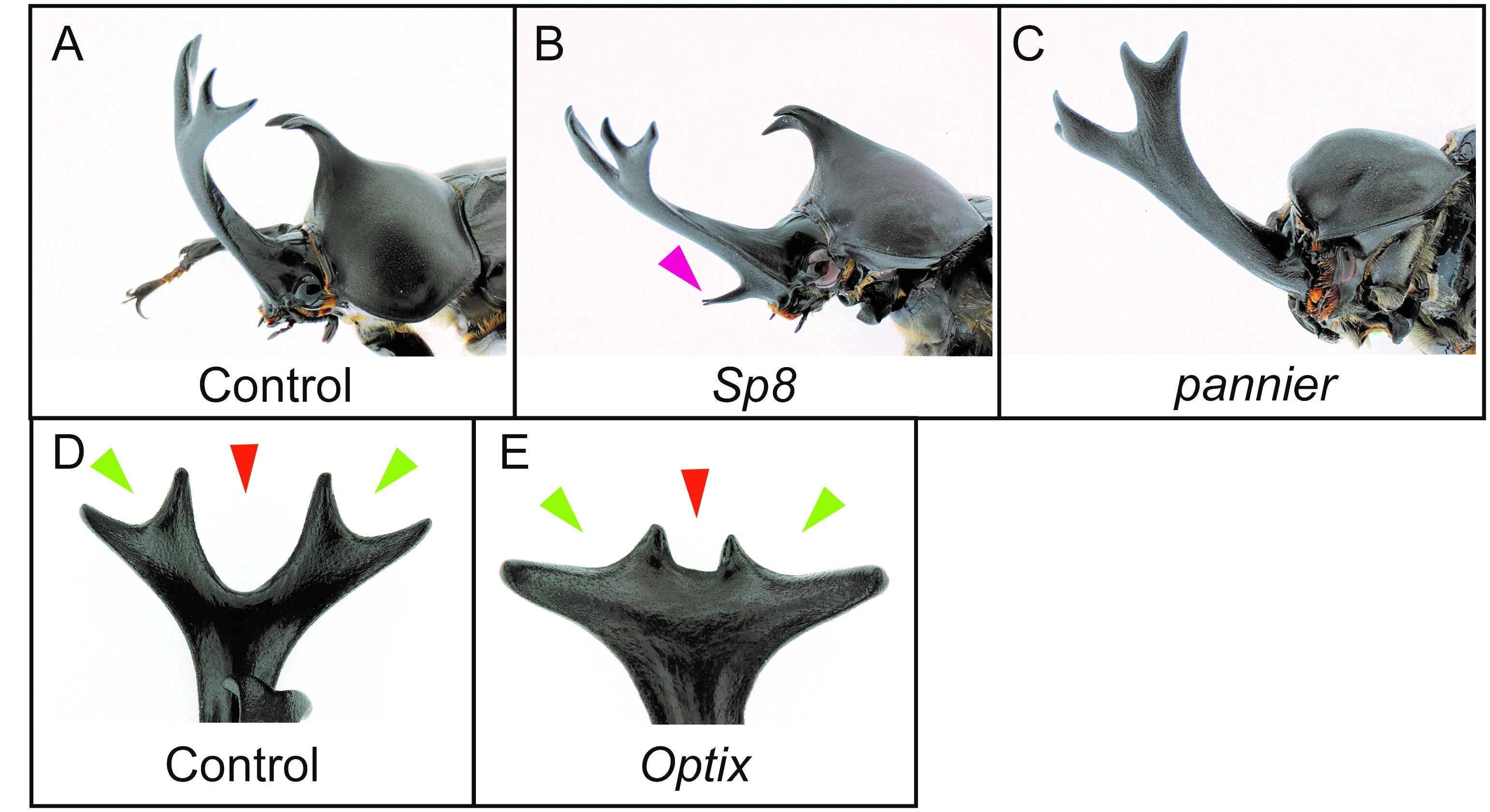
The sexual dimorphism of horns first appears in the horn primordia during the prepupal stage (Fig.3, Prepupa). One of my main research aims is to find out which genes control horn formation, so we dissected horn primordia in males and females at this stage and performed comparative transcriptome analysis by RNA-seq. We performed an intersexual comparison between male and female horn primordial transcriptome datasets, and tried to identify genes driving the development of different morphologies between male and female. In addition, we also compared transcriptome data between different horn types (head and prothoracic horns) intersexually in males and females. As a result, we identified 1,553 differentially expressed genes (DEGs) in total. To identify the essential genes for horn formation, we focused on genes encoding 38 transcription factors and 11 signaling molecules included in the 1,553 DEGs, and performed larval RNA interference (RNAi) screening. In beetles including T. dichotomus, larval RNAi experiments is extremely efficient to analyze the function of genes during postembryonic development. Double-stranded RNA (10 – 50 µg) was injected laterally into the T1 segment of each last instar larva before the prepupal stage using a 1 ml syringe with a 30 gauge needle. As a result, 11 genes were newly identified as horn formation genes (Fig. 5A-E). Interestingly, these 11 genes are mostly categorized as larval head- and appendage-patterning genes.
Fig. 5 Larval RNAi experiments (A) Control RNAi phenotype (EGFP). (B) Sp8 RNAi phenotype. (C) pannier RNAi phenotype. (D) Dorsal view of a head horn tip in control RNAi phenotype (EGFP). (E) Dorsal view of a head horn in Optix RNAi.
Future perspective of beetle horn studies
In addition to the above 11 genes, we have identified a number of other horn formation genes, but the gene regulatory network for horn formation is still unknown. So far, we have set up various resources and tools necessary for beetle horn research (precise staging of beetle horn formation, larval RNAi, whole mount in situ hybridization, immunostaining etc.). Therefore, it has become possible to analyze the horn formation gene regulatory network through developmental biological approaches. In addition, we are going to test whether novel cis-regulatory elements play a crucial role in acquisitions of horns during evolution. To this end, we are planning to perform ATAC-seq (Assay for Transposase-Accessible Chromatin sequencing), a technique to assess genome-wide chromatin accessibility, as a project to estimate cis-regulatory elements involved in horn formation and acquisition. Through the above approaches, we believe a part of evolutionary mechanisms associated with acquisition of animal diversity will be unravelled at the molecular level.
If you would like any more information about rhinoceros beetles and evo-devo questions in Scarab beetles, please get in touch with me (shinichi@nibb.ac.jp) or Teruyuki Niimi (niimi@nibb.ac.jp).
References
Ohde, T., Morita, S., Shigenobu, S., Morita, J., Mizutani, T., Gotoh, H., Zinna, RA., Nakata, M., Ito, Y., Wada, K., Kitano, Y., Yuzaki, K., Toga, K., Mase, M., Kadota, K., Rushe, J., Lavine, LC., Emlen, DJ. and Niimi, T. (2018) Rhinoceros beetle horn development reveals deep parallels with dung beetles. PLOS Genetics, 14: e1007651.
Location: The Francis Crick Institute, Midland Road, London
Contract: Fixed-term, (3-4 months)
Full time
Salary: Competitive with benefits, subject to skills and experience
Vacancy ID: 9385
Short summary
We are seeking a highly motivated and collaborative Laboratory Research Scientists in the area of human embryology and stem cell biology to join Dr. Kathy Niakan’s laboratory. Our lab seeks to understand gene function and to characterise gene expression during human pre-implantation development. This foundational information not only informs our understanding of human biology but also has clinical importance for infertility treatment and the therapeutic use of embryonic stem cells to treat various diseases.
The successful candidate is likely to be collaborative, energetic, focused, and productive individual. Excellent organisational, analytical, and communication skills are essential.
Project scope
Dr Niakan’s laboratory focuses on understanding the mechanisms of lineage specification in human embryos and the derivation of novel human stem cells. The post holder will report directly to the Group Leader, Kathy Niakan. Details of research projects currently being undertaken can be seen at: http://www.crick.ac.uk/kathy-niakan
The pluripotent epiblast of the early human embryo has the unique potential to give rise to the entire fetus in vivo and can self-renew indefinitely as embryonic stem cells (hESCs) in vitro. Understanding how this lineage is established is of fundamental biological importance and has significant clinical implications for both infertility treatment and the use of hESCs to treat various diseases.
The aim of this project is to further understand fundamental aspects of early human development and to use this knowledge to inform our understanding of existing stem cells and to establish novel alternative stem cells. Research techniques used in the laboratory include: molecular biology, advanced microscopy, human and mouse preimplantation embryo culture and micromanipulation, CRISPR/Cas9-mediated genome editing, genome-wide techniques including single-cell RNA-sequencing combined with DNA-sequencing.
About us
The Francis Crick Institute is a biomedical discovery institute dedicated to understanding the fundamental biology underlying health and disease. Its work is helping to understand why disease develops and to translate discoveries into new ways to prevent, diagnose and treat illnesses such as cancer, heart disease, stroke, infections, and neurodegenerative diseases.
An independent organisation, its founding partners are the Medical Research Council (MRC), Cancer Research UK, Wellcome, UCL (University College London), Imperial College London and King’s College London.
The Crick was formed in 2015, and in 2016 it moved into a new state-of-the-art building in central London which brings together 1500 scientists and support staff working collaboratively across disciplines, making it the biggest biomedical research facility under in one building in Europe.
The Francis Crick Institute will be world-class with a strong national role. Its distinctive vision for excellence includes commitments to collaboration; developing emerging talent and exporting it the rest of the UK; public engagement; and helping turn discoveries into treatments as quickly as possible to improve lives and strengthen the economy.
If you are interested in applying for this role, please apply via our website.
The closing date for applications is 21 December 2018 at 23:30.
All offers of employment are subject to successful security screening and continuous eligibility to work in the United Kingdom.

 (No Ratings Yet)
 (1 votes)


 (14 votes)
 A position for a postdoctoral fellow is available in the lab of Dr. David Parichy at University of Virginia to study functions of thyroid hormone and other circulating factors during the development and homeostasis of adult neural crest stem cells and their derivatives in zebrafish.
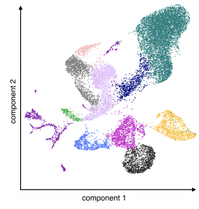 maintenance and fate specification during ontogeny and regeneration, and consequences of hormonal signaling for cancer initiation and metastasis.