CNIC conference: Cardiovascular Development, Disease and Repair
Posted by nmercader, on 10 September 2013
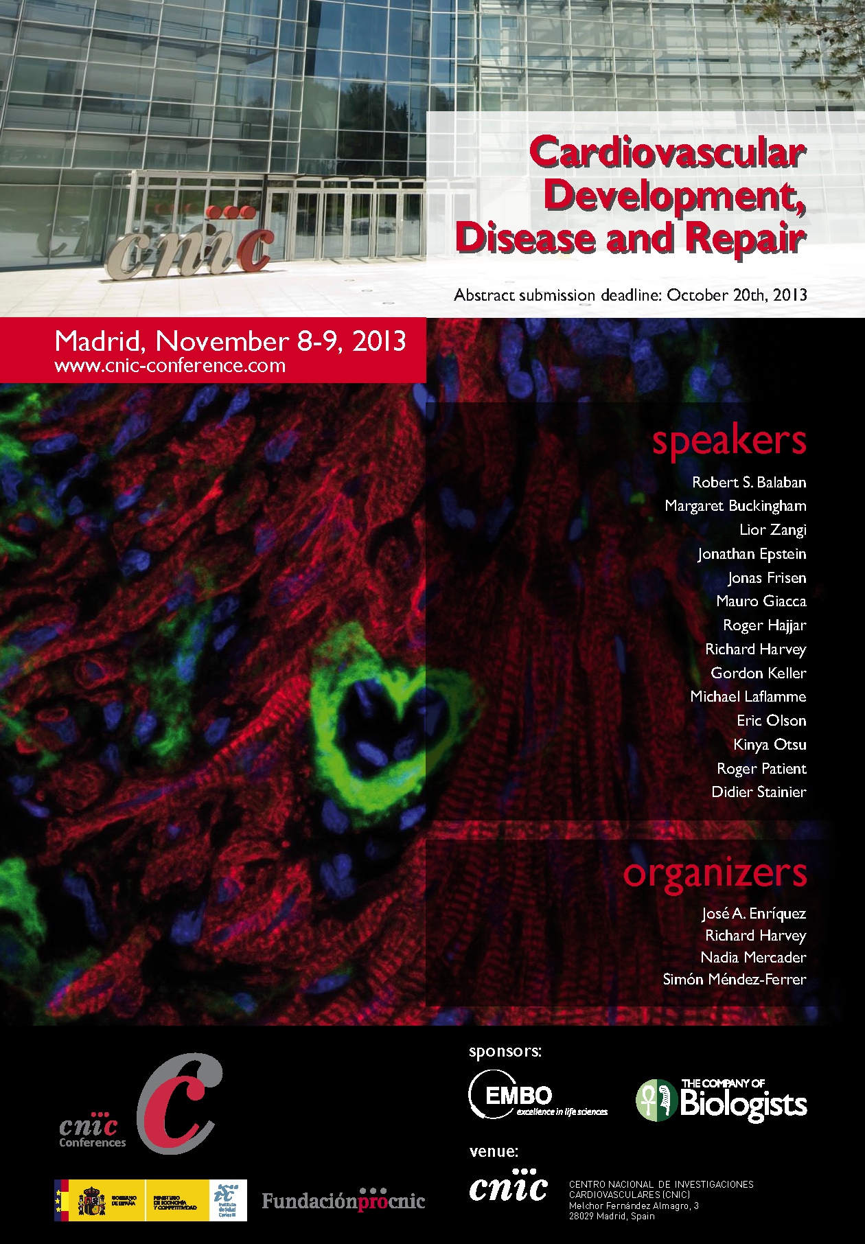
CNIC conference: Cardiovascular Development, Disease and Repair
8-9th November 2013
Centro Nacional de Investigaciones Cardiovasculares CNIC,
Madrid, Spain.
Abstract submission deadline: 20th October 2013
For more information and registration please visit the following webpage:
http://www.cnic-conference.com/
Cardiovascular diseases are the leading cause of death worldwide and their treatment is associated with an enormous economic burden. Cardiovascular diseases impact the whole organism, and as such affect cognitive function and accelerate aging. In recent years it has become increasingly clear that the causes of cardiovascular diseases are multifactorial, involving complex interactions between different genes, cells, organs and the environment. Combinations of gene therapy, cell therapy (including cell reprogramming and reactivation of endogenous progenitor cells) and tissue engineering represent promising strategies for the repair and regeneration of the heart and the vasculature. The development and future clinical application of these approaches requires a thorough understanding of the biology of adult stem cells. Similarly, knowledge of cardiovascular development is crucial for understanding reparative processes, since regeneration recapitulates some aspects of the formation of the embryonic cardiovascular system. Research in model organisms with a high regenerative capacity is providing important insights into the mechanisms controlling cardiovascular regeneration.
In this CNIC Conference we will present and discuss research at the frontiers of stem cell biology, cardiovascular development, regulation and metabolism, and the mechanisms of cardiac repair.
Topics
• Cardiovascular regeneration as a recapitulation of embryonic development.
• Shifting the balance from repair to regeneration.
• Molecular mechanisms of cardiovascular development.
• The emerging role of metabolism in cardiovascular development, homeostasis and repair.
• Origin and fate of cardiovascular progenitor cells.
• The role of stem cells during homeostasis and injury.
• Translating basic research to the clinic: how can basic science impact human cardiovascular health?
 (No Ratings Yet)
 (5 votes)